Spalteholz HANDATLAS DER ANATOMIE DES MENSCHEN VON WERNER SPALTEHOLZ
メニューは解剖学(TA)にリンクしてあります。図の番号をクリックすると下記の説明へ、右側の用語をクリックすると解剖学(TA)にジャンプします。
1028


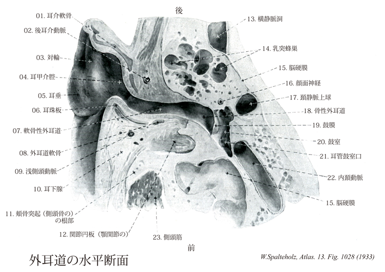
- 1028_00【External acoustic meatus; External auditory meatus外耳道 Meatus acusticus externus】
→(外耳道は側頭骨の鼓室部を耳介から鼓膜へ至る通路で骨性部分。軟骨性外耳道からなる。)
- 1028_01【Auricular cartilage耳介軟骨 Cartilago auriculae】 Supporting framework of the auricle that is composed of elastic cartilage.
→(弾性軟骨よりなり、耳介の骨格構造を支持する。)
- 1028_02【Posterior auricular artery後耳介動脈 Arteria auricularis posterior; Arteria retroauricularis】 Third branch exiting dorsally from the external carotid artery. It runs beneath the parotid gland and over the stylohyoid posterior to the auricle. It also supplies the muscles attached to the mastoid process and styloid process.
→(外頚動脈の背側へ出る第三枝。乳様突起の外側面から耳介の後ろを後上方に向かって走り乳様突起と耳介の間に分布する。)
- 1028_03【Antihelix対輪 Antihelix】 Curved elevation in front of the tail of helix.
→(耳輪後部の前にある弓状の隆起。(Feneis))
- 1028_04【Cavity of concha耳甲介腔 Cavitas conchae; Cavum conchae】 Main part of the concha situated below the crus of helix and behind the tragus.
→(耳輪脚の下方で、耳珠の後方にある耳甲介の主部。 (Feneis))
- 1028_05【Lobule of auricle; Lobe of ear耳垂 Lobulus auricularis】 Noncartilaginous, inferior end of the auricle.
→(耳介の最も低い部分で脂肪と線維性組織からなり、耳介軟骨で補強されていない。)
- 1028_06【Tragal lamina耳珠板 Lamina tragi】 Lateral part of the cartilage of acoustic meatus. It lies in front of the external acoustic pore.
→(外耳道軟骨の外側部。外耳道の外開口部の前方にある。 (Feneis))
- 1028_07【Cartilaginous external acoustic meatus軟骨性外耳道 Meatus acusticus externus cartilagineus】 Lateral, cartilaginous one-third of the external acoustic meatus.
→(外耳道の外側1/3。 (Feneis))
- 1028_08【Cartilage of acoustic meatus外耳道軟骨 Cartilago meatus acustici; Cartilago meatus acustici externi】 Cartilage connected with that of the auricle. It forms a furrow that opens superiorly and posteriorly.
→(耳介の軟骨と関連があり、上方と後方の開いた溝をなす。 (Feneis))
- 1028_09【Superficial temporal artery浅側頭動脈 Arteria temporalis superficialis】 Superficial terminal branch of the external carotid artery. It ascends between the external auditory canal and temporomandibular joint, accompanying the auriculotemporal nerve, anterior to the auricle, to the temporal region where it distributes branches.
→(浅側頭動脈は外頚動脈の終枝。耳介の前を上行枝、頬骨弓の後端の上で皮下にあらわれ、前頭枝と頭頂枝とに分かれ、前頭部から側頭部にわたって分布する。)
- 1028_10【Parotid gland耳下腺 Glandula parotidea; Glandula parotis】 It occupies the retromandibular fossa, extending to the temporomandibular joint and the ramus of mandible.
→(耳下腺はヒト最大の唾液腺で、左右の耳の前下方にあり、下は下顎角まで、上は頬骨弓まで、後方は胸鎖乳突筋まで、内側は側頭下窩の下顎骨下顎枝まで広がっている。その分泌管の耳下腺管によって上顎第2大臼歯の頬粘膜に開口する。終末部(線房)は純漿液性の分泌物からなる(これは他の大唾液腺との大きな違いである)。介在部および線条部もよく発達している。小葉内(腺の実質内)に多数の脂肪細胞が散在するもの、大きな特徴の一つで他の唾液腺と容易に区別できる点である。Parotisという語は、para(傍)とotis(耳)との複合語で、耳の傍らにあるものという意味である。17世紀のフランスの解剖学者リオランの命名である。)
- 1028_11【Zygomatic process of temporal bone頬骨突起(側頭骨の) Processus zygomaticus (Ossis temporale)】 Projection from the temporal bone that contributes to the zygomatic arch.
→(側頭面の下部で外耳孔の前上方にあたる所から前方に向かって長い頬骨突起を出す。この突起の前端は頬骨の側頭突起に達して、やや外方に張り出した頬骨弓を形成する。)
- 1028_12【Articular disc of temporomandibular joint関節円板(顎関節の) Discus articularis temporomandibularis; Discus articulationis temporomandibularis】 Biconcave disc composed of layers of tough fibrous tissue and fibrocartilage located between the head of the mandible and the mandibular fossa. It is attached around its periphery to the joint capsule, dividing the joint into two cavities, and together they form the disco-capsular system, a single functional unit.
→(関節円板は下顎頭のための可動性関節臼の役割を果たしている。この関節円板は前部では散在性の軟骨細胞を含む線維軟骨から成っている。その後部は2層性を示す。下顎窩の後壁に固着する上層は弾性線維を含む粗線線維性結合組織から成るが、下顎頭の後壁に付着している下層は非常に丈夫な密線線維結合組織からなるが、下顎頭の後壁に付着している下層は非常に丈夫な密線線維結合組織からなっている。前方で関節円板は関節包ならびに外側翼突筋と強固に結びついている。一般的には関節円板または関節半月は膠原線維の多い線維軟骨性の結合組織から成っている。円板は関節腔を完全に、半月はそれを不完全に分けている。円板および半月は誘導機能をもち、関節面の接触を良くし、場合によっては、顎関節または胸鎖関節などに見られるように、完全に分かれた2つの関節腔をつくりだすことさえある。このような関節円板は疾患時に、あるいは摘出されても新生が起こりうる。)
- 1028_13【Transverse sinus横静脈洞;横洞 Sinus transversus】 It commences at the confluence of sinuses and passes laterally to the sigmoid sinus.
→(二つの横静脈洞は静脈洞交会から起こり後頭骨の横洞溝の中を、外側に向かってから前方に走る。そして左右のそれぞれの横静脈洞は、後頭骨と側頭骨の岩様部との縫合部でS状静脈洞となって下方に曲がり後方に向かう。横静脈洞には上錐体静脈洞、下大脳静脈、下小脳静脈、板間静脈などが注ぐ。)
- 1028_14【Mastoid cells; *Mastoid air cells乳突蜂巣 Cellulae mastoideae】 Pneumatized cells that, like the tympanic cavity, are lined with squamous or cuboidal epithelium.
→(側頭骨乳様突起内にある多数の小さな相通じている腔。乳様突起洞あるいは鼓室洞に連なる。鼓室と同様、扁平または立方上皮で被われる。)
- 1028_15【Cranial dura mater; Pachymeninx; Dura mater脳硬膜;硬膜 Dura mater cranialis; Dura mater encephali; Pachymeninx; Dura mater】 Membrane forming a protective capsule around the brain. In the growing body it is firmly attached to the periosteum of the cranial bones. The meningeal and periosteal layers always remain divided at the dural venous sinuses. After growth stops, the periosteal layer separates slightly from the bone, remaining firmly attached at only a few sites, e.g., the crista galli.
→(脳硬膜は脊髄硬膜と異なり1枚の膜をなし、脳の被膜であると同時に頭蓋骨内面の骨膜である。これは小児では骨と密着しているが、成人では頭蓋底、頭蓋縫合以外は骨から離れている。脳硬膜は内外の2層からなり、両層は通常癒着しているが、硬膜静脈洞のある所や三叉神経節のある所などでは2層が離れている。脳硬膜の外面は頭蓋骨との間を結合する突起のために粗で、両者の間には不完全に内皮細胞でおおわれたリンパ腔隙があり、これは硬膜上腔と呼ばれる。脳硬膜の内面は平滑で、これとクモ膜との間には連続して内皮細胞で覆われた狭い硬膜下腔がある。脳硬膜は内方に向かって強靱な突起を出して、脳を固定するのを助けている。これには大脳鎌、小脳鎌、小脳テントがある。)
- 1028_16【Facial nerve [VII]顔面神経[脳神経VII] Nervus facialis [VII]】 Nerve arising from the second pharyngeal arch. It emerges from the brain at the pontocerebellar angle between the pons and inferior olive and passes with the vestibulocochlear nerve to the petrous part of the temporal bone, which it exits via the stylomastoid foramen. It supplies the muscles of facial expression.
→(顔面神経は第七脳神経である。狭義の顔面神経と中間神経とを合わせたもので、混合神経である。その主部をなす狭義の顔面神経は運動神経で、起始核たる顔面神経核は延髄上部から橋背部にかけてあり、これから出る神経は橋の後縁で脳を去り、内耳神経とともに内耳道に入り、その底で内耳神経と分かれ、内耳神経と分かれ、顔面神経管孔を経て顔面神経管に入り、間もなく殆ど直角をなして後外側に曲がる。この曲がるところは鼓室前庭窓の後上で顔面神経膝といい、ここに膝神経節がある。ついで弓状に後下方へ走り、茎乳突孔を通って頭蓋底外面に出て耳下腺中に入り、耳下腺神経叢を作った後、つぎつぎに多くの枝を出して広頸筋およびこれから分化したすべての浅頭筋(表情筋)、茎突舌骨筋、顎二腹筋後腹、アブミ骨筋などに分布する。以上の運動神経線維とは別に、膝神経節中の神経細胞から出る味覚神経線維が集まって、舌下腺および顎下腺に至る副交感性の分泌線維とともに中間神経を作り、広義の顔面神経の一部をなす。膝神経節細胞は偽単極性で、神経細胞より出る一条の突起はただちに分かれて、末梢および中枢の2枝となる。中枢枝は顔面神経に密接しつつ内耳道を経て脳に入って孤束核に終わり、末梢枝は、いわゆる上唾液核から出て舌下腺、顎下腺に至る副交感性の分泌腺にとともにいわゆる鼓索神経を作り、途中で再び分泌線維と分かれて舌神経に入り、舌体に分布して味覚を司る。)
- 1028_17【Superior bulb of jugular vein頚静脈上球 Bulbus superior venae jugularis; Bulbus venae jugularis superior】 Dilatation at the beginning of the vein in the jugular foramen.
→(頚静脈孔で静脈が起こる部分での膨大。(Feneis))
- 1028_18【Osseous external acoustic meatus骨性外耳道 Meatus acusticus externus osseus】
→()
- 1028_19Rivinus' membrane【Tympanic membrane鼓膜 Membrana tympanica】 Membrane stretched diagonally at the end of the external acoustic meatus. It has a diameter of 9-11 mm.
→(鼓膜は外耳道と中耳すなわち鼓室との境にある直径約1cmのほぼ卵円形薄い膜。鼓膜は外耳道に対して傾斜し、外面を前下方に向けている。鼓膜の外面は平面でなく、内方に向かってやや陥凹している。鼓膜は生体で耳鏡を外耳道に挿入して観察すると、やや透明で、白色の線条がみられツチ骨柄によってできるツチ骨条といわれる。ツチ骨条の上端には外側突起の部分がツチ骨隆起である。その前後に前ツチ骨ヒダと後ツチ骨ヒダとがあり、緊張部と弛緩部との境界である。緊張部ではその前上方より鼓膜中央まで内面にツチ骨柄が付着するために鼓膜痔帯が内方に向かって漏斗状に陥入し、鼓膜臍を形成する。鼓膜は外側の皮膚面、固有層、内側の粘膜面の三者からなる。皮膚面は外耳道の皮膚のつづきで重層扁平上皮を有する。固有層は線維性結合組織からなり、その線維の走向により内外の2層を区別することができる。粘膜面は鼓室表面の粘膜のつづきであって、単層扁平上皮によりおおわれている。鼓膜の皮膚面には外耳道神経の枝が、また粘膜面には鼓室神経の枝が、それぞれ分布する。)
- 1028_20【Tympanic cavity鼓室 Cavitas tympani; Tympanum】 Cleftlike pneumatized space between the bony labyrinth and the tympanic membrane.
→(側頭骨の錐体の中にあり、外耳道とは鼓膜によって境され、咽頭腔と耳管をもって交通する腔所である。鼓室の中には3個の耳小骨とその付属器があり、これらは鼓膜の振動を内耳に伝える役割を果たす。鼓室は臨床的に故障が起こりやすい場所で、中耳炎の炎症がひろくなると乳突洞を経て乳頭蜂巣へ波及し、または錐体尖の方にも及ぶ。鼓室の各壁(各面)が、どのような構造物に接しているかまとめると:上壁(骨壁を隔てて中頭蓋窩に接する)、下壁(骨壁を隔てて内頚静脈の頚静脈上丘に接する)、前壁(耳管の鼓室口がある)、内側壁(蝸牛の骨壁が岬角を作る)。)
- 1028_21【Tympanic opening of pharyngotympanic tube耳管鼓室口 Ostium tympanicum tubae auditivae; Ostium tympanicum tubae auditoriae】 Opening of the auditory tube in the anterior wall of the tympanic cavity. It usually lies slightly above the floor of the tympanic cavity.
→(耳管の鼓室前壁の開口部。鼓室底よりやや上方である。 (Feneis))
- 1028_22【Internal carotid artery内頚動脈 Arteria carotis interna】 It passes from the carotid bifurcation, without any branches, to the cranial base, continuing in the carotid canal to its terminal division into the middle and anterior cerebral arteries.
→(内頚動脈は、総頚動脈から起こり、頚部では頭蓋底にいたるまでは枝を出さない。ついで頚動脈管をへて中大脳動脈と前大脳動脈に分枝するまでをいう。内頚動脈は頚部、側頭骨錐体部(岩様部)、海綿静脈洞部、大脳部の4つの部分に分けられる。この内頚動脈の海綿静脈洞部と大脳部とは、特別な形態を呈するので、「頚動脈サイフォン」とよばれている。内頚動脈の主な枝として、眼動脈、後交通動脈、前脈絡叢動脈がでる。内頚動脈は、視交叉の外側で小さな前大脳動脈と大きな中大脳動脈とに分岐する。中大脳動脈は内頚動脈の直接の続きで終枝と考えられる。)
- 1028_23【Temporalis muscle; Temporal muscle側頭筋 Musculus temporalis】 o:Inferior temporal line, infratemporal crest, temporal fascia [temporal fossa], i: Its fibers converge at the coronoid process and continue inferiorly to the level of the occlusal plane and near the pterygomandibular raphe. It raises and retracts the mandible, and fixes the pharynx during swallowing. I: Mandibular nerve.
→(側頭筋は扇状になって側頭窩および側頭筋膜から起始する。筋線維は収斂して、頑丈な腱をもって下顎骨筋突起に付着する。付着腱は上方へ伸びて筋肉内にまで達する。側頭筋は頬骨弓下を通過して、その付着部に達する。その筋線維がかなりの長さであるので、筋はかなりの収縮可能性を有するし、かつ純粋な“咬むための筋”である。歯をかみ合わせると、側頭筋の収縮を耳介の上方で触れることができる。側頭部をコメカミというのは、コメをカムときに動くからである。)