Spalteholz HANDATLAS DER ANATOMIE DES MENSCHEN VON WERNER SPALTEHOLZ
メニューは解剖学(TA)にリンクしてあります。図の番号をクリックすると下記の説明へ、右側の用語をクリックすると解剖学(TA)にジャンプします。
339


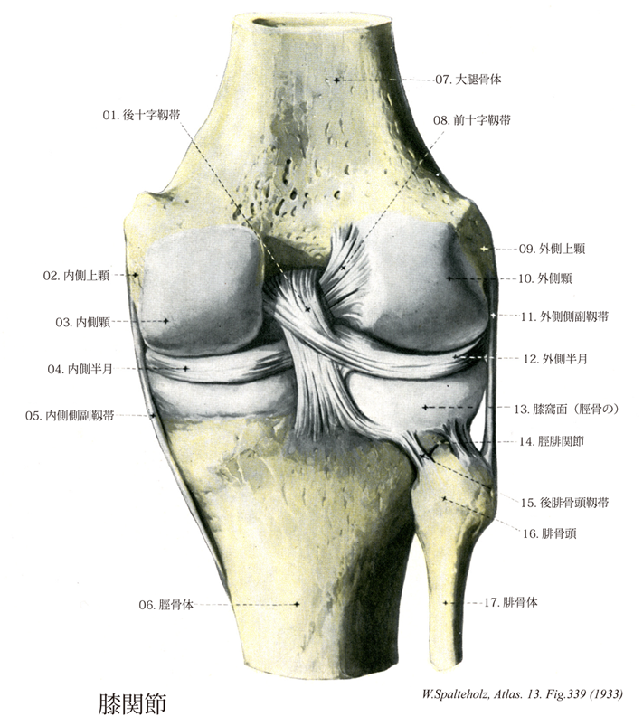
- 339_00【Knee joint膝関節 Articulatio genus】
→(膝関節は大腿骨下端の内側顆および外側顆と、脛骨上面の同名部分との間の関節で、これに関節包の前壁にある膝蓋骨が構成に加わる。腓骨は関与しない。脛骨上面の関節面には、内側顆と外側顆の表面に線維軟骨の関節円板があって、大腿骨下端の関節面に対する。関節円板は周辺が厚く、中心部は薄いから、断面ではクサビ形を呈している。内側半月は半円形であるが、外側半月はほぼ完全な円形で内側半月に比べて小さい。膝関節は屈伸運動のみを行う蝶番関節とみなさえるが、膝を曲げた状態では下腿の内旋(10°)、外施(40°)が可能である。また膝を十分に伸ばすとき、その最終段階では下腿のわずかな外施(5°)がみられ(これを終末回旋という)、この状態から再び膝をまげるときには、その最初の段階として下腿の内旋がおこなわれる。この意味で純粋な蝶番関節ではない。直立位(膝を伸ばした状態)では、大腿骨の内側顆と外側顆は、それぞれ下面の比較的平面的な部分で広く脛骨に接するが、膝をまげたときには、大腿骨の内側顆、外側顆の後面にある弯曲の強い局面によって脛骨に接する。膝蓋骨の関節面は、この屈伸に際して大腿骨下端の前面にある膝蓋面を上下に移動する。関節内には、膝蓋骨の下端から大腿骨の顆間窩に向かって滑膜のヒダが前後に走り、これを膝蓋下滑膜ヒダという。このヒダから内外両側に向かって内部に脂肪組織(膝蓋下脂肪体という)を含んだ滑膜のヒダがのびて関節腔のすきまをみたしている。これを翼状ヒダという。関節の付属靱帯として次のものがある。(1)膝十字靱帯:関節腔内のほぼ中央でX字状に交差する二つの強力な靱帯で、脛骨に対する付着部の位置的関係によってそれぞれ前十字靱帯、後十字靱帯という。前者は脛骨の前顆間区anterior intercodylar areaに付着し、膝関節腔内を上後外側方に走り、大腿骨の外側顆内面後部に付着する。この靱帯は膝関節屈曲時にゆるみ、膝関節完全伸展時に緊張する。前十字靱帯は、脛骨上で大腿骨が後方に偏位するのを防ぐ。膝関節屈曲時における前十字靱帯は、脛骨上端が前に倒れようとするのを妨げる。後者は脛骨の後顆管区posterior intercondylar areaに付着し、膝関節腔内を上前内側方に走り、大腿骨の内側顆外側面前部に付着する。この靱帯の前線維はは、膝関節伸展時にゆるみ、膝関節屈曲時に緊張する。また、この靱帯の後線維は膝関節伸展時にゆるみ、膝関節屈曲時に緊張する。後十字靱帯は、脛骨上で大腿骨が前方に変異するのを防ぐ。膝関節時における後十字靱帯は、脛骨上端が後ろに倒れようとするのを妨げる。(2)前半月大腿靱帯・後半月大腿靱帯:外側半月の脛骨上面における後端部から出て上外方に走り、後十字靱帯のすぐ後方で大腿骨内側顆の外面につく強い線維束が後半月大腿靱帯で一名Wrisbergの靱帯という。一方、同じく外側半月の後端部から出て前方に走り、前十字靱帯の外側部につく弱い線維束を前半月大腿靱帯という。これは欠如することがしばしばある。(3)膝横靱帯:強さに個体差が大きい。両関節半月の前面を結んで脛骨上面の前端部を横走する。(4)斜膝窩靱帯:半膜様筋の停止腱からつづいて関節包の後面を上外方へ走り、大腿骨外側顆の後面の付近へ放散する。(5)弓状膝窩靱帯:脛骨頭よりおこり、関節包の後面を上内方へ向かって膝窩筋の起始部の表層をおおう。明瞭でないこともある。(6)外側側副靱帯:強い棒状の線維束で、大腿骨の外側顆よりおこり、関節包の外側を下方に走って腓骨頭へつく。この靱帯と関節包との間にはせまい隙間があって、ここを外側下膝動脈が通る。(7)内側側副靱帯:内側半月の表層に接して関節包を補強する幅の広い薄い靱帯で、大腿骨内側顆と脛骨の内側顆を結ぶ。外側側副靱帯とともに蝶番関節に特徴的な縦走靱帯であるが、膝を伸ばした状態では緊張して関節の小手に役立ち、膝をまげた状態では弛緩して、下腿の回旋を可能にする。(8)膝蓋靱帯:本体は大腿四頭筋の停止腱ともみなすべきもので、膝蓋骨の下端から脛骨粗面にのびる。強い扁平な靱帯で長さ約8cm、幅は膝蓋骨下端の起始部で約3cmで、膝関節方の前面下部を補強する。表層の線維は膝蓋骨の前面をこえて大腿四頭筋の腱からつづく。(9)内側膝蓋靱帯・外側膝蓋支帯:大腿四頭筋停止腱のうち、膝蓋骨を介して膝蓋靱帯となる中央の部分を除いてその両側の部分をいう。これは膝蓋骨の両側を通って下方にのび、脛骨粗面の両側で脛骨上端部につく。内外両側から膝蓋骨を支えて、屈伸運動にあたって膝蓋骨の左右への動揺を防ぐ。)
- 339_01【Posterior cruciate ligament後十字靱帯;後交叉靱帯 Ligamentum cruciatum posterius; Ligamentum decussatum posterius】 Band that extends posteriorly from the posterior intercondylar area to anterosuperior to the medial aspect of the medial femoral condyle.
→(後十字靱帯は前十字靱帯に反して脛骨後顆間区の後のうちから起こり、外側半月から補助線維を受けて斜めに前内方に上り、前十字靱帯の後側を通ってやや拡がりながら大腿骨内側顆の顆間窩に向かう面の前部で軟骨との境に至る。後十字靱帯の方が垂直に近い走行を示す。)
- 339_02【Medial epicondyle of femur内側上顆;脛側上顆(大腿骨の) Epicondylus medialis; Epicondylus tibialis】 Bony prominence on the medial aspect of the medial condyle.
→(内側顆の内側面には輪郭がやや不明瞭な内側上顆が突出する。)
- 339_03【Medial condyle of femur内側顆;脛側顆(大腿骨の) Condylus medialis; Condyus tibialis】 Rounded projection on the medial aspect of the femur forming part of the knee joint.
→(大腿骨体の下部(遠位部)はことに著しく厚く大きく、その下端は左右の肥厚した内側顆および外側顆となる。内側顆は狭く長く、凸面の張り出しが強い。)
- 339_04【Medial meniscus内側半月;脛側半月(膝関節の) Meniscus medialis; Meniscus tibialis】 The crescent-shaped medial meniscus lies beneath the medial femoral condyle and is attached to the tibial collateral ligament. It is highly susceptible to injury.
→(内側半月は半月状で内側側副靱帯と癒着している。その付着部は比較的たがいに離れている。この半月は前よりも後の方が広い。つまり、前脚は後脚よりも狭い。内側半月はその付着状態によって外側半月よりもはるかに可動性が少ない。下腿の外旋のさい、内側半月はもっとも強くずれ動き、無理にひっぱられる。しかし内旋時にはこの半月は負荷を免れる。)
- 339_05【Tibial collateral ligament内側側副靱帯;脛側側副靱帯 Ligamentum collaterale tibiale】 Medial collateral ligament that extends from the medial femoral epicondyle to the tibia. It is attached to the joint capsule and the medial meniscus.
→(内側側副靱帯は内側上顆から起こり、脛骨内側顆の内側縁と後縁に着き、また内側半月の周縁に強固につながる。この側副靱帯は強力で幅広いが外側側副靱帯よりも弱く激しくぶつかる競技においてよく断裂する。)
- 339_06【Shaft of tibia; Body of tibia脛骨体;脛骨幹 Corpus tibiae】
→(脛骨体は脛骨の近位端と遠位端の間にある断面がほぼ三角柱状の骨幹部で、下3分の2あたりがもっとも細くなっており(脛骨骨折の起こりやすい部位)、少し外方、すなわち腓骨の側に近づき、全体としてごくかるいS状の曲がりを示す。これに前内外の3縁と内外後の3面がある。)
- 339_07【Shaft of femur; Body of femur大腿骨体;大腿骨幹 Corpus femoris】
→(大腿骨体は長い柱状の骨幹をなす部分で、前方に軽く凸弯している。その中央4分の2はほぼ円柱状であるが、上4分の1と下4分の1は楕円柱状に近い。下4分の1は下方にいくにしたがって幅が広くなっている。)
- 339_08【Anterior cruciate ligament前十字靱帯;前交叉靱帯 Ligamentum cruciatum anterius; Ligamentum decussatum anerius】 Band that extends from the anterior intercondylar area superiorly and posteriorly to the medial aspect of the lateral femoral condyle.
→(大腿骨および脛骨の相対する関節面は、関節腔内を上下に走る強い2靱帯によって固く結ばれる。これらはほぼ十字形に交わり、全体として膝十字靱帯と呼ばれ、その前方のものを前十字靱帯、後方のものは後十字靱帯である。前十字靱帯は脛骨の前顆間区の内側部(内側顆の上関節面に接して)から起こり、後外方に上ってやや拡がりながら大腿骨外側顆の顆間窩に向かう面の後部で軟骨との境に着く。大腿骨に対して脛骨が前方へすべり出るのを防いでいる。)
- 339_09【Lateral epicondyle of femur外側上顆;腓側上顆(大腿骨の) Epicondylus lateralis; Epicondylus fibularis】 Bony prominence on the lateral aspect of the lateral condyle.
→(外側顆の外側面には外側上顆が突隆し、ここに粗線が外側唇の下方への延長線が終わる。膝窩筋が起こる。)
- 339_10【Lateral condyle of femur外側顆;腓側顆(大腿骨の) Condylus lateralis; Condylus fibularis】 Rounded projection on the lateral aspect of the femur.
→(外側顆は幅が広くやや平坦で、その前端は少し前上方に突出する。)
- 339_11【Fibular collateral ligament of knee joint外側側副靱帯;腓側側副靱帯(膝関節の) Ligamentum collaterale fibulare】 Lateral collateral ligament that extends from the lateral femoral epicondyle to the head of the fibula. It lacks secure attachment to the joint capsule and meniscus.
→(外側側副靱帯は外側上顆より起こる円柱形の線維束で、下端は腓骨頭に着くため、下部は関節包から離れている。(この間隙を膝窩筋腱、大腿二頭筋腱の一部が通る)。)
- 339_12【Lateral meniscus外側半月;腓側半月(膝関節の) Meniscus lateralis; Meniscus fibularis】 Nearly circular ring below the lateral femoral condyle with close-together attachments. It is not anchored to the fibular collateral ligament and thus is relatively mobile.
→(外側半月はほぼ環状を呈する。その付着部はたがいに近接しており、半月の幅はだいたい同じである。外側半月は内側半月よりも可動性が大きい。その前方端の脛骨への付着部は顆管隆起intercondylar eminenceの直前にあたる前顆間区の部分にあり、後方端の脛骨への付着部は顆管隆起の直後にあたる後顆管区の部分にある。外側半月の後方端から小線維束が出て、後十字靱帯に沿って大腿骨の内側顆にまで達するのが普通である。外側半月の辺縁部は膝関節の外側側副靱帯から、膝窩筋腱で隔てられており(膝窩筋腱の一部が外側半月の辺縁部から起こる)、このために外側半月の基部の機械力に対する安定性は比較的乏しい状態となる。)
- 339_13【Popliteal surface of tibia膝窩面(脛骨の) Facies poplitea; Planum popliteum (Tibia)】
→(")
- 339_14【Tibiofibular joint; Superior tibiofibular joint脛腓関節;脛腓連結 Articulatio tibiofibularis; Juctura tibiofibularis】 Articulation between the head of the fibula and the lateral condyle of the tibia.
→(脛骨の外側顆と腓骨頭との間の平面関節。関節面は小さい卵円形で、ほとんど動きはない。関節包の外面には次の靱帯がある。(1)前腓骨頭靱帯:腓骨頭より脛骨外側顆の前面へ。(2)後腓骨頭靱帯:腓骨頭より脛骨外側顆の後面へ。)
- 339_15【Posterior ligament of fibular head後腓骨頭靱帯 Ligamentum capitis fibulae posterius】 Weaker group of fibers passing posteriorly from the head of the fibula to the tibia. See 20 for function.
→(腓骨頭から後側を脛骨へ張る弱い線維群。)
- 339_16【Head of fibula腓骨頭;腓骨小頭 Caput fibulae; Capitulum fibulae】 Proximal end of the fibula.
→(腓骨の上端の膨らみは腓骨頭と呼ばる。腓骨頭の前面から長趾伸筋、長腓骨筋、後面からヒラメ筋の一部が起こり、外側面に大腿二頭筋が付着く。)
- 339_17【Shaft of fibula; Body of fibula腓骨体;腓骨幹 Corpus fibulae】
→(腓骨体は腓骨全長の大部分を占める棒状の部分。細長い三角柱状で、かるくねじれている。前縁、後縁、骨間縁があって、それにより3面が区切られる。腓骨体は下腿の筋の中に埋まっているので、それぞれの面はそれらの筋の付くところであり、面と面を区切る縁は、いずれも下腿の筋、ないし筋群を分ける結合組織性の膜が着くところである。したがって、各筋の発達の程度により、縁の強さや面の大きさ、また、ねじれの強さにかなりの個人差がある。)