Spalteholz HANDATLAS DER ANATOMIE DES MENSCHEN VON WERNER SPALTEHOLZ
メニューは解剖学(TA)にリンクしてあります。図の番号をクリックすると下記の説明へ、右側の用語をクリックすると解剖学(TA)にジャンプします。
519 


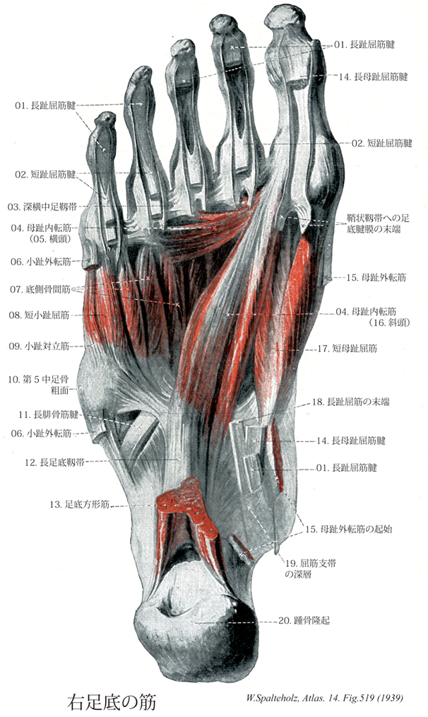
- 519_01【Flexor digitorum longus tendon長趾屈筋腱 Tendo musculus flexor digitorum longus】
→()
- 519_02【Flexor digitorum brevis tendon短趾屈筋腱 Tendo musculus flexor digitorum brevis】
→()
- 519_03【Deep transverse metatarsal ligament深横中足靱帯;横中足骨小頭靱帯 Ligamentum metatarsale transversum profundum; Ligamenta capitulorum ossium metatarsi transversa】 Band that extends horizontally, connecting the heads of the metatarsals.
→(深横中足靱帯は各中足骨頭を横に結ぶ靱帯で、前方では底側靱帯と癒合する。足底面は溝状にくぼみ、ここを趾屈筋の腱が通る。またこの靱帯の背側を骨間筋が通り、底側を虫様筋が通る。)
- 519_04【Adductor hallucis muscle母趾内転筋;母指内転筋(足の) Musculus adductor hallucis】 Muscle comprised of the following two heads. Supports the arch of the foot. Plantar flexion of proximal phalanx. Adduction of great toe. I: Lateral plantar nerve.
→(母趾内転筋の斜頭は立方骨、外側楔状骨、深靱帯および第2~4中足骨底から起こる。横頭は第3~5中足趾節関節および深横中足靱帯から起こる。これら2頭の総停止腱は外側種子骨を介して中足指節関節包および母趾基節骨に付く。母趾内転筋は長趾屈筋と短趾屈筋の腱によってほとんど完全におおわれている。母趾の筋を容れる部にあるのは停止と斜頭内側縁部にすぎない。母趾内転筋は外側足底神経の深枝による支配を受ける。母趾内転筋の斜頭の収縮により中足趾節関節における第1趾の屈曲(短母趾屈筋の作用を助ける)が得られる。母趾内転筋の横頭は中足骨群を寄せ集める作用を示し、横足弓の維持のうえでの重要な役割を演じる。)
- 519_05【Transverse head of adductor hallucis muscle横頭(母趾内転筋の) Caput transversum (Musculus adductor hallucis)】 o: Joint capsules of third to fifth metatarsophalangeal joints, i: Together with the transverse head on the lateral sesamoid bone and proximal phalanx of great toe. Mainly supports transverse arch of foot.
→(母趾内転筋の横頭は第二~五趾の基節関節の関節包に起こり、外側種子骨に停止する。とくに横方向に足弓を保持する。)
- 519_06【Abductor diditi minimi muscle of foot小趾外転筋;小指外転筋(足の) Musculus abductor digiti minimi pedis】 o:Pisiform, flexor retinaculum. i: Proximal phalanx of little finger. Abduction. I: Ulnar nerve.
→(小趾外転筋は踵骨の足底面、特に踵骨隆起の外側突起、足底腱膜および第5中足骨粗面から起こる。その停止は第5の基節骨底に停止する。外側足底神経の支配を受ける。この筋は体重を支えない下肢においては第5趾を屈曲、外転させる作用を示し、足に体重がかかる場合には外側縦足弓を上方に引き、外側縦足弓を維持するのに役立つ。)
- 519_07【Plantar interosseous muscles底側骨間筋 Musculi interossei plantares】 o: Muscle arising from a single head on third through fifth metatarsals. i: Bases of proximal phalanges. Adduction and flexion at metatarsophalangeal joints. I: Lateral plantar nerve.
→(3つの底側骨間筋は第3~5中足骨底側面と長足底靱帯から起こり、第3~5趾基節骨底内側面へ付着する。普通、指背腱膜には達しない。外側足底神経の支配をうけるこれらの筋の収縮により、足の第2趾に向かうような各趾の内転、各趾の中足趾節関節の屈曲、および趾節間関節の伸展が得られる。)
- 519_08【Flexor digiti minimi brevis muscle of foot短小趾屈筋;短小指屈筋(足の) Musculus flexor digiti minimi brevis pedis】 o: Base of fifth metatarsal and long plantar ligament, i: Proximal phalanx of little toe. Flexion and abduction of little toe. 1: Lateral plantar nerve.
→(短小趾屈筋と小趾対立筋は第5中足骨底、長足底靱帯および長腓骨筋腱鞘から共通腱をもって起こる。小趾の短屈筋は第5趾の基節骨底に、小趾の対立筋は第5中足骨外側面に停止する。人では対立筋は短小趾屈筋の弱い分束としてしか出現せず、その停止部でしか同定できない。外側足底神経の浅枝による支配を受け、中足趾節関節で第5趾を屈曲させる作用を示す。)
- 519_09【Opponens digiti minimi of foot小趾対立筋;小指対立筋(足の) Musculus opponens digiti minimi pedis】 Part of the flexor digiti minimi brevis that is occasionally present.o:Distal half of fifth metatarsal.
→(短小趾屈筋と小趾対立筋は第5中足骨底、長足底靱帯および長腓骨筋腱鞘から共通腱をもって起こる。小趾の短屈筋は第5趾の基節骨底に、小趾の対立筋は第5中足骨外側面に停止する。人では対立筋は短小趾屈筋の弱い分束としてしか出現せず、その停止部でしか同定できない。)
- 519_10【Tuberosity of fifth metatarsal bone [V]第5中足骨粗面 Tuberositas ossis metatarsi quinti [V]】 Bony protuberance on the lateral aspect of the base of the fifth metatarsal. Attachment site of the fibularis brevis muscle.
→(第5中足骨底の外側には第5中足骨粗面(短腓骨筋の着くところ)があり、体表から骨の突起として触れる。これが独立した小骨(Vesaliusの骨)となることがある。)
- 519_11【Fibularis (peroneus) longus tendon長腓骨筋腱 Tendo musculus peroneus longus; Tendo musculus fibularis longus】
→()
- 519_12【Long plantar ligament長足底靱帯 Ligamentum plantare longum】 Firm band that passes from the calcaneus just anterior to the calcaneal tuberosity to the cuboid and the bases of metatarsals II-V. It supports the longitudinal arch of the foot.
→(長足底靱帯は足底の靱帯のうち最も表層にありまた最も長い。そのほかの底側足根靱帯はこれにより下方から被われ、その間に粗な結合組織が介在する。距骨隆起の下面から起こって前方に広がり、その深層の線維は立方骨の長腓骨筋腱溝の後の立方骨粗面に着く。浅層の線維は同腱の表面を越えて少なくも3束に分かれて中足骨底に着く。)
- 519_13【Quadratus plantae muscle; Flexor accessorius muscle足底方形筋;副趾屈筋 Musculus quadratus plantae; Musculus flexor accessorius】 o: Calcaneus. i: Lateral border of tendon of flexor digitorum longus. Toe flexion and support of longitudinal arch of foot. I: Lateral plantar nerve.
→(足底方形筋は踵骨底側面に起こり、長趾屈筋の腱に停止する。同筋は副趾屈筋とも呼ばれるが、それは長趾屈筋の停止腱が趾を引く方向を矯正するからである(趾の底屈時)。外側部の趾へ行く腱は、線維性の腱鞘によって長軸方向に固定される前に、中足骨上を斜走する。この腱の斜走は足底方形筋の索引によって中足骨長軸に沿った方向となる。外側足底神経の支配を受ける。この筋の収縮により長趾屈筋腱は後方へ引っ張られるために、第2~5趾の屈曲が得られる。 踵骨からおこって長母趾屈筋腱に停止し、その補助に働く。神経支配:外側足底神経。(イラスト解剖学))
- 519_14【Flexor hallucis longus tendon長母趾屈筋腱 Tendo flexor hallucis longus; Tendo musculus flexor hallucis longus】
→()
- 519_15【Abductor hallucis muscle母趾外転筋;母指外転筋(足の) Musculus abductor hallucis】 o: Calcaneal tuberosity. i: Medial sesamoid bone and proximal phalanx of great toe. Medial abduction, supports longitudinal arch of foot. I: Medial plantar nerve.
→(母趾外転筋は踵骨隆起の内側突起、屈筋支帯および足底腱膜から起始する。腱となり内側種子骨を介して母趾の基節骨底内側面および短母趾屈筋の内側腱に停止する。内側足底神経の支配を受ける。この筋の収縮は母趾の屈筋と外転とをもたらす(体重を支えていない下肢の場合)。また、体重を支えている下肢においては、この筋の収縮が内側縦足弓の維持に役立つ。 )
- 519_16【Oblique head of adductor hallucis斜頭(母趾内転筋の) Caput obliquum (Musculus adductor hallucis)】 o: Second to fourth metatarsals, lateral cuneiform bone and cuboid, i: Together with the transverse head on the lateral sesamoid bone and proximal phalanx of great toe.
→(母趾内転筋の斜頭は第二~四中足骨、外側楔状骨および立方骨に起こり、横頭と友に、外側種子骨および第一趾基節骨に停止する。横方向および縦方向の足弓の保持に重要。)
- 519_17【Flexor hallucis brevis muscle短母趾屈筋;短母指屈筋(足の) Musculus flexor hallucis brevis】 o: Cuneiform (I), long plantar ligament, tendon of tibialis posterior, plantar aponeurosis. Forms the groove for the flexor hallucis longus. Plantar flexion of great toe. I: Medial plantar nerve.
→(短母趾屈筋は楔状骨、底側踵立方靱帯および後脛骨筋の腱から起始する。その内側頭は母趾外転筋の腱とともに内側種子骨を介して中足指節関節に停止する。その外側頭は母趾内転筋の腱とともに外側種子骨を介して基節骨に停止する。短母趾屈筋は内側足底神経の支配を受ける。この筋の収縮により第1趾の中足趾節関節における屈曲が得られる。また、この筋は内側縦束裂を維持する役割も果たす。)
- 519_18【Flexor digitorum longus muscle長趾屈筋;長指屈筋(足の) Musculus flexor digitorum longus】 o: Tibia, i: Distal phalanges of the second through fifth toes. Plantar flexion, supination, flexion of toes. I: Tibial nerve.
→(長趾屈筋はヒラメ筋線より遠位の脛骨後面およびヒラメ筋腱弓の一部から起こる。その停止腱は基節骨の領域で短趾屈筋の腱を貫通し(第2~5)趾の末節骨に停止する。長趾屈筋の腱は腱鞘に包まれて、内果溝を後脛骨筋腱の背外側に走り、屈筋死体の下を載距突起内側縁に沿って足底に至る。舟状骨粗面のレベルでは長母趾屈筋腱の浅層を通る。この際、長母趾屈筋の健束が長趾屈筋の腱に混じる。この腱交叉位遠では足底方形筋が長趾屈筋の腱に付く。この付加的な屈筋は長趾屈筋停止腱の牽引方向を趾放線の長軸方向と関連させる。同一趾へ向かう長趾屈筋(「貫通筋」と短趾屈筋「被貫通筋」)の停止腱は腱鞘(滑液鞘に包まれる。腱鞘は第1中足骨頭の上方から始まり、末節骨までのびている。これらの滑液鞘は線維鞘に包まれる。線維鞘は手指におけると同じように横走線維と交織する線維(輪状および十字部)からなる。)
- 519_19【Flexor retinaculum of foot屈筋支帯[足の];破裂靱帯 Retinaculum musculorum flexorum pedis; Ligamentum laciniatum】 Multilayered band situated over the long flexor tendons passing from the medial malleolus to the calcaneus. Its superficial portion invests the tibial nerve and posterior tibial artery and veins. Its deep portion forms an osteofascial canal with compartments containing the posterior tibial flexor muscles, flexor digitorum longus, and flexor hallucis longus.
→(足の屈筋支帯は下腿筋膜の厚くなったもので、内果の下部から扇状に広がって、前部は舟状骨に後部は踵骨につき中間部は足底腱膜に移行する。屈筋支帯は後脛骨筋と長趾屈筋の腱を被い、またその間の隔壁を骨に送ったのち、載距突起についてさらに長母指屈筋腱溝を被う深葉と、脛骨神経および後脛骨動静脈を被う浅葉とに分かれる。)
- 519_20【Calcaneal tuberosity踵骨隆起 Tuber calcanei】 Tuberosity on the posterior aspect of the calcaneus.
→(踵骨の後半部は大きな骨塊となって後方に飛び出している。この部分は踵骨隆起と呼ばれ、いわゆるかかとの主要部を成している。その後面には表面にギザギザした稜線が横に走っているが、ここはアキレス腱がつく場所である。)