Spalteholz HANDATLAS DER ANATOMIE DES MENSCHEN VON WERNER SPALTEHOLZ
メニューは解剖学(TA)にリンクしてあります。図の番号をクリックすると下記の説明へ、右側の用語をクリックすると解剖学(TA)にジャンプします。
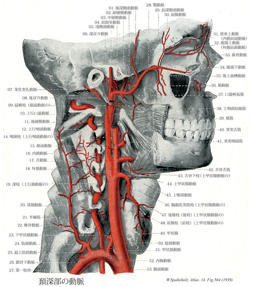
564 


- 564_01【Posterior deep temporal artery後深側頭動脈 Arteria temporalis profunda posterior】 Branch ascending in the temporal fossa to the temporal muscle.
→()
- 564_02【Accessory branch of middle meningeal artery; Accessory meningeal artery中硬膜動脈の副硬膜枝;副硬膜動脈 Ramus accessorius (Arteria meningea media); Ramus meningicus accessorius】 Accessory branch arising from the meningeal or maxillary artery that supplies surrounding muscles and the auditory tube. It sometimes passes through the foramen ovale to the middle cranial fossa, supplying the dura mater up to the trigeminal ganglion.
→(中硬膜動脈の副硬膜枝は側頭下窩で中硬膜動脈または上顎動脈から分枝し、上行して卵円孔を通り抜け三叉神経節、硬膜、骨膜、骨内面に分布する。)
- 564_03【Middle meningeal artery中硬膜動脈 Arteria meningea media】 Artery passing medial to the lateral pterygoid and through the foramen spinosum into the middle cranial fossa, where it distributes vessels between the dura mater and bone.
→(中硬膜動脈は顎動脈より起こり外側翼突筋の内側を通り棘孔から中頭蓋窩に入り、そこで岩様部枝、腹硬膜枝、上鼓室動脈、前頭枝、頭頂枝に分枝する。上記の部位と終末枝を通って前頭蓋窩と中頭蓋窩に分布し、後頭動脈の硬膜枝、上行咽頭動脈、眼動脈、涙腺動脈、茎乳突孔動脈、顎動脈の腹硬膜枝、深側頭動脈と吻合する。)
- 564_04【Anterior tympanic artery前鼓室動脈 Arteria tympanica anterior】 Artery accompanying the chorda tympani through the petrotympanic fissure into the tympanic cavity.
→(前鼓室動脈は顎動脈の基部より起こり、中耳に分布する。内頚動脈・上行咽頭動脈の鼓室枝と吻合する。)
- 564_05【Superficial temporal artery浅側頭動脈 Arteria temporalis superficialis】 Superficial terminal branch of the external carotid artery. It ascends between the external auditory canal and temporomandibular joint, accompanying the auriculotemporal nerve, anterior to the auricle, to the temporal region where it distributes branches.
→(浅側頭動脈は外頚動脈の終枝。耳介の前を上行枝、頬骨弓の後端の上で皮下にあらわれ、前頭枝と頭頂枝とに分かれ、前頭部から側頭部にわたって分布する。)
- 564_06【Deep auricular artery深耳介動脈 Arteria auricularis profunda】 Artery passing backward and upward to the temporomandibular joint, external auditory canal, tympanic membrane, and mucosa of the tympanic cavity.
→(深耳介動脈は顎動脈より起こり、顎関節、耳下腺、外耳道、鼓膜外面に分布する。浅側頭動脈、後耳介動脈の耳介枝と吻合する。)
- 564_07【Stylomastoid artery茎乳突孔動脈 Arteria stylomastoidea】 Thin vessel accompanying the facial artery. It runs with the facial artery from the stylomastoid foramen to the hiatus for greater petrosal nerve, where it supplies the dura mater. Before reaching the hiatus, it distributes branches to the middle and inner ear.
→(茎乳突孔動脈は後耳介動脈より起こり、外耳道、乳突蜂巣、半規管、アブミ骨筋、前庭に分布する。内頚動脈・上行咽頭動脈の鼓室枝、迷路動脈と吻合する。)
- 564_08【Posterior auricular artery後耳介動脈 Arteria auricularis posterior; Arteria retroauricularis】 Third branch exiting dorsally from the external carotid artery. It runs beneath the parotid gland and over the stylohyoid posterior to the auricle. It also supplies the muscles attached to the mastoid process and styloid process.
→(外頚動脈の背側へ出る第三枝。乳様突起の外側面から耳介の後ろを後上方に向かって走り乳様突起と耳介の間に分布する。)
- 564_09【Tonsillar branch of facial artery扁桃枝(顔面動脈の) Ramus tonsillaris (Arteria facialis)】 Branch frequently arising from the ascending palatine artery. It penetrates the wall of the pharynx and supplies the palatine tonsil and posterior part of tongue.
→(扁桃枝はしばしば上行口蓋動脈よりでて口蓋扁桃への主たる血液を供給し、他の扁桃枝とも広範囲に吻合している。)
- 564_10【Ascending palatine artery上行口蓋動脈 Arteria palatina ascendens】 It ascends on the lateral wall of the pharynx beneath the styloglossus to the palatoglossal and palatopharyngeal arches, soft palate, and palatine tonsil. It can replace or be replaced by the ascending pharyngeal artery.
→(上行口蓋動脈は顔面動脈より起こり、咽頭外側壁、口蓋扁桃、耳管、軟口蓋に分布する。顔面動脈の扁桃枝、舌背動脈、下行口蓋動脈と吻合する。)
- 564_11【Posterior meningeal artery後硬膜動脈;後頭硬膜動脈 Arteria meningea posterior; Arteria meningea occipitalis】 Artery lying lateral to the internal carotid artery. It usually passes through the jugular foramen to the dura mater and diploe of the posterior cranial fossa.
→(後硬膜動脈は上行咽頭動脈より起こり、後頭蓋窩の硬膜に分布する。中硬膜動脈、椎骨動脈の枝と吻合する。)
- 564_12【Ascending pharyngeal artery上行咽頭動脈 Arteria pharyngea ascendens】 It usually arises from the posterior side of the external carotid artery above the superior thyroid artery. It ascends along the lateral wall of the pharynx, passing medial to the stylohyoid and continuing to the cranial base.
→(上行咽頭動脈は外頚動脈より起こり、咽頭と茎状突起の筋の間を頭蓋底まで上行し咽頭壁、軟口蓋後頭窩に分布する。)
- 564_14【Pharyngeal branches of ascending pharyngeal artery咽頭枝(上行咽頭動脈の) Rami pharyngeales (Arteria pharyngea ascendens)】 Branches supplying the wall of the pharynx. They occasionally pass to the auditory tube and palatine tonsil.
→(上行咽頭動脈の咽頭枝は口腔咽頭・鼻咽頭の壁に分布する。)
- 564_15【Facial artery顔面動脈 Arteria facialis】 Third anterior branch of the external carotid artery. It lies behind the posterior belly of digastric muscle, stylohyoid, and submandibular gland. It crosses the mandible along the anterior border of the masseter and supplies the muscles of facial expression.
→(顔面動脈は舌動脈のやや上方で、外頚動脈の前側から起こり、下顎角の内側で顎下腺の上面を前方に走り、化学体の下縁をまわって顔面に現れる。顔面に出ると、蛇行しながら口角を経て鼻の側縁に沿って上場し内眼角(メガシラ)に至る。顔面動脈が下顎骨の下縁をまたがって顔面に出るところで体表から脈動を触れる。この部位は咬筋の前縁(歯を強くかみ合わせると触れる)にあたる。)
- 564_16【Internal carotid artery内頚動脈 Arteria carotis interna】 It passes from the carotid bifurcation, without any branches, to the cranial base, continuing in the carotid canal to its terminal division into the middle and anterior cerebral arteries.
→(内頚動脈は、総頚動脈から起こり、頚部では頭蓋底にいたるまでは枝を出さない。ついで頚動脈管をへて中大脳動脈と前大脳動脈に分枝するまでをいう。内頚動脈は頚部、側頭骨錐体部(岩様部)、海綿静脈洞部、大脳部の4つの部分に分けられる。この内頚動脈の海綿静脈洞部と大脳部とは、特別な形態を呈するので、「頚動脈サイフォン」とよばれている。内頚動脈の主な枝として、眼動脈、後交通動脈、前脈絡叢動脈がでる。内頚動脈は、視交叉の外側で小さな前大脳動脈と大きな中大脳動脈とに分岐する。中大脳動脈は内頚動脈の直接の続きで終枝と考えられる。)
- 564_17【Lingual artery舌動脈 Arteria lingualis】 Second anterior branch of the external carotid artery. It enters the tongue behind the greater horn of hyoid bone, where it is covered by the hyoglossus, and runs near the inferior surface of the tongue to its tip.
→(舌動脈は外頚動脈第二前方枝で、外側は舌骨舌筋に被われて舌面下を走り舌深動脈となる。舌骨上枝、舌背枝、舌下動脈に分枝する。)
- 564_18【External carotid artery外頚動脈 Arteria carotis externa】 It extends from the carotid bifurcation to its terminal division into the superficial temporal and maxillary arteries posterior to the neck of mandible.
→(外頚動脈は主として前頚部と顔面に分布する動脈で、甲状軟骨上縁の高さで総頚動脈から分かれておこり、顎二腹筋後腹と茎突舌骨筋の内側を通り、耳下腺におおわれて下顎後窩を上行し、下顎頚の高さで顎動脈と浅側頭動脈の2終枝に分かれる。分枝は次のとおりである。①上甲状腺動脈、②上咽頭動脈、③舌動脈、④顔面動脈、⑤後頭動脈、⑥後耳介動脈、⑦浅側頭動脈、⑧顎動脈)
- 564_19【Deep branch of ascending cervical artery深枝(上行頚動脈の) Ramus profundus (Arteria cervicales ascendens)】
→()
- 564_20【Deep cervical artery深頚動脈 Arteria cervicalis profunda】 Artery running posteriorly between the transverse processes of C7 and Tl, then ascending ventrally on the semispinalis. It supplies the neck muscles.
→(深頚動脈は第7頚椎横突起と第1肋骨の間を後走して固有背筋に侵入し、頭半棘筋と頚半棘筋の間を上行する。)
- 564_21【Semispinalis muscle半棘筋 Musculus semispinalis】 Superficial layer of the transversospinal muscles. Its fibers span as many as five or more vertebrae. Absent in the lumbar region.
→(半棘筋は3層からなる横突棘筋系の最表層のものである。これは腰部では欠損する。筋走行は横突起から棘突起または後頭骨の相同部へ走る。筋束は少なくとも5個、通常6~7個の椎骨を越える。起始域は第(12)11胸椎から第3頚椎に、停止域は第(4)3胸椎から第2頚椎と後頭平面である。頭部のもの(頭半棘筋)は環椎後頭関節を越えて正中近くの後頭骨の鱗状部にわたり、関係する共同筋に応じて頭部の強力な回旋、静止、または強度の背屈に協力する。)
- 564_22【Vertebral artery; VA椎骨動脈 Arteria vertebralis】 Artery arising posterior to the anterior scalene muscle and usually passing from the sixth cervical vertebra through the foramina transversaria, then over the arch of the atlas behind its lateral mass, passing anteriorly through the posterior atlantooccipital membrane and foramen magnum into the cranial cavity.
→(椎骨動脈(VA)は鎖骨下動脈から最初に出る枝であり、前斜角筋の後面に沿って上行し、6番目の頚椎(ときには5番目の頚椎)の横突孔を通って上行するが、そのさい、椎間孔から出てくる脊髄神経の腹側方に位置する。やがて、椎骨動脈は外側方に曲がり、孔環椎後頭膜を貫通し、大後頭孔を通り、硬膜を貫いて後頭蓋窩にはいる。頭蓋窩にはいる少し前に椎骨動脈が示す弯曲は「予備」のループであって、頭部の運動時に動脈に張力が加わるのをふせいでいる。橋の下縁のレベルで、両側の椎骨動脈が1本になって脳底動脈が形成される。形態学的にみて椎骨動脈と内頚動脈はよく似ている。すなわち、外形動脈を分枝する以外には重要な枝を出さずに両者とも垂直に上行する。また、両者ともに特徴的な曲がりくねったコース(「頚動脈サイフォン」、「椎骨動脈サイフォン」)をとって脳底に達する。両者の主な差異は、左右の椎骨動脈が合して1本の脳底動脈になるのに対して、内頚動脈の方は左右のものがそれぞれ独立に走る点である。しかし、流体力学的に見ると、左右の椎骨動脈から脳底動脈に流入する血液は混合することはなく、左側椎骨動脈からの血液は脳幹の左側を流れ、右側椎骨動脈からの血液は脳幹の右側を流れる。)
- 564_23【Inferior thyroid artery下甲状腺動脈 Arteria thyroidea inferior】 Artery passing along the anterior border of the anterior scalene muscle to the level of the sixth cervical vertebra and then behind the common carotid artery to the thyroid gland.
→(下甲状腺動脈は沿う頚動脈の後方を内側へ走り、甲状腺下部にいたる。甲状腺に分布する腺枝のほかに、周囲気管への分枝として下喉頭動脈、咽頭枝、食道枝、気管枝を分岐する。)
- 564_24【Costocervical trunk肋頚動脈 Truncus costocervicalis】 Origin: posterior wall of subclavian artery, behind the anterior scalene muscle. Trunk of deep cervical artery and supreme intercostal artery.
→(肋頚動脈は鎖骨下動脈の後側でおこり、まもなく2枝に分かれる。①深頚動脈、②最上肋間動脈)
- 564_25【Supreme intercostal artery; Highest intercostal artery最上肋間動脈 Arteria intercostalis suprema】 Common trunk for the first two intercostal arteries.
→(最上肋間動脈は肋骨頚の前を下行枝、第1および第2肋間動脈となる。他の肋間動脈と同様に、背枝と脊髄枝を分枝する。)
- 564_26【Subclavian artery鎖骨下動脈 Arteria subclavia】 Artery that passes with the roots of brachial plexus between the anterior and middle scalene muscles through the scalene space, over the first rib in the groove for the subclavian artery. From the lateral border of the first rib, it continues as the axillary artery.
→(鎖骨下動脈は上肢の主幹動脈の根部をなし、右側は腕頭動脈から、左側は大動脈弓からそれぞれ分かれてはじまり、前斜角筋の後方を通って第1肋骨外側縁で腋窩動脈につづく。胸・頚・上肢移行部の動脈として、多彩な分枝と変異に富むことを特徴とする。分枝はつぎの通りである。椎骨動脈、内胸動脈、甲状頚動脈、肋頚動脈、下行肩甲動脈に分枝し、第一肋骨を越えたところで腋窩動脈となる。)
- 564_27【First rib [I]第一肋骨 Costa prima [I]】 The only rib that is only bent around the edge.
→(第1肋骨は強い弓状の弯曲を示すが、ねじれがほとんどないので、上下に扁平である。肋骨頚は細いが、肋骨体は幅広い。第1肋骨での上面と下面の区別は他の肋骨ほど容易ではない。しかし上面では肋骨体の中央で内側知覚に小さな突出物(前斜角筋が付くところ)があり、この小突出物のすぐうしろには、肋骨体を斜めに横切る幅1cm弱の浅い溝(肋骨下動脈が接するための溝)が見える。小突出物の前方にも更に幅の広い溝(鎖骨顆上脈が接するためのくぼみ)があるが、その輪郭ははっきりしないことが多い。第1肋骨の肋骨頭関節面もクサビ形でなく、丸い凸面を示す。また第1肋骨ではその弯曲が急に変わる点(すなわち他の肋骨での肋骨角に相当する部分)が肋骨結節に一致している。)
- 564_28【Maxillary artery顎動脈;上顎動脈 Arteria maxillaris】 Thicker terminal branch of the external carotid artery. It lies beneath the temporomandibular joint and behind the ramus of mandible, running laterally or medially from the lateral pterygoid to the pterygopalatine fossa.
→(顎動脈は外頚動脈の最大の終枝である。下顎頚の後で起こり、咀嚼筋を通り、下顎枝の内側(側頭下窩)を前に走って翼口蓋窩に入る。顎動脈は顔面・頭部の深部(脳硬膜・鼓室・咀嚼筋・上顎骨・下顎骨・歯・歯肉・口蓋・鼻腔など)に広く分布する動脈で、その経過中に多くの枝を出している。顎動脈は外側翼突筋の外側(すなわち表層)を走る場合が多いが(94%)、外側翼突筋の内側(すなわち下層)を走る例も6%の頻度で見られる。また、顎動脈が頬神経の下層を通る例も24%にみられる。顎動脈に伴行するべき静脈が、太い単一の血管ではなく、静脈叢の形になっているのは、咀嚼運動の際の咀嚼筋の収縮瘤によって静脈壁が圧迫されて「欝血」congestionを起こすのを防ぐためである。)
- 564_29【Anterior deep temporal artery前深側頭動脈 Arteria temporalis profunda anterior】 Branch ascending in the temporal fossa to the temporal muscle.
→(前深側頭動脈は側頭筋内を上行し、この筋に分布する。)
- 564_30【Lacrimal artery涙腺動脈 Arteria lacrimalis】 Artery branching off the ophthalmic artery laterally and passing with the lacrimal nerve along the upper margin of the lateral rectus muscle to the lacrimal gland.
→(涙腺動脈は眼動脈より側方に出て外側直筋の上縁に沿って起こり、涙腺、外直筋、上直筋、上眼瞼、前額、側頭窩に分布する。)
- 564_31【Supratrochlear artery滑車上動脈;内側前頭動脈 Arteria supratrochlearis; Arteria frontalis medialis; Arteria frontalis】 Ascending terminal branch of the ophthalmic artery traversing the frontal notch to supply the forehead. It anastomoses with the artery from the opposite side, the supra-orbital artery, and superficial temporal artery.
→(眼動脈の上行最終枝。眼窩内から眼窩上縁の前頭切痕を経て、前頭部に現われ、上行する小動脈。対側、眼窩上動脈、浅側頭動脈と吻合する。)
- 564_32【Supra-orbital artery眼窩上動脈;外側前頭動脈 Arteria supraorbitalis; Arteria frontalis laterallis】 Artery running beneath the roof of the orbit on the levator palpebrae superioris and through the supra-orbital notch to supply the muscles and skin of the forehead.
→(眼動脈は2本の終枝、すなわち滑車上動脈と眼窩上動脈にわかれる。眼窩上動脈は眼窩内を上壁に沿って前進し、眼窩上縁の眼窩上切痕を通って、前頭部に出て上行枝分布する。)
- 564_33【Dorsal nasal artery鼻背動脈 Arteria dorsalis nasi】 Descending terminal branch of the ophthalmic artery. It exits the orbit between the trochlea of superior oblique and medial palpebral ligament. It gives off a branch to the lacrimal sac, penetrates the nerve of the orbicularis oculi, anastomoses with the angular artery of the facial artery, and proceeds to the dorsum of nose.
→(鼻背動脈は眼動脈より起こり、尾根の横の皮膚に分布する。眼角動脈と吻合する。)
- 564_34【Infra-orbital artery眼窩下動脈 Arteria infraorbitalis】 It enters the orbit through the inferior orbital fissure and travels in the infra-orbital groove and canal to the infraorbital foramen.
→(眼窩下動脈は翼口蓋窩から下眼窩裂を経て眼窩に入り、さらに眼窩下管を通って眼窩下孔から顔面に出て、その近くに分布する。眼窩下動脈は上顎洞・上顎の歯・歯肉にも動脈(前上歯槽動脈)を出している。)
- 564_35【Posterior superior alveolar artery後上歯槽動脈 Arteria alveolaris superior posterior】 Artery that enters the maxilla at the maxillary tuberosity. Site of hemorrhage if the tuberosity is injured during molar extraction.
→(後上歯槽動脈は翼口蓋窩の中で顎動脈第三部より起こり、上顎臼歯およびその歯肉と上顎洞粘膜に分布する。)
- 564_36【Buccal artery頬動脈;頬筋枝(顎動脈の) Arteria buccalis】 Branch running anteriorly and inferiorly on the buccinator muscle, supplying the buccal mucosa and gingiva. It anastomoses with the facial artery.
→(頬動脈は頬筋の上縁に沿い、頬筋および頬粘膜へ分布。)
- 564_37【Tensor veli palatini muscle口蓋帆張筋 Musculus tensor veli palatini】 o: Spine of sphenoid bone, scaphoid fossa, and anterior (lateral) lip of cartilaginous part of auditory tube, i: After changing direction at the pterygoid hamulus, its fibers merge with the palatine aponeurosis, stiffening the anterior (lateral) wall of the membranous lamina of auditory tube and tensing the soft palate. I: Mandibular nerve.
→(口蓋帆張筋は舟状窩、蝶形骨大翼下面の細い帯および耳管の膜性外壁から起始する。翼突窩のレベルで口蓋帆張筋はすでに腱に移行し、腱は翼突鈎をめぐって方向を転じて、水平に口蓋腱膜へ放射する。口蓋帆張筋は燕下swallowing or deglutionの時に耳管を開く作用がある。)
- 564_38【Superior constrictor muscle; Superior pharyngeal constrictor muscle上咽頭収縮筋;頭咽頭筋 Musculus constrictor pharyngis superior; Musculus cephalopharyngicus】 Superior constrictor muscle that consists of the following four parts, which insert on the pharyngeal raphe. I: Pharyngeal plexus.
→(上咽頭収縮筋には正中の方へわずかに上行する線維束があり、その起始する部位によってさらに細かく区分される(翼突咽頭部、頬咽頭部、顎咽頭部、舌咽頭部)。)
- 564_39【Buccinator muscle頬筋 Musculus buccinator】 Muscle arising from the pterygomandibular raphe and adjacent areas of the maxilla and mandible to the height of the first molar teeth, and inserting into the orbicularis oris at the angle of the mouth. It forms the cheek, moves food from the oral vestibule between the dental arcades during mastication, prevents entrapment of the mucous membrane of the mouth, and is active during laughing and crying. I: Facial nerve.
→(頬筋は頬の筋性土台に該当し、口角部で口輪筋に付着する。頬筋は弓状に上顎骨歯槽突起の臼歯部、かつ下顎骨歯槽突起から起こる。上および下顎間は腱性の翼突下顎放線によって橋渡しされ、この放線もまた頬筋の起始である。上咽頭収縮筋の一部がこの放線の後部で起始する。口角付近で、線維索が交叉するので、頬の上方に位置する部分は下唇に広範囲わたって達することもあるし、達しないこともある頬筋は上顎の第2大臼歯のレベルで耳下腺管によって貫通され、しかも本筋は脂肪体からこれを隔てる浅筋膜(頬咽頭筋膜)を有する唯一の顔面筋である。頬筋は上・下歯列弓および頬粘膜間に入り込んだ植物片を再度歯列弓間に押し戻し、咀嚼および植物片のかたちづくりに重要な役割を果たしている。本筋は口腔前庭を圧縮して、空気あるいは液体を口裂を通してふき出す(泡をふき出す、口笛をふく、吐き出す:“トランペット吹きの筋”)。両側の頬筋の収縮はは口角の外側部をくぼませる。参考:この筋は頬粘膜に密に結合しているが、皮膚との間は脂肪組織で隔てられている。上顎第2大臼歯の高さで耳下腺管に貫かれる。)
- 564_40【Styloglossus muscle茎突舌筋 Musculus styloglossus】 o: Styloid process, i: Radiates from posterosuperior into the lateral part of the tongue and merges with the hyoglossus. It draws the tongue backward and upward. I: Hypoglossal nerve.
→(茎突舌筋は外舌筋の1つ。茎状突起(および茎突下顎靱帯)から放射して口蓋咽頭弓のレベルで舌に至る。茎突舌筋の線維の主部は舌縁で舌尖に向かって走り(筋の縦索)、個々の線維束は内側へ曲がり、横舌筋(内舌筋)の線維に付着する。)
- 564_41【Stylopharyngeus muscle茎突咽頭筋 Musculus stylopharyngeus】 o: Styioid process, i: It extends medially between the superior and middle constrictor muscles and reaches the wall of the pharynx, thyroid cartilage, and epiglottis. I: Glossopharyngeal nerve.
→(茎突咽頭筋は茎状突起の頭蓋底近くから起こり、上および中咽頭収縮筋の間を通って筋の内面に至り、口蓋咽頭筋の線維束とともに甲状軟骨に停止する。一部は咽頭蓋の粘膜下に終わる。)
- 564_42【Hyoglossus muscle舌骨舌筋 Musculus hyoglossus】 o: Body of hyoid bone and greater horn of hyoid bone, i: It attaches from inferior to the lateral parts of the tongue and extends anteriorly to the lingual aponeurosis. It draws the postsulcal part of tongue posteriorly and inferiorly. I: Hypoglossal nerve.
→(舌骨舌筋は長方形筋板として舌骨大角、ならびに舌骨体小部、および舌腱膜の外側縁の間に広がる。舌骨舌筋は下縦舌筋(内舌筋)および(存在する場合は)小角舌筋によってオトガイ舌筋から隔てられる。外側では、舌骨舌筋は顎舌骨筋、顎二腹筋及び茎突舌骨筋によって被われている。)
- 564_43【Infrahyoid branch of superior thyroid artery舌骨下枝;舌骨枝(上甲状腺動脈の) Ramus infrahyoideus; Ramus hyoideus】 Branch coursing on the hyoid bone and anastomosing with its counterpart from the opposite side.
→(上甲状腺動脈の舌骨下枝は上甲状腺動脈の派出部からの小枝で甲状舌骨筋に深く進入し、反対側の対称枝と吻合する。)
- 564_44【Superior thyroid artery上甲状腺動脈 Arteria thyroidea superior】 Usually the first branch of the external carotid artery. It divides into the following seven branches.
→(上甲状腺動脈は外頚動脈より起こり、上咽頭動脈、舌骨下枝、胸鎖乳突筋枝、輪状甲状筋枝と2本の終末枝に分布する。)
- 564_45【Superior laryngeal artery上喉頭動脈 Arteria laryngea superior】 It penetrates the thyrohyoid membrane and lies beneath the mucosa of the piriform recess. It supplies the upper portion of the mucosa and the inner laryngeal muscles. Principle laryngeal artery. It is connected with the inferior laryngeal artery.
→(上喉頭動脈は上甲状腺動脈より起こり、咽頭の筋肉と粘膜に分布する。上甲状腺動脈の輪状甲状枝、下咽頭動脈の終末枝と吻合する。)
- 564_46【Sternocleidomastoid branch of superior thyroid artery胸鎖乳突筋枝(上甲状腺動脈の) Ramus sternocleidomastoideus】 Branch supplying the sternocleidomastoid muscle.
→(上甲状腺動脈の胸鎖乳突筋枝は同名の筋にいたる枝。)
- 564_47【Posterior glandular branch of superior thyroid artery後腺枝;後枝(上甲状腺動脈の) Ramus glandularis posterior; Ramus posterior (Arteria thyreoidea superior)】 Branch that mainly supplies the upper portion, and to a lesser extent, the posterior portion of the thyroid gland.
→(上甲状腺動脈の後腺枝は下行して同側の甲状腺頂部に分布し、後縁を下行して下甲状腺動脈と吻合する。)
- 564_48【Anterior glandular branch of superior thyroid artery前腺枝;前枝(上甲状腺動脈の) Ramus glandularis; Ramus anterior (Arteria thyreoidea superior)】 Branch that mainly supplies the anterior portion of the thyroid gland.
→(上甲状腺動脈の前枝は胸骨甲状筋の深部を通り、外側葉上端の内側に沿って上行し、峡部上縁で対側の動脈枝と吻合する。主に甲状腺の前面に分布する。)
- 564_49【Thyroid gland甲状腺 Glandula thyroidea】 Produces thyroxine and triiodothyronine, hormones that increase metabolic processes. Pathological enlargement is known as goiter.
→(甲状腺は前頚部の後頭前側にある不規則な楕円形嚢胞からなる内分泌腺で、成人で25~40gである。甲状腺は舌根の上皮が落ち込んで生じた原基が、下の方へ伸びだして出して現在見る位置に落ち着いたものである。舌根の陥入部の名残りが盲孔であり、移動経路に尾を引いて残った原基が発達したものが錐体葉である。この一部が筋組織から成るmのを甲状腺挙筋という(出現率20~30%)。甲状腺挙筋が存在する場合は、舌骨または甲状軟骨から起こって甲状腺に停止している。甲状腺は2種類のまったく異なったホルモンを分泌する。主な甲状腺ホルモンはヨウ素を含むアミノ酸誘導体で全身の物質代謝を亢進させる。1分子に含まれるヨウ素原子の数によって、T4(チロキシン)とT3(3-ヨードチロニン)を区別する。もう一種の甲状腺ホルモンはポリペプチドでカルチトニン(またはチロカルチトニン)という、血中カルシウムイオンの濃度を低下させるホルモンである。ヨウ素をふくむホルモンは甲状腺濾胞を形成する濾胞細胞から分泌され、カルチトニンは濾胞の間あるいは濾胞の周辺に存在する濾胞傍細胞から分泌される。濾胞は甲状腺の構造単位であって中空球状の細胞集団であるが、細胞はその周辺に1層にならんでいるだけで、内腔はコロイドという濃厚な蛋白溶液で満たされている。この蛋白はチログロブリンとよばれ、ヨウ素を含む糖蛋白である。濾胞[上皮]細胞は機能状態によって形が異なり、単層立方または単層円柱上皮が普通であるが、コロイドが極端にたまっているときは、単層扁平上皮となる。この細胞はよく発達した粗面小胞体とGolgi装置をふくみ、糸球体も被い。分泌物はGolgi装置で径150~200nmの小果粒あるいは小胞につめこまれて、濾胞内腔に近い細胞表面の知覚に運ばれる。これは細胞先端部あるいはそのやや下方に集まっていることが多いので、subapical granule(またはvesicle)とよばれる。この果粒は開口分泌によって、その内容を濾胞腔に放出すると思われる。甲状腺が下垂体前葉ホルモンの一種であるTSH(甲状腺刺激ホルモン)によって刺激されると、細胞表面に偽足状の突起が現れて、コロイドを貪食する。そのようにして貪食されたコロイドをふくむ空胞を、コロイド滴という。これに水解小体が融合して、加水分解酵素を得ると、コロイド滴内でチログロブリンが分解され、甲状腺ホルモンであるT4およびT3が生ずる。これらのホルモンは低分子であるから、細胞内を拡散して、基底側に運ばれ、濾胞に近接して分布している毛細血管に吸収されるのである。濾胞傍細胞は動物によって発達が異なり、ヒトでは非常に少ない。細胞質が明るくみえるので、clear cellの略としてC-cellとよばれることがある。これは鰓後体に由来する細胞で、血中カルシウムを低下させるホルモンを分泌する。濾胞細胞のやや外方に位置するが、共通の基底膜で包まれる。しかし、この細胞は濾胞腔に面することはない。径200nm前後の小果粒を多数含んでおり、動物にカルシウムを注射するとこの果粒が著明に減少することから、カルチトニン産生細胞であることがわかった。この果粒は一般のペプチドホルモン産生細胞であることがわかった。この果粒は一般のペプチドホルモン産生細胞と同様に、Golgi装置で産生されて、細胞基底部(基底膜に面する表面)から、開口分泌の様式で放出される。)
- 564_50【Common carotid artery総頚動脈 Arteria carotis communis】 Artery of the neck without any branches. It runs on both sides of the trachea and larynx and passes deep to the sternocleidomastoid. It arises on the right from the brachiocephalic trunk and on the left from the aortic arch.
→(総頚動脈は頭部に血液を送る血管の主幹。右は腕頭動脈の枝、左は大動脈弓の上行部より出る。そのため左総頚動脈は右のものよりも4~5cm長い。総頚動脈は枝を出さず、気管・喉頭の両側を上行し、甲状軟骨上縁の高さで音叉のような形をなし内・外頚動脈に分かれる。分岐部の後側には頚動脈小体が存在する。また分岐部のないし内頚動脈始部の壁はやや薄く膨隆しており(頚動脈洞)、舌咽神経の枝を介し血圧を感受するという。)
- 564_51【Thyrocervical trunk甲状頚動脈 Truncus thyrocervicalis】 Varying common trunk of the inferior thyroid artery, transverse cervical artery, and suprascapular artery.
→(甲状頚動脈は前斜角筋内側縁で起発し、次の諸枝にわかれる。①下甲状腺動脈、②上行頚動脈、③頚横動脈、④肩甲上動脈)
- 564_52【Internal thoracic artery内胸動脈 Arteria thoracica interna】 Artery arising from the subclavian artery and descending along the anterior inner side of the thorax to the diaphragm.
→(内胸動脈は胸骨縁に沿い前胸壁内面を下行し、横隔膜前端を貫いて上腹壁動脈に移行し、腹直筋内で下腹壁動脈と吻合して、前正中線に沿う縦走動脈路を形成する。異常の経過からして縦隔と前胸壁に分布するのに適している。縦隔への枝としては、縦隔枝、胸腺枝、気管支枝さらに横隔神経に伴走する心膜横隔動脈がある。前胸壁への枝としては、胸骨枝、肋間隙を外側に走り肋間動脈と吻合する前肋間枝、肋間隙を貫き乳腺枝を分岐しうる貫通枝、ならびに横隔膜と胸壁下部に分布する筋横隔動脈などがある。なお側胸壁内面を下行する外側肋骨枝がまれに内胸動脈初部からおこることがある。)
- 564_53【Brachiocephalic trunk腕頭動脈 Truncus brachiocephalicus】 It arises at the beginning of the aortic arch and divides behind the right sternoclavicular joint into the right subclavian artery and right common carotid artery.
→(大動脈弓から最初にでる動脈で、右胸鎖関節の後ろで鎖骨下動脈と右総頚動脈に分れる。しばしば最下甲状腺動脈を出す。)