Spalteholz HANDATLAS DER ANATOMIE DES MENSCHEN VON WERNER SPALTEHOLZ
メニューは解剖学(TA)にリンクしてあります。図の番号をクリックすると下記の説明へ、右側の用語をクリックすると解剖学(TA)にジャンプします。
565 


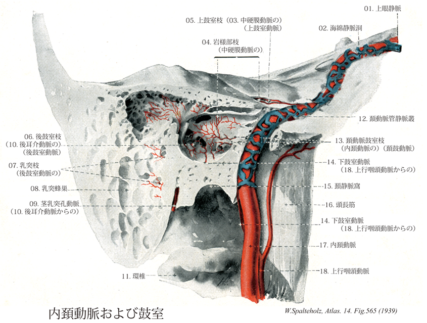
- 565_01【Superior ophthalmic vein上眼静脈 Vena ophthalmica superior】 Vein arising medially above the eyeball with the nasofrontal vein and passing through the superior orbital fissure to the cavernous sinus.
→(上眼静脈は眼動脈に沿って走る。すなわち内眼角でおこり、この部で顔面静脈や眼角静脈と吻合し、眼窩上壁を内側に沿って走行する。おおよそ眼動脈の分布域からの静脈を集める。上眼窩裂を通って頭蓋腔に入り海綿静脈洞に注ぐ。)
- 565_02【Cavernous sinus海綿静脈洞 Sinus cavernosus】 Spongy structure of expanded veins on both sides of the sella turcica into which the ophthalmic veins and other veins empty. The carotid artery and abducent nerve lie within it and cranial nerves III, IV, VI, and V2 travel in its lateral side wall.
→(海綿静脈洞は静脈間が網目に吻合して大きい不規則な網状構造をしている。この海綿静脈洞は蝶形骨洞、トルコ鞍、下垂体などの両側にある静脈洞、上眼窩裂から錐体乳突部の岩様部まで広がっている。海綿静脈洞は、内頚動脈と外転神経をとり囲む。静脈洞の外側壁には動眼神経、滑車神経、三叉神経の枝である眼神経と上顎神経が存在する。左右の海綿静脈洞は脳底静脈叢および下垂体前面にある前海綿間静脈叢と後面にある後海綿間静脈叢により対側の静脈洞と連絡する。眼静脈と蝶形骨頭頂静脈洞は、海綿静脈洞に注ぎ込む。海綿静脈洞は、後方に向かい上錐体静脈洞と下錐体静脈洞に入り、上錐体動脈洞は横静脈洞に、下垂体静脈洞は、短い静脈網によって翼突筋静脈叢や喉頭静脈叢とも連絡する。)
- 565_03【Middle meningeal artery中硬膜動脈 Arteria meningea media】 Artery passing medial to the lateral pterygoid and through the foramen spinosum into the middle cranial fossa, where it distributes vessels between the dura mater and bone.
→(中硬膜動脈は顎動脈より起こり外側翼突筋の内側を通り棘孔から中頭蓋窩に入り、そこで岩様部枝、腹硬膜枝、上鼓室動脈、前頭枝、頭頂枝に分枝する。上記の部位と終末枝を通って前頭蓋窩と中頭蓋窩に分布し、後頭動脈の硬膜枝、上行咽頭動脈、眼動脈、涙腺動脈、茎乳突孔動脈、顎動脈の腹硬膜枝、深側頭動脈と吻合する。)
- 565_04【Petrosal branch of middle meningeal artery岩様部枝(中硬膜動脈の) Ramus petrosus; Ramus pyramidis superficialis (Arteria meningea media)】 Small branch to the petrous part of temporal bone. It anastomoses with the stylomastoid artery via the hiatus for greater petrosal nerve.
→(中硬膜動脈の岩様部枝は中硬膜動脈の頭蓋内に最初の枝で顔面神経管裂を通って茎突乳突動脈動脈と吻合する。)
- 565_05【Superior tympanic artery上鼓室動脈;上鼓室枝(中硬膜動脈の) Arteria tympanica superior】 It arises near the petrosal branch and travels with the lesser petrosal nerve to the tympanic cavity.
→(上鼓室動脈は中硬膜動脈より岩様部枝に近接して起こり、小錐体神経とともに中耳に分布する。他の鼓室動脈と吻合する。)
- 565_06【Posterior tympanic artery後鼓室動脈;後鼓室枝(後耳介動脈の) Arteria tympanica posterior】 Artery traveling in the facial canal together with the chorda tympani to the tympanic membrane.
→(後鼓室動脈は茎乳突孔孔動脈より起こり、鼓索神経とともに顔面神経管からでて中耳に分布する。他の鼓室動脈と吻合する。)
- 565_07【Mastoid branch of posterior tympanic artery乳突枝(後鼓室動脈の) Rami mastoidei (Arteria tympanicae posterioris)】 Branches supplying the mastoid cells.
→(後鼓室動脈の乳突枝は後耳介動脈の茎突乳突の枝で顔面神経神経管内から出て乳突蜂巣に分布する。)
- 565_08【Mastoid cells; *Mastoid air cells乳突蜂巣 Cellulae mastoideae】 Pneumatized cells that, like the tympanic cavity, are lined with squamous or cuboidal epithelium.
→(側頭骨乳様突起内にある多数の小さな相通じている腔。乳様突起洞あるいは鼓室洞に連なる。鼓室と同様、扁平または立方上皮で被われる。)
- 565_09【Stylomastoid artery茎乳突孔動脈 Arteria stylomastoidea】 Thin vessel accompanying the facial artery. It runs with the facial artery from the stylomastoid foramen to the hiatus for greater petrosal nerve, where it supplies the dura mater. Before reaching the hiatus, it distributes branches to the middle and inner ear.
→(茎乳突孔動脈は後耳介動脈より起こり、外耳道、乳突蜂巣、半規管、アブミ骨筋、前庭に分布する。内頚動脈・上行咽頭動脈の鼓室枝、迷路動脈と吻合する。)
- 565_10【Posterior auricular artery後耳介動脈 Arteria auricularis posterior; Arteria retroauricularis】 Third branch exiting dorsally from the external carotid artery. It runs beneath the parotid gland and over the stylohyoid posterior to the auricle. It also supplies the muscles attached to the mastoid process and styloid process.
→(外頚動脈の背側へ出る第三枝。乳様突起の外側面から耳介の後ろを後上方に向かって走り乳様突起と耳介の間に分布する。)
- 565_11【Atlas; First cervical vertebra; [CI]環椎[C1];第1頚椎 Atlas [CI]】 First cervical vertebra. It does not have a body.
→(第一頚椎(環椎)は、ほかの頚椎と比べて特殊な形をしていしている。環椎(第一頚椎)には椎体と棘突起は存在せず、短い前弓と長い後弓および外側塊の三つの部分が大きな椎孔を囲んでいる。前弓は椎体の前縁部に相当し、前面中央には前結節が、後面の中央には歯突起窩がある。後弓は椎弓に相当する部分で、後面の中央には棘突起に相当する部分で、後面の中央には棘突起に相当する後結節がある。外側塊は前弓と後弓を結合する分で著しく肥厚している。外側塊からは外側へ向かってかなり大きい横突起が出ており、横突起の基部には比較的内頚の大きな横突孔がある。外側塊の上面には長楕円形の上関節窩が、下面には平らな下関節窩があって、それぞれ後頭骨の後頭顆、軸椎の歯突起がおさめられてりう。後半の部分は本来の椎孔に相当し、三角形状である。頭上に天空を支えるギリシャの神Atlas(Titan)にちなんで命名された。)
- 565_12【Internal carotid venous plexus頚動脈管静脈叢;内頚動脈静脈叢 Plexus venosus caroticus internus】 Venous plexus in the carotid canal between the cavernous sinus and pterygoid plexus.
→(内頚動脈神経叢は海綿静脈洞と内頚動脈とに結合する、側頭骨頚動脈管内にある内頚動脈周囲の静脈網。)
- 565_13【Caroticotympanic arteries頚鼓動脈;頚動脈鼓室枝;頚鼓小管枝(内頚動脈の) Arteriae caroticotympanicae; Ramus caroticotympanicus(Arteria carotis interna)】 Branches to the tympanic cavity.
→(内頚動脈鼓室枝は頚動脈管通過中に派出する数本の小枝であり、頚鼓小管を通って鼓室に入る。)
- 565_14【Inferior tympanic artery下鼓室動脈 Arteria tympanica inferior】 Artery passing via the tympanic canaliculus into the tympanic cavity, reaching the mucosa of the medial wall. It is accompanied by the tympanic nerve.
→(下鼓室動脈は上行咽頭動脈より起こり、中耳に分布する。他の動脈の鼓室枝と吻合する。)
- 565_15【Jugular fossa頚静脈窩;頚窩 Fossa jugularis】 Widening of the jugular foramen that contains the superior bulb of the jugular vein.
→(錐体下面の後縁に近い中部には弓状の大きく深い頚静脈窩がる。頚静脈上球を容れる。)
- 565_16【Longus capitis muscle頭長筋 Musculus longus capitis】 o: Anterior tubercles of C3-C6. i: Basilar part of occipital bone. Anterior and lateral flexion of the head and cervical vertebral column. I: Cervical plexus (C1-C3).
→(頭長筋と頚長筋はおうおうにして互いの境界が明瞭に定められない。この筋は縦あるいは斜めに走る線維束を持つ複合羽状構造をもつ。筋腹には通常不完全ながら腱画が挿入されている。頭長筋の起始は第3,4,5,6頚椎横突起の前結節。停止は後頭骨底部の下面。機能として頚椎と糖を屈曲しかつ回旋の補助をする。神経支配は第1,2,3,4頚神経の筋枝。動脈は下甲状腺動脈の上行頚枝、上行咽頭動脈の前脛骨枝、脛骨動脈の筋枝から受ける。)
- 565_17【Internal carotid artery内頚動脈 Arteria carotis interna】 It passes from the carotid bifurcation, without any branches, to the cranial base, continuing in the carotid canal to its terminal division into the middle and anterior cerebral arteries.
→(内頚動脈は、総頚動脈から起こり、頚部では頭蓋底にいたるまでは枝を出さない。ついで頚動脈管をへて中大脳動脈と前大脳動脈に分枝するまでをいう。内頚動脈は頚部、側頭骨錐体部(岩様部)、海綿静脈洞部、大脳部の4つの部分に分けられる。この内頚動脈の海綿静脈洞部と大脳部とは、特別な形態を呈するので、「頚動脈サイフォン」とよばれている。内頚動脈の主な枝として、眼動脈、後交通動脈、前脈絡叢動脈がでる。内頚動脈は、視交叉の外側で小さな前大脳動脈と大きな中大脳動脈とに分岐する。中大脳動脈は内頚動脈の直接の続きで終枝と考えられる。)
- 565_18【Ascending pharyngeal artery上行咽頭動脈 Arteria pharyngea ascendens】 It usually arises from the posterior side of the external carotid artery above the superior thyroid artery. It ascends along the lateral wall of the pharynx, passing medial to the stylohyoid and continuing to the cranial base.
→(上行咽頭動脈は外頚動脈より起こり、咽頭と茎状突起の筋の間を頭蓋底まで上行し咽頭壁、軟口蓋後頭窩に分布する。)