Spalteholz HANDATLAS DER ANATOMIE DES MENSCHEN VON WERNER SPALTEHOLZ
メニューは解剖学(TA)にリンクしてあります。図の番号をクリックすると下記の説明へ、右側の用語をクリックすると解剖学(TA)にジャンプします。
566 


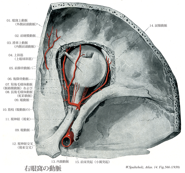
- 566_01【Supra-orbital artery眼窩上動脈;外側前頭動脈 Arteria supraorbitalis; Arteria frontalis laterallis】 Artery running beneath the roof of the orbit on the levator palpebrae superioris and through the supra-orbital notch to supply the muscles and skin of the forehead.
→(眼動脈は2本の終枝、すなわち滑車上動脈と眼窩上動脈にわかれる。眼窩上動脈は眼窩内を上壁に沿って前進し、眼窩上縁の眼窩上切痕を通って、前頭部に出て上行枝分布する。)
- 566_02【Anterior meningeal branch of anterior ethmoidal artery; Anterior meningeal artery前硬膜動脈;前硬膜枝;前頭硬膜動脈(前篩骨動脈の) Ramus meningeus anterior (Arteria ethmoidalis anterior); Arteria meningea anterior】 Branch supplying the dural portion of the anterior cranial fossa.
→(前篩骨動脈の前硬膜動脈は頭蓋腔内の前篩骨動脈より起こり、前頭蓋窩の髄膜に分布する。中硬膜動脈の分枝、内頚動脈の硬膜枝、涙腺動脈と吻合。)
- 566_03【Supratrochlear artery滑車上動脈;内側前頭動脈 Arteria supratrochlearis; Arteria frontalis medialis; Arteria frontalis】 Ascending terminal branch of the ophthalmic artery traversing the frontal notch to supply the forehead. It anastomoses with the artery from the opposite side, the supra-orbital artery, and superficial temporal artery.
→(眼動脈の上行最終枝。眼窩内から眼窩上縁の前頭切痕を経て、前頭部に現われ、上行する小動脈。対側、眼窩上動脈、浅側頭動脈と吻合する。)
- 566_04【Superior oblique muscle上斜筋;上眼球斜筋 Musculus obliquus superior; Musculus obliquus bulbi superior】 o:Medial to the common tendinous ring on the body of sphenoid, i: After a hook-shaped course, obliquely behind the equator. Its tendon passes through the trochlea. Action: Abduction, intorsion, and depression of the eye. I: Trochlear nerve.
→(上斜筋は眼窩傍結合組織すなわち視神経鞘と(おもに)蝶形骨体の結合組織である総腱輪の内側から起こる。上斜筋は眼窩錐体の内側直近の上を前方に走行する。眼球の縁で上斜筋の丸みのある腱は結合組織性の吊り索(滑車)を通過し鋭角で後方に曲がる。さらに上斜筋の腱は上直筋の下でこれと交差し眼球上後側頭部の強膜に停止する。目の動き:視線を内側かつ下方に向ける。)
- 566_05【Anterior ethmoidal artery前篩骨動脈 Arteria ethmoidalis anterior】 Together with the anterior ethmoidal nerve it emerges from the anterior ethmoidal foramen, ascends to beneath the dura mater of the anterior cranial fossa, descending through the cribriform plate of the ethmoid into the frontal sinus and nasal cavity as well as anterior and middle ethmoidal cells.
→(前篩骨動脈と後篩骨動脈は同名孔を通過して鼻腔上部に分布する。前篩骨動脈から前硬膜動脈が派出する。)
- 566_06【Posterior ethmoidal artery後篩骨動脈 Arteria ethmoidalis posterior】 Artery coursing together with the posterior ethmoidal nerve beneath the superior oblique muscle through the posterior ethmoidal foramen. It supplies the dura mater overlying the cribriform plate, and passes into the nasal cavity to the mucosa of the posterosuperior part.
→(後篩骨動脈は眼動脈より起こり、鼻腔外側壁の上後部と後篩骨洞に分布する。)
- 566_07【Short posterior ciliary arteries短後毛様体動脈;脈絡膜動脈 Arteriae ciliares posteriores breves; Arteriae chorioideae】 Between 10 and 15 arteries that penetrate the sclera around the optic nerve, supplying the choroid, ciliary body, and passing to the major circulus arteriosus of iris.
→(短後毛様体動脈は脈絡膜動脈とも呼ばれ6~15本あり、視神経の眼球進入部の付近で強膜を貫いて脈絡膜に分布する。)
- 566_08【Long posterior ciliary arteries; Iridic arteries長後毛様体動脈;虹彩動脈 Arteriae ciliares posteriores longae; Arteriae iridicae】 One lateral and one medial artery. They pass from posterior between the sclera and choroid, supply the ciliary body, and terminate at the major circulus arteriosus of iris.
→(長後毛様体動脈は虹彩動脈ともよばれ内側と外側の2本ある。視神経の眼球進入部の内外両側で眼球に入り、強膜と脈絡膜との間を前進して虹彩に達する。虹彩では、毛様体縁で鱗状の大虹彩動脈輪をつくり、その枝はさらに瞳孔縁のまわりで小虹彩動脈輪をつくる。)
- 566_09【Ophthalmic artery眼動脈 Arteria ophthalmica】 Artery arising from the anteriorly convex arch of the internal carotid artery and passing under the optic nerve within the optic canal into the orbit.
→(眼動脈は内頚動脈の枝で、頭蓋腔から視神経管を通って眼窩に入る幹動脈である。眼動脈は中頭蓋窩において前床突起の内側で内頚動脈から起こり、視神経管の中を視神経の下を通るが眼窩に入ると、視神経の上を斜めに横切って前内方にすすむ。眼窩でいろいろの枝(①網膜中心動脈、②涙腺動脈、③筋枝、④毛様体動脈、⑤前篩骨動脈・後篩骨動脈、⑥眼窩上動脈・眼窩下動脈、⑧鼻背動脈)に分かれ、眼窩の内容(眼球・副眼器)および前頭部・鼻腔壁の一部に分布する。)
- 566_10【Muscular arteries; Muscular branches of ophthalmic artery筋枝;筋動脈(眼動脈の) Arteriae musculares; Rami musculares (Arteria ophthalmica)】 Branches supplying the extraocular muscles.
→()
- 566_11【Optic nerve [II]視神経;視束[脳神経II] Nervus opticus; Fasciculus opicus [II]】 Nerve emerging medial to the posterior pole of the eyeball and extending to the optic chiasma.
→(視神経は脳神経の1つとして扱われてはいるが、実は前脳胞の延長部である。眼球網膜の第8層である神経細胞層中にある多極神経細胞から出る神経線維が集まって出来る神経である。すなわち杆状体細胞および錐体状細胞よりの興奮は網膜の内顆粒層の双極細胞に伝わり、それがさらに神経細胞層の細胞に連絡し、この神経細胞の出す神経突起である線維はまず眼球の後極よりやや内下方の一ヶ所に集まって、視神経円板を作り、強大な神経幹となり、網膜の続きである視神経鞘に囲まれて後内側に向かう。眼球から約15~20mm隔ったたところで、眼動脈の枝である網膜中心動脈およびこれに伴う静脈が外側から入り込み、その中軸を通って網膜に分布する。左右両側の視神経は眼窩後端の視神経管を通って頭蓋腔に入り、次第に相近づいて蝶形骨体上の視神経溝でほぼ半交叉をして視交叉を作り、そのつづきは視索と名が変わって間脳の外側膝状体および中脳の上丘などの第一次視覚中枢に達して、ここで終わる。網膜が眼胚から発達するので経路に相応する。ヒトの視神経は眼球網膜の神経細胞層中にある多極神経細胞から出る100万本以上の神経線維からなる。すなわち、杆状体細胞および錐体状細胞よりの興奮は網膜の内顆粒層の双極細胞に伝わり、それがさらに神経細胞層の細胞に連絡し、この神経細胞の出す神経突起である線維はまず眼球の後極よりやや内下方の一ヶ所に集まって、視神経円板を作り、強大な神経幹となり、網膜の続きである視神経鞘に囲まれて後内側に向かう。眼球から約15~20mm隔ったたところで、眼動脈の枝である網膜中心動脈およびこれに伴う静脈が外側から入り込み、その中軸を通って網膜に分布する。左右両側の視神経は眼窩後端の視神経管を通って頭蓋腔に入り、次第に相近づいて蝶形骨体上の視神経溝でほぼ半交叉をして視交叉を作り、そのつづきは視索と名が変わって間脳の外側膝状体および中脳の上丘などの第一次視覚中枢に達して、ここで終わる。)
- 566_12【Optic chiasm; Optic chiasma視神経交叉;視交叉;視束交叉 Chiasma opticum; Chiasma fasciculorum opiticorum】 Decussation of medial optic nerve fibers between the optic tract and optic nerve.
→(視神経交叉は視床下部の漏斗の吻側にある扁平な線維板で、視神経線維が交叉しているところ。視交叉の背側から両側に開いて出る線維束は視索である。第三脳室の終板と灰白隆起の間で視交叉は第三脳室の底の一部を成す(視交叉陥凹)。視交叉はその上面で(終板の前方)前交連動脈と接し、下面はトルコ鞍の鞍隔膜の上に乗っている。眼球網膜の鼻側半からの線維は交叉して対側へ行き、側頭半からの線維は同側を交叉せずに後方へすすむ。下垂体前葉から発生する腫瘍が視交叉を圧迫することがある。)
- 566_13【Internal carotid artery内頚動脈 Arteria carotis interna】 It passes from the carotid bifurcation, without any branches, to the cranial base, continuing in the carotid canal to its terminal division into the middle and anterior cerebral arteries.
→(内頚動脈は、総頚動脈から起こり、頚部では頭蓋底にいたるまでは枝を出さない。ついで頚動脈管をへて中大脳動脈と前大脳動脈に分枝するまでをいう。内頚動脈は頚部、側頭骨錐体部(岩様部)、海綿静脈洞部、大脳部の4つの部分に分けられる。この内頚動脈の海綿静脈洞部と大脳部とは、特別な形態を呈するので、「頚動脈サイフォン」とよばれている。内頚動脈の主な枝として、眼動脈、後交通動脈、前脈絡叢動脈がでる。内頚動脈は、視交叉の外側で小さな前大脳動脈と大きな中大脳動脈とに分岐する。中大脳動脈は内頚動脈の直接の続きで終枝と考えられる。)
- 566_14【Lacrimal artery涙腺動脈 Arteria lacrimalis】 Artery branching off the ophthalmic artery laterally and passing with the lacrimal nerve along the upper margin of the lateral rectus muscle to the lacrimal gland.
→(涙腺動脈は眼動脈より側方に出て外側直筋の上縁に沿って起こり、涙腺、外直筋、上直筋、上眼瞼、前額、側頭窩に分布する。)
- 566_15【Anterior clinoid process前床突起;小翼突起 Processus clinoideus anterior; Processus alae parvae】 Projection from the lesser wing of the sphenoid bone that is directed posteriorly toward the middle and posterior clinoid processes.
→(蝶形骨小翼の後縁は遊離縁をなし、その内側端に視神経管の後外側から後内側に向かう前床突起がある。)