Spalteholz HANDATLAS DER ANATOMIE DES MENSCHEN VON WERNER SPALTEHOLZ
メニューは解剖学(TA)にリンクしてあります。図の番号をクリックすると下記の説明へ、右側の用語をクリックすると解剖学(TA)にジャンプします。
591 


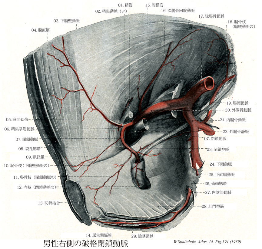
- 591_01【Ductus deferens; Deferent duct精管 Ductus deferens; Vas deferens】 The course of the ca. 50 cm long ductus deferens is initially tortuous, then becomes straight. It is a continuation of the duct of epididymis, opening into the urethra.
→(精巣上体からはじまる精巣の分泌管で、精巣上体尾につづく精子を送る通路。精索中にある。全長約30cm(延ばせばその2倍)、膀胱底で紡錘状に膨れ、精管膨大部といい、内部に膨大部憩室を含む。膨大部の下端で、精嚢が精嚢排出管を経て合流し、これより遠位では精管は射精管と呼ばれ、尿道前立腺部後壁にある精丘の上で、尿道に開く。)
- 591_02【Testicular artery♂精巣動脈(♂) Arteria testicularis; A. spermatica♂】 Artery arising at the level of the second lumbar vertebra. It crosses over the ureter and passes on the ductus deferens through the inguinal canal into the testes.
→(精巣動脈は大動脈より起こり、尿管枝、精巣挙筋動脈、精巣上体枝に分布し、精巣、尿管、精巣挙筋、精巣上体に分布する。腎動脈、下腹壁動脈、精管動脈の枝と吻合する。)
- 591_03【Inferior epigastric artery下腹壁動脈 Arteria epigastrica inferior】 It arises dorsally from the inguinal ligament and ascends to the inner surface of the rectus abdominis. producing the lateral umbilical fold. It anastomoses with the superior epigastric artery.
→(下腹壁動脈は鼡径靱帯のすぐ上方で外腸骨動脈よりおこり、壁側腹膜におおわれながら深鼠径輪の内側に沿って上方に走って前腹壁に入る。まもなく横筋筋膜を貫き、弓状線の前を通って腹直筋と腹直筋鞘後葉との間を上行し、この筋に枝を与えながら筋中で上腹壁動脈と吻合しておわる。深鼠径輪の内側を通るときに、鼡径管の内容物である精管または子宮円索の内側を経て上行する。)
- 591_04【Rectus abdominis muscle腹直筋 Musculus rectus abdominis】 o: Fifth to seventh costal cartilages, xiphoid process, i: Pubic crest and pubic symphysis. Anterior flexion of the trunk, lowering of the thorax, and elevation of the pelvis. 1: Thoracic nerves T7-T12.
→(前腹壁の筋で白線の両脇にあり腱画によって筋腹がいくつかに仕切られているのが特徴である。起始は、内側腱は恥骨結合から、外側腱は恥骨稜から起こる。停止は剣状突起の前面、第5,6,7肋骨の肋軟骨の表面。機能として、腹部の圧縮、腹部内臓の保護、強い呼気時に働く、骨盤と脊柱の屈曲。神経支配は下部6本の肋間神経の前枝、腸骨下腹神経と腸骨鼡径神経。動脈は上下腹壁動脈の筋枝から受ける。断面が楕円形のこの筋の停止腱から分かれた線維は、正中線を越え、白線の尾側への続きとして恥骨結合から陰茎(陰核)の背側面に向かう陰茎(陰核)提靱帯に加わる。腹直筋が強く働くのは背臥位から状態を起こすとき、ボートを漕ぐときなどである。腹筋の発達した人では腱画の位置が皮膚の上からくぼんで見える。)
- 591_05Hesselbach's ligament【Interfoveolar ligament窩間靱帯 Ligamentum interfoveolare】 Craniocaudal band of thickened fascia posterior to the inguinal canal.
→(鼡径管の入口である深鼡径輪の内側縁の所では、下腹壁静脈のすぐ前面で横筋筋膜が肥厚して、上下に走っている。この横筋筋膜の漠然とした肥厚が窩間靱帯とよばれるものである。窩間という意味はその場所が内側鼡径窩と外側鼡径窩の境の部位に相当するからである。)
- 591_06【Cremasteric artery♂精巣挙筋動脈;挙睾筋動脈 Arteria cremasterica; Arteria musculi cremasteris♂】 Branch supplying the cremaster and spermatic cord. It corresponds to the artery of round ligament of uterus.
→(精巣挙筋動脈は精管に伴行して鼡径管を通り、精巣挙筋および他の精索の皮膜へ分布する。)
- 591_07【Obturator artery閉鎖動脈 Arteria obturatoria】 Artery running in the lateral wall of the pelvis and passing through the obturator foramen to the adductors.
→(閉鎖動脈は内腸骨動脈の前枝より起こり、骨盤側壁を走り、閉鎖孔をへて腸骨、恥骨、閉鎖筋、内転筋に分布する。腸腰動脈、下腹壁動脈、内側大腿回旋動脈と吻合し、恥骨枝、寛骨臼枝、前枝、後枝に分枝する。)
- 591_08Gimbernat's ligament【Lacunar ligament; Lacunar inguinal ligament裂孔靱帯 Ligamentum lacunare】 Arched connective-tissue band that extends inferiorly from the medial attachment of the inguinal ligament to the pubis.
→(鼡径靱帯の内側部は後方に向かって広がり、恥骨筋膜と癒着しながら恥骨櫛内側部に至る。これを裂孔靱帯といい、血管裂孔の内側縁をなし、外方にすこしくぼんで鋭い。ギムベルナト靱帯とも呼ばれる。ギムベルナト Gimbernat, Don Mannuel Louise Antonio de (1734-1790)スペインの外科医、解剖学者1762年から1774年までバルセロナ大学教授。カルロス3世の侍医。ギベルナト靱帯(1768年)を起始、女性のヘルニア手術法を開発(""Nuevo metodo de operar en la hernia crural"", 1793)。)
- 591_09Henle's ligament【Inguinal falx; Conjoint tendon鼡径鎌;結合腱 Falx inguinalis; Tendo conjunctivus】 Tendinous fibers forming an arch that extends from the aponeurosis of the transverse abdominal muscle into the pectineal ligament.
→(鼡径鎌(結合腱)は恥骨稜、恥骨櫛に停止する腹横筋と内腹斜筋の共通の腱。腹横筋腱膜から恥骨櫛靭帯へ弓状に入る線維。鼡径管の後壁を助成する。)
- 591_10【Pubic branch of inferior epigastric artery恥骨枝(下腹壁動脈の) Ramus pubicus (Arteria epigastricae inferioris)】 Branch extending to the pubis.
→(下腹壁動脈の恥骨枝は鼡径靱帯に沿って内方へ走り、次いで大腿輪に沿って下行し、恥骨の内面で閉鎖動脈の恥骨枝吻合する。この吻合枝が発達すると、あたかも閉鎖動脈が下腹動脈よりおこるような外観を呈する。これを副閉鎖動脈または死冠という。)
- 591_11【Pubic branch of obturator artery恥骨枝(閉鎖動脈の) Ramus pubicus (Arteria obturatoria)】 It anastomoses with the obturator branch of the inferior epigastric artery.
→(閉鎖動脈の恥骨枝は閉鎖管にはいる直前部から分枝して恥骨後面を上行する。同名の反対側の枝や下腹壁動脈の恥骨枝と吻合する。)
- 591_12【Internal branch of obturator artery内枝(閉鎖動脈の) Ramus internus (Arteria obturatoria)】
→()
- 591_13【Pubic symphysis; Pubis symphysis; Symphysis pubis恥骨結合 Symphysis pubica】 Cartilaginous joint between the rami of the pubis.
→(恥骨結合は骨盤前面で左右の恥骨が正中線上で向かい合ってできる連結。両側の恥骨結合面がうすい硝子軟骨に被われ、その間に線維軟骨でできる恥骨間円板が連結する。その構造は椎間円板の線維輪に似る。女性では妊娠時にこの結合は弱められ、またこのことは分娩時における新生児の頭の産道通過を助ける。モルモットなどでは、女性ホルモンの投与によって、実験的にこの結合を弱めることができる。付属する靱帯に次のものがある。(1)上恥骨靱帯:恥骨結合の上縁で左右の恥骨を結ぶ。(2)恥骨弓靱帯:恥骨結合の下縁で、左右の恥骨下枝を結び、恥骨弓をつくる。下面で尿生殖膜との間隙を陰茎静脈が通る。 Symphysisははsyn(一緒に)physis(生える)、すなわち「自然に癒合したもの」という意味である。解剖学用語としてのsymphysisは線維軟骨結合という一般名詞であるが、慣用的にはpubicaという形容詞なしでも恥骨結合を指すことが多い。正しい読み方はスィンフィスィスである。)
- 591_14【Urogenital diaphragm尿生殖隔膜 Diaphragma urogenitale】 A term that has been replaced. What was previously conceived of as a unit is now divided into separate terms: perineal membrane, transverse perineal ligament, deep transverse perineal muscle.
→(尿生殖隔膜は骨盤隔膜の前部の下側(浅側)にあり、恥骨弓の間に張っている三角形の線維性膜である。尿生殖膜は上・下2枚の筋膜からなる。この筋膜を、それぞれ、上・下尿生殖隔膜筋膜という。とくに下尿生殖隔膜筋膜は比較的厚く強靱で、会陰膜ともいわれる。上生殖隔膜筋膜は明瞭でないことも多い。上尿生殖隔膜筋膜と下尿生殖隔膜筋膜とは後縁では癒合し、会陰腱中心に付着する。上下の隔膜筋膜は前上縁でも癒合し肥厚して、恥骨結合のすぐ下で会陰横靱帯をつくる。尿生殖隔膜は、男性では尿道によって、女性では尿道と腟とで貫かれる。)
- 591_15【Transversus abdominis muscle; Transverse abdominal muscle腹横筋 Musculus transversus abdominis】 Inner surface of the seventh through twelfth costal cartilages, thoracolumbar fascia, iliac crest, anterior superior iliac spine, inguinal ligament, i: Rectus sheath, linea semilunaris. I: Intercostal nerves 7-12, iliohypogastric nerve, ilioinguinal nerve, genitofemoral nerve.
→(腹横筋の起始は下位6本の肋骨の肋軟骨内面、腰筋膜の内層、腸骨稜の内唇の前2/3、鼡径靱帯の外方1/3。停止は腱膜鞘につつまれて両腹斜筋とともに白線の中へ。機能としては腹部の圧縮、腹部内臓の保護、強い呼気時に働く。神経支配は下位6本の肋間神経の前枝、腸骨下腹神経と腸骨鼡径神経。動脈は深腸骨回旋動脈、下腹壁動脈。腹横筋は胸横筋の尾側に隣接している。この筋は、第7(6,5)から第12肋軟骨の内面、腰椎の肋骨突起(胸腰筋膜の深葉を介して)、腸骨稜の内唇および鼡径靱帯の外側部から起こる。この筋線維は、ほぼ水平に(腹直筋に直角)に走り、半月状の外側に凸の線、半月線を越えて腱膜となる。腹横筋の腱膜は腹直筋鞘の形成に関わる。その腱膜の線維は、白線で内腹斜筋の腱膜の線維と連結している。)
- 591_16【Deep circumflex iliac artery; Deep iliac circumflex artery深腸骨回旋動脈 Arteria circumflexa iliaca profunda】 Artery that curves posterolaterally beneath the transversalis fascia along the iliac crest.
→(深腸骨回旋動脈は下腹動脈とほぼ同じ高さで、外腸骨動脈の外側面よりでて、横筋筋膜におおわれて鼡径靱帯の内面に沿って上前腸骨棘に向けて外上方へ走り、次いで腸骨稜に沿ってそのほぼ中央部に達し、その間に側副筋に分布する。)
- 591_17【Common iliac artery総腸骨動脈 Arteria iliaca communis】 It extends from the aortic bifurcation at the fourth lumbar vertebra to its division into the internal and external iliac arteries in front of the sacroiliac joint. Its branches are insignificant.
→(総腸骨動脈は腹部大動脈の第四腰椎の前で大動脈から分かれる左右1対の終枝。仙骨岬角のレベルにおける仙腸関節の前で、内・外腸骨動脈に分枝する。とくに記載するほどの枝はない。)
- 591_18【Iliacus branch of iliolumbar artery; Iliac branch of iliolumbar artery腸骨枝(腸腰動脈の) Ramus iliacus (Arteria iliolumbalis)】 Branch to the iliacus that lies parallel to the pelvis and extends to the iliac fossa. It anastomoses with the deep circumflex iliac artery.
→(腸腰動脈の腸骨枝は腰動脈の枝を含む腸腰動脈の最終枝で、腸骨窩のなかを腸骨筋・腸骨そして腸骨稜に付着する近くの筋肉に分布する枝で骨盤と平行して走る。深腸骨回旋動脈と吻合。)
- 591_19【Iliolumbar artery腸腰動脈 Arteria iliolumbalis】 It passes below the psoas and internal iliac artery into the iliac fossa.
→(腸腰動脈は内腸骨動脈より起こり、骨盤および骨盤筋に分布する。新腸骨回旋動脈、腰動脈と吻合する。)
- 591_20【External iliac artery外腸骨動脈 Arteria iliaca externa】 Second branch of the common iliac artery, which continues as the femoral artery.
→(外腸骨動脈は総腸骨動脈からつづいて、仙腸関節の前面で内腸骨動脈とわかれたあと、大腰筋の内側縁に沿って下行し、鼡径靱帯のほぼ中央でその下を通過して大腿前面出て、大腿動脈に移行する。内腸骨動脈から分かれて、鼡径靱帯の下を通過するまでの部分を指す。)
- 591_21【Internal iliac artery; Hypogstric artery内腸骨動脈;下腹動脈 Arteria iliaca interna】 Artery beginning at the division of the common iliac artery, passing from here into the lesser pelvis and extending to the upper border of the greater sciatic foramen. Its branches are highly variable.
→(内腸骨動脈は総腸骨動脈より起こり、腸腰動脈、外側仙骨動脈、閉鎖動脈、上臀動脈、下臀動脈、臍動脈、上膀胱動脈、下膀胱動脈、中直腸動脈、内陰部動脈に分岐する。)
- 591_22【External iliac vein外腸骨静脈 Vena iliaca externa】 It arises at the superior end of the femoral vein below the inguinal ligament and ends where it unites with the internal iliac vein to form the common iliac vein.
→(外腸骨静脈は下肢の静脈を集める本幹で、そのほか一部は前腹壁の下部からも血液を集める。大腿静脈の続きとして鼡径靱帯の下で血管裂孔にはじまり、大腰筋の内側に沿って上行して、仙腸関節の前面で内腸骨静脈と合して総腸骨静脈をつくっておわる。)
- 591_23【Obturator nerve閉鎖神経 Nervus obturatorius】 It arises from L2-L4 and travels beneath the psoas, posterior to the internal iliac artery, lateral to the ureter, continuing through the obturator canal to the adductors and the skin of the medial aspect of the thigh.
→(閉鎖神経は第二・第三・第四腰神経からなる腰神経叢から起こり、垂直に下行し、大腿筋の内側縁から出て、総腸骨動脈の後側を通って骨盤孔に入る。骨盤の側壁内面に沿って走り、閉鎖動静脈とともに閉鎖管を抜けて大腿上部の内側部に出る。前枝は長内転筋と薄筋の間に現れ、大腿皮膚の下3分の2へ分布。 筋枝は大腿の内転筋に分布する。大腿の内側部皮膚と内転筋分に分布する。皮枝は大腿内側の皮膚に分布する。関節枝は股関節と膝関節とに分布する。)
- 591_24【Inferior gluteal artery下殿動脈;下臀動脈 Arteria glutea inferior】 After passing through the greater sciatic foramen, it runs beneath the piriformis, distributing branches beneath the gluteus maximus. It anastomoses with the superior gluteal artery, obturator artery, and circumflex femoral arteries.
→(下臀動脈は内腸骨動脈より起こり梨状筋の下で大坐骨孔を通り(梨状筋下孔)、それは大臀筋の下を通過して股関節、臀部に分布する。内陰部動脈の枝、外側仙骨動脈、上臀動脈、閉鎖動脈、内側・外側大腿回旋動脈と吻合する。)
- 591_25【Inferior rectal artery下直腸動脈;肛門動脈 Arteria rectalis inferior; Arteria analis】 Artery passing transversely through the ischioanal fossa and supplying both sphincters as well as the skin below the anal valves.
→(下直腸動脈は内陰部動脈の枝で、同名神経に伴行し、肛門管下半部に分布する。)
- 591_26【Sacrospinous ligament; Sacrospinal ligament仙棘靱帯 Ligamentum sacrospinale】 Band that extends from the sacrum and the coccyx to the ischial spine, dividing the greater and lesser sciatic foramina.
→(仙棘靱帯は坐骨棘から起こり、仙結節靱帯の前面でこれと交叉して内後方に進み、やや拡がって仙骨下部および尾骨の側縁につく。骨盤の後外側の、仙骨と寛骨の間に出来る大きな仙坐切痕は、後下方から仙結節靱帯によって閉ざされて上下に長い孔となり、これは仙棘靱帯によって上方の大坐骨切痕を含む大坐骨孔と、下方の小坐骨切痕を含む小坐骨孔とに分かれる。)
- 591_27【Internal pudendal artery内陰部動脈 Arteria pudenda interna】 Artery exiting the pelvis through the greater sciatic foramen and passing through the lesser sciatic foramen to the lateral wall of the ischioanal fossa.
→(内陰部動脈は内腸骨動脈の枝であり、大坐骨孔(梨状筋下孔)を通って骨盤からでて、小坐骨孔を経て坐骨直腸窩側壁にゆく。陰部神経との伴行を示す。)
- 591_28【Levator ani muscle肛門挙筋 Musculus levator ani】 Principle muscle of the pelvic diaphragm. It is derived from the abdominal wall musculature and permeated by smooth-muscle cells. I: Sacral plexus, S2-S5. It consists of the following parts.
→(肛門挙筋の丈夫な前部(恥骨尾骨筋)は分界線直下の恥骨の内面から起こり、薄い後部(腸骨尾骨筋)は腸骨から起こる。その起始腱は内閉鎖筋筋膜に接して移行し、閉鎖筋膜から発する腱束を受ける。これらの線維の起始部では腱性の係留物(肛門挙筋腱弓)により強化されている。左右両側で恥骨尾骨筋の内側線維束は挙筋脚を形成している。それらの線維束は背方と尾方、また直腸の前では外側を通り、それぞれ会陰の中心腱へ放散する薄い前直腸線維束や前立腺挙筋として前立腺筋膜(あるいは恥骨腟筋として腟壁)へと分かれる。それより鼻側にある肛門挙筋の線維束は恥骨直腸筋として直腸の背側を取り囲み、反対側の線維と共にループを形成する。恥骨尾骨筋の外側束は尾骨と仙骨の背側に広がる。腸骨尾骨筋の筋線維は尾骨と仙骨に付き、また肛門と尾骨の間では強靱な線維束である肛門尾骨靱帯に付いている。)
- 591_29【Artery of penis陰茎動脈 Arteria penis】
→()