Spalteholz HANDATLAS DER ANATOMIE DES MENSCHEN VON WERNER SPALTEHOLZ
メニューは解剖学(TA)にリンクしてあります。図の番号をクリックすると下記の説明へ、右側の用語をクリックすると解剖学(TA)にジャンプします。
603 


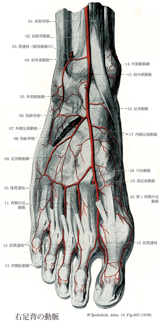
- 603_01【Tibialis anterior muscle前脛骨筋 Musculus tibialis anterior】 o:Lateral surface of tibia, interosseous membrane, deep fascia of leg. i: Medial aspects of medial cuneiform and first metatarsal. Dorsiflexion and supination of foot. I: Deep fibular nerve.
→(前脛骨筋は脛骨外側顆、脛骨外側面(近位2/3)、下腿筋膜および筋間膜から起始する。第1中足骨と第1楔状骨あたりの足底部に停止する。収縮中に筋腹は脛骨近位1/3の骨縁上に突出する。その腱は脛骨遠位1/3にかけて形成され、伸筋支帯の下を通って足の内側縁へ至る。その腱鞘は伸筋支帯より近位に始まり、距腿関節の関節腔のレベルにまで伸びている。腱鞘は前脛骨筋腱の遠位部および近位部浅層をおおい、中間部を包んでいる。前脛骨筋と長趾伸筋に対する近位の筋枝は深腓骨神経から同神経がまだ腓骨筋群を容れる部位を通っている内に分かれる。深腓骨神経が長趾伸筋を貫通してから遠位の筋枝が両筋の各々に行き(通常2条の)筋枝が母趾の伸筋へ行く。)
- 603_02【Anterior tibial artery前脛骨動脈 Arteria tibialis anterior】 It extends from its origin at the inferior border of the popliteus to the inferior border of the inferior extensor retinaculum. After penetrating the interosseous membrane, it lies between the tibialis anterior and extensor digitorum longus, then between the tibialis anterior and extensor hallucis longus.
→(前脛骨動脈は、腋窩の遠位部、すなわち膝窩筋の下縁の高さで、膝窩動脈が二分して生ずる枝の一つ。分岐後、下腿骨間膜の上部を越え、骨間膜の前面に出て下行する。経過中に下腿の前外側にある筋とその付近に枝を送る。動脈はとくに上部と下部で膝関節と足関節との周囲の膝蓋動脈網に小枝を送り吻合する。前脛骨動脈は、足関節のすぐ上方で表層に現れ、足関節の前側で前脛骨筋の腱の外側に沿って走り、足背で足背動脈となる。)
- 603_03【Perforating branch of fibular artery貫通枝;穿通枝(腓骨動脈の) Ramus perforans (Arteria fibularis)】 It pierces the interosseous membrane immediately above the malleolus and passes to the lateral malleolar network and dorsum of foot.
→(腓骨動脈の貫通枝は外果の上方約5cmの付近で分岐し、ただちに下腿骨間膜を貫いて下腿の前面に出て、前外果動脈と吻合する。)
- 603_04【Anterior lateral malleolar artery前外果動脈;前外踝動脈;前腓側踝動脈 Arteria malleolaris anterior lateralis; Arteria melleolaris fibularis anterior】 Artery passing beneath the tendon of the extensor digitorum longus to the lateral malleolar network.
→(前外果動脈は距腿関節の少し上方で本幹より分かれ、長趾伸筋の下を下方へ走り、外果動脈網へ。途中、腓骨動脈の貫通枝と腓骨か端部の前面で吻合する。この吻合枝はきわめて細いが、ときに発達して前脛骨動脈の弱小を補い、脛骨動脈から足背動脈へと接続することがある(約7%)。)
- 603_05【Lateral malleolar network; Lateral malleolar arterial network外果動脈網;外踝動脈網 Rete malleolare laterale; Rete malleolare fibulare】 Arterial plexus overlying the lateral malleolus.
→(外果動脈網は外果の表層の皮下にある動脈網で、前外果動脈、外側足根動脈、腓骨動脈の貫通枝、外果枝、踵骨枝によりつくられる。)
- 603_06【Fibularis brevis muscle; Peroneus brevis muscle短腓骨筋 Musculus fibularis brevis; Musculus peroneus brevis】 o: Distal two-thirds of fibula, i: Tuberosity of fifth metatarsal bone. Pronation and plantar flexion. I: Superior fibular nerve.
→(短腓骨筋は腓骨遠位1/2と両方の筋間中隔から起こり、長腓骨筋とともに外果のうしろを通って第5中足骨粗面に付着する。長深伸筋群の系統発生上の名残は下等哺乳類によく発達しており、弱い停止腱が第5趾の足背筋膜へ伸びている。)
- 603_07【Lateral tarsal artery外側足根動脈;腓側足根動脈 Arteria tarsalis lateralis; Arteria tarsea fibularis】 It arises at the level of the head of talus and passes under the short extensors of the toes toward the cuboid. It anastomoses with the arcuate artery.
→(外側足根動脈は舟状骨上面の付近で分岐し、短趾伸筋におおわれて足根骨の上を外方へ走り、付近の骨、関節、筋へ。)
- 603_08【Extensor digitorum brevis muscle短趾伸筋;短指伸筋(足の) Musculus extensor digitorum brevis】 o: Dorsal aspect of calcaneus. i: Dorsal aponeuroses of second through fourth toes. I: Deep fibular nerve.
→(短趾伸筋は踵骨の上端および下伸筋の支帯より起こり第2~4趾、ときには第2~5趾(約8%)の背側腱膜へ至る。この筋の4本の腱はいずれも前内側に進むが、そのうち最内側のもの(ときに短母指伸筋腱extensor hallucis brevis tendonともよばれる)は第1趾の基節底に停止する残りの3本の腱はそれぞれ第2,第3、第4趾に向かう長指伸筋腱への合流を示す。短趾伸筋は深腓骨神経の支配を受ける。この筋は第1~4趾を伸展させる。この筋の作用は距腿関節で足が背屈して長指伸筋が動けない状態下で特に顕著となる。)
- 603_09【Dorsal network of foot足背動脈網 Rete dorsale pedis】
→(")
- 603_10【Posterior perforating branches (to dorsal metatarsal arteries)後貫通枝;穿通枝;穿通枝(背側中足動脈に行く) Rami perforantes (posteriores)】
→()
- 603_11【Dorsal metatarsal arteries背側中足動脈 Arteriae metatarsales dorsales】 Four branches that pass distalward over the intermetatarsal spaces and divide into two dorsal digital arteries each.
→(背側中足動脈は第1背側中足動脈は足背動脈の2終枝の一つとして分岐し、第2、第3、第4背側中足動脈は弓状動脈または足底の動脈より分岐する。それぞれの中足骨間隙で背側骨間筋の表層を前進し、各趾の基部で2本の背側趾動脈に分かれる。各動脈は中足骨間隙において足底の動脈から貫通枝を受ける。)
- 603_12【Anterior perforating arteries to dorsal metatarsal arteries前貫通枝(背側中足動脈へ行く) Rami perforantes aneriores】
→()
- 603_13【Dorsal digital arteries of foot背側趾動脈;背側指動脈(足の) Arteriae digitales dorsales】 Interdigital arteries that arise from the metatarsal arteries.
→(背側趾動脈は背側中足動脈が趾の基部で分岐した2本の背側趾動脈は、隣接趾の対向縁を前進して趾先に達する。計10本の背側指動脈のうち、母趾の内側縁に分布する枝は第1背側中足動脈から、小指の外側縁に達するものは、第4背側中足動脈または外側足根硬脈より分布する。しかし実際には背側趾動脈は貫通枝を介して底側中足動脈より血液をうけることが多い。)
- 603_14【Medial malleolar network; Medial malleolar arterial network内果動脈網;脛骨踝動脈網 Rete malleolare mediale; Rete malleolare tibiale】 Arterial plexus overlying the medial malleolus.
→(内果動脈網は内果の表層に皮下にある動脈網で前内か動脈、内側足根動脈、後脛骨動脈の内果枝と踵骨枝によってつくられる。)
- 603_15【Anterior medial malleolar artery前内果動脈;前脛側踝動脈 Arteria malleolaris anterior medialis; Arteria melleolaris tibialis anterior】 It passes beneath the tendon of the tibialis anterior to the medial malleolar network.
→(前内果動脈は前外果動脈とほぼ同じ高さで分岐し、長母指伸筋と前脛骨筋の腱の下を内方へ走り、内果動脈網へ。)
- 603_16【Dorsalis pedis artery; Dorsal artery of foot足背動脈 Arteria dorsalis pedis】 Continuation of the anterior tibial artery on the dorsum of foot. After crossing under the tendon of the extensor hallucis longus and passage of the extensor retinaculum, it lies lateral to the tendon where it is palpable.
→(足背動脈は前脛骨動脈よりつづいて、距腿関節の前面から起こり、足背の内側縁を下行して、母趾と第2趾の間にある第1中足骨間隙の近位で、第1背側中足動脈と深足底枝に分かれておわる。前脛骨動脈が弱小化して、代償的に発達した腓骨動脈の貫通枝が、前外果動脈を経て足背動脈に接続することが約7%に出現する。)
- 603_17【Medial tarsal arteries内側足根動脈 Arteriae tarsales mediales; Arteriae tarsales tibiales】 Several free branches passing to the medial border of the foot.
→(内側足根動脈は足の内側縁で分岐する二、三の小枝。足の内側縁と内果動脈網へ。)
- 603_18【Arcuate artery of foot弓状動脈(足の) Arteria arcuata】 It curves laterally over the bases of metatarsals beneath the extensor digitorum brevis.
→(足の弓状動脈は外側足根動脈よりも少し遠位で分岐し、短趾伸筋に被われ中足骨の基部を外側方に弓状をなして走り、足の外側縁で外側足根動脈と吻合する。途中、これからは前方に向かって順次に第2,第3,第4背側中足動脈を分岐する。しかし弓状動脈が充分に発達して上記全ての背側中足動脈は足底の動脈から供給される。日本人では弓状動脈が10%以上の頻度で欠如し、また存在する場合でも細いことが多い。)
- 603_19【Deep plantar artery; Deep plantar branch of dorsal artery of foot深足底動脈;穿通中足動脈;深足底枝(背側中足動脈の) Arteria plantaris profundus; Ramus plantaris profundus】 Especially thick, perforating branch of a dorsal metatarsal artery that anastomoses with the plantar arch.
→(深足底動脈は足背動脈の2終枝の一つ。第1中足骨間隙で、第1背側骨間筋両頭の間を通って足底に出て、足底動脈弓に内側端に接続する。)
- 603_20【First dorsal metatarsal artery第1背側中足動脈 Arteria metatarsea dorsalis (I); Arteria metatarsea dorsalis prima】
→()