Spalteholz HANDATLAS DER ANATOMIE DES MENSCHEN VON WERNER SPALTEHOLZ
メニューは解剖学(TA)にリンクしてあります。図の番号をクリックすると下記の説明へ、右側の用語をクリックすると解剖学(TA)にジャンプします。
796


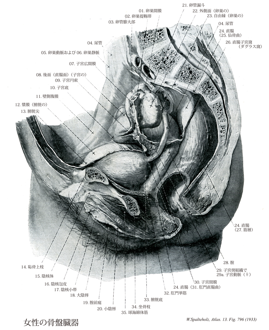
- 796_01【Mesovarium♀卵巣間膜 Mesovarium♀】 Mesentery of the ovary. Posteriorly directed fold of the broad ligament of uterus.
→()
- 796_02【Suspensory ligament of ovary; Infundibulopelvic ligament♀卵巣提靱帯;卵巣提索;骨盤漏斗靱帯 Ligamentum suspensorium ovarii♀】 Band derived from the superior gonadal fold that passes between the tubal extremity of ovary and the lateral wall of the pelvis.
→()
- 796_03【Ampulla of uterine tube; Ampulla of oviduct卵管膨大部 Ampulla tubae uterinae】 Lateral enlargement of the uterine tube. Its lumen tapers to form the isthmus of uterine tube.
→(卵管膨大部は漏斗につづく太い部。長さ7~8cmで、卵管全長の約2/3を占める。卵巣の前上方をアーチ状に走る。膨大部は太いが、壁は薄い。粘膜には、きわめて複雑なヒダが発達し、内腔のほとんどを占めている。)
- 796_04【Ureter尿管 Ureter】 Urinary duct situated in the retroperitoneum. It connects the renal pelvis with the urinary bladder, measures 25-30 cm in length and is about 3 mm thick.
→(尿管は全長約25~27cmで、上半分は腹腔内を走り腹部といわれ、下半分は骨盤内にあり骨盤部といわれる。腎盂につづき、腎臓から膀胱に至る管。輪層と縦層の平滑筋に囲まれた移行上皮によって裏打ちされ、外部は外膜でおおわれている。腎門の内下側から出て、大腰筋の前面を斜めに内下方に向かい、精巣(卵巣)動脈の後ろで、これと交叉して下行する。第四腰椎の高さで、総腸骨動・静脈の前を横切って骨盤内に入る。ついで、骨盤の側壁に沿って走り、最後に前内方にまたがって骨盤邸の上面を走り膀胱に開く。尿管はつぎの3箇所にやや細い狭窄部をもつ。すなわち、1.腎盂から尿管への移行部(上端部)、2.腹部から骨盤部への移行部(この部は総腸骨動・静脈と交叉し、尿管は腹膜と癒着している、3.膀胱壁を貫く部(尿管は膀胱壁を斜めに貫き、長さは約2cm)の3箇所である。)
- 796_05【Ovarian artery♀卵巣動脈(♀) Arteria ovarica♀】 Artery arising at the level of the second lumbar vertebra. It passes in the suspensory ligament of ovary to the ovary. It anastomoses with the uterine artery.
→(卵巣動脈は大動脈より第二腰椎の高さで起こり、尿管、卵巣、卵巣索、卵管に分布する。子宮動脈と吻合する。)
- 796_06【Ovarian vein卵巣静脈 Vena ovarica】
→()
- 796_07【Broad ligament of uterus♀子宮広間膜;子宮広ヒダ;子宮広靭帯 Ligamentum latum uteri; Plica lata uteri♀】 Anteriorly situated sheet of connective tissue that is covered with peritoneum and passes between the lateral surface of the uterus and lateral pelvic wall. It divides the female pelvis into two spaces, a vesico-uterine pouch and a rectouterine pouch.
→(子宮と外側骨盤壁の間にある神経、血管をいれた腹膜の重複膜。 (Feneis))
- 796_08【Intestinal surface of uterus; Posterior surface of uterus後面;直腸面(子宮の) Facies intestinalis uteri; Facies rectalis】 Posterosuperior surface that is in contact with the intestine.
→(後上方へ向かった面で腸が接触する。 (Feneis))
- 796_09Hunter's ligament【Round ligament of uterus子宮円索;子宮鼡径索 Ligamentum teres uteri; Chorda uteroinguinalis (teres)】 Ligament derived from the caudal gonadal fold during development. It passes from the tubal angle through the parametrium and inguinal canal into the labia majora.
→(卵管開口部の前下方、左右両側で子宮に付着している筋線維を含む線維帯。鼡径管から大陰唇に至る。男性の精索に相当し、これも鼡径管を通り同じような腓腹をもつが相同ではなく精巣導帯と相同である。スコットランドの解剖学者William Hunter (1718-1783)によって記載された。WilliamはJohn Hunterの兄で、彼の業績はグラスゴーのハンター博物館に残されている。)
- 796_10【Fundus of uterus子宮底 Fundus uteri】 Rounded end of the uterus above the openings of the uterine tubes.
→(卵管侵入部より上方にある子宮の頂部。 (Feneis))
- 796_11【Parietal peritoneum壁側腹膜 Peritoneum parietale】 Peritoneum lining the abdominal wall.
→(壁側腹膜は腹壁の腹膜。)
- 796_12【Serosa of bladder; Bladder serosa漿膜(膀胱の) Tunica serosa vesicae】 Peritoneal covering that mostly surrounds the body of bladder.
→(膀胱漿膜は膀胱上面や外側面をおおう臓側腹膜)
- 796_13【Apex of bladder; Vertex of urinary bladder膀胱尖;膀胱頂 Apex vesicae; Vertex vesicae】 Tip of the bladder pointing anterosuperiorly and attached to the anterior abdominal wall by the median umbilical ligament.
→()
- 796_14【Superior pubic ramus; Ramus of pubis恥骨上枝;寛骨臼部(恥骨枝の) Ramus superior ossis pubis; Pars acetabularis ramus ossis pubis】 The part of the pubis above the obturator foramen.
→(恥骨上枝は、寛骨臼の前下部と、そこから前下方に伸びて恥骨体の上部につづく三角柱状の部分をいう。)
- 796_15【Body of clitoris陰核体 Corpus clitoridis】 Union of the two crura of clitoris below the pubic symphysis.
→(陰核体は左右1対の陰核海綿体が結合性の陰核筋膜で包まれてできる。陰核は陰茎と異なって尿道で貫かれていないので、陰核対には尿道海綿体もない。陰核対には恥骨結合から起こる陰核提靱帯がつく。)
- 796_16【Prepuce of clitoris陰核包皮 Preputium clitoridis】 Union of the two labia minora above the glans of clitoris.
→(小陰唇は陰核の部分で前後2葉に分かれ、前葉は陰核包皮として陰核亀頭を包む。)
- 796_17【Frenulum of clitoris陰核小帯 Frenulum clitoridis】 Small fold that gives attachment to the two labia minora below the glans of clitoris.
→(小陰唇から陰核の下面へと走る二重のヒダ。(Feneis))
- 796_18【Labium majus; Greater lip大陰唇 Labium majus pudendi】 Longitudinal prominence overlying a pad of fat. It is covered with hair on its external surfaces. It extends from the mons pubis to the perineum and borders with the pudendal cleft.
→(大陰唇は男性の陰嚢に相当する。その皮膚の正常も陰嚢に似て色が浅黒く、陰毛も生えているけれども、陰嚢と違って左右のものが正中で癒合せずに、分かれている。内面は粘膜様の皮膚の隆起。この左右の大陰唇の間の裂け目を陰裂という。左右の大陰唇と、その間の陰裂を総称して陰門という。)
- 796_19【Vestibule of vagina腟前庭 Vestibulum vaginae】 It is enclosed mainly by the labia minora. Site of opening of the female urethra, vagina, and greater and lesser vestibular glands.
→(腟前庭は、胎生期の尿生殖洞の名残である。左右の小陰唇の間にある裂隙で、ここに尿道・腟および大前庭腺の導管が開いている。尿生殖洞は尿直腸中隔の発達により直腸から分離した後の排泄腔の腹側部。男女両性の膀胱下部、男性の尿道前立腺部、女性胃の尿道と腟前庭を生じる。)
- 796_20【Labium minus; Lesser lip小陰唇 Labium minus pudendi】 Cutaneous fold that is devoid of fat and hair and contains sebaceous glands. It forms the boundary of the vestibule of vagina.
→(小陰唇は大陰唇の内側にある2つの皮膚とヒダである。皮膚は毛や脂肪を欠き平滑で、粘膜に似ている。陰毛はないが豊富な皮脂腺をもつ扁平なヒダ。成人では小陰唇は大陰唇で被われている。小児では、大陰唇の発達が悪いので、小陰唇の大部分は陰核とともに露出する。後方で左右が連絡する部分に陰唇小体というヒダをつくる。)
- 796_21【Infundibulum of uterine tube; Infundibulum of oviduct卵管漏斗;卵管漏斗部 Infundibulum tubae uterinae】 Funnel-shaped beginning of the uterine tube.
→(漏斗状の卵管起始部で卵巣に接する。 (Feneis))
- 796_22【Lateral surface of ovary外側面(卵巣の) Facies lateralis ovarii】 Surface of the ovary resting against the wall of the pelvis.
→()
- 796_23【Free border of ovary; Free margin of ovary自由縁(卵巣の) Margo liber ovarii】 Free border located opposite to the hilum of ovary.
→()
- 796_24【Rectum直腸 Rectum; Intestinum rectum】 Tenia-free 15 cm long segment extending between the sigmoid colon and anus.
→(直腸は消化管の末端部でS状結腸につづく大腸の一部である。結腸から直腸への移行はゆるやかで、仙骨中央部あたりがほぼ両者の境界となる。直腸は腸間膜を欠き、直腸ヒモを示さない部分である。直腸の下端は、骨盤隔膜を貫く寸前までで、それ以下は肛門管である。肛門管の直上部にあたる直腸窩部はふくらみ、ここを直腸膨大部という。膨大部上方には横走するヒダが2~3本認められ、直腸横ヒダといい、最も恒常的なものは右壁にあって、コールラウシュのヒダという。直腸ははじめ仙骨の曲がりに沿って前方に凹の間借りを示し、これを仙骨曲といい、下端近くでは前方に凸の曲がりを示し、これを会陰曲という。直腸壁の平滑筋の筋層のうち、重筋層と一部の輪筋層は周辺の臓器へとのび、直腸尾骨筋、直腸膀胱筋、直腸尿道筋などとよばれる筋束をなす。直腸が内容をいれて拡張すると、壁の伸展刺激は求心性神経線維によって仙髄に伝えられ、反射的に内容の排出、すなわち排便が起こる。このような排便中枢の中枢は仙髄(S2~4)にあり、肛門脊髄中枢anospinal centerといわれる。排便defecationのさいには、交感神経が抑制されるとともに、副交感神経の興奮が高まって、大腸の蠕動・収縮がおこり、内肛門括約筋は弛緩し、さらに陰部神経を介して外肛門括約筋も随意的に緩められる。そのほかに、腹壁の筋・横隔膜・骨盤隔膜を作る肛門挙筋の収縮によって腹圧が高められ排便を助ける。)
- 796_25【Sacral flexura of rectum; Rectal sacral flexure; Sacral rectal flexure仙骨曲(直腸の) Flexura sacralis recti】 Anteriorly concave curvature of the rectum that conforms to the sacrum.
→(直腸の仙骨曲は前方へ凹の、仙骨に一致した直腸弯曲。)
- 796_26Douglas, Pouch of【Rectouterine pouch; Rectouterine fossa♀直腸子宮窩;ダグラス窩 Excavatio rectouterina♀】 Deepest point in the female peritoneal cavity, located between the uterus and rectum. The peritoneal cavity is readily accessible from externally for puncture through the posterior vaginal fornix.
→(ダグラス窩とも呼ばれる。直腸、子宮および両直腸子宮ヒダの間で、腹腔の最深部。S状結腸や回腸などが入り込む。腹膜炎でここに膿が貯留したり(ダグラス窩膿瘍、Douglas' abscess)、子宮外妊娠で血液が貯留したりする。1730年、スコットランドの内科医・解剖学者James Douglas (1675-1742)によって報告された。腹直筋鞘後面の弓状線をダグラス線ともいうが、これも彼の名である。)
- 796_27【Muscular layer of rectum筋層(直腸の) Tunica muscularis recti】 Muscular wall of the rectum.
→(直腸の筋層は内輪筋層は、移行部付近で最も肥厚し、内肛門括約筋を形成して終わっている。外縦筋層は、徐々に、肛門挙筋(縦走する骨格筋)に置き換えられていき、やがて結合組織中に終わる。また、内肛門括約筋の外側には、輪走する横紋筋があり、これを外肛門括約筋とよぶ。)
- 796_28【Vagina腟 Vagina】 Fibromuscular canal about 10 cm long that is flattened frontally and appears H-shaped in cross-section.
→(腟は女性の交接器であり、産道の下部をなし、月経による産物を排出する経路となる。上方は子宮口を通じて子宮腔に、下方は腔口を通じて腟前庭で外界に開く。腟口を不完全に閉じる粘膜のヒダが処女膜で、その遺物が残るものを処女膜痕という。子宮の腟部が腟後壁の方を向くため、後壁は長く(7cm以上)、前腟はやや短い(6cm)。子宮腟部と腟壁の間で、腟部を輪状に取りまく陥凹を腟円蓋という。腟壁には横皺が多く、腟粘膜皺といい、前・後壁には縦の隆起があって、前皺柱、後皺柱という。前皺柱の下部は尿道により生ずる腟の尿道隆起に連続する。粘膜上皮は重層扁平上皮、筋層は平滑筋からなる。腟の刺激によって、性感特に絶頂反応(オルガスム)が起こるメカニズムについたは、ほとんど何もわかていなかった。産婦人科医の話によると、腟口の一部分を除けば、腟は機械的な刺激には鈍感らしい。メスで切っても鉗子でつまんでも、患者は痛みを訴えないという。組織学的にも、腟壁に豊富な知覚神経が分布している様子はなく、終末装置のようなものは全く見られない。それでは性交による腟の快感はどこからくるのだろうか?最近の研究によると、その答えは、腟の前方を走る尿道にありそうだ。免疫組織化学の発達によって、尿道の固有層と更に上皮の中に、知覚神経(Substance PとCGPRという、いずれも知覚神経に特徴的な伝達物質を含む)が、想像を絶するほど密な網を作っていることが分かってきた。それに加えて上皮の中には、セロトニン(5HT)というアミンを分泌する、神経細胞に似た正常の細胞(paraneuron)がたくさんに分布している。このパラニューロンは、上皮の中に樹枝状に突起を伸ばし、その分布と形態から見て、圧迫される、引き延ばされる、といった機械的な刺激を受ける機械受容細胞mechano-receptorと推定される。)
- 796_29【Parametrium子宮傍組織 Parametrium】 Subperitoneal connective tissue on both sides of the uterus.
→()
- 796_29a【Uterine artery♀子宮動脈(♀) Arteria uterina♀】 Corresponds to the artery of the ductus deferens, passing in the base of the broad ligament of uterus to the cervix of uterus and ascending in a very tortuous course along the side of the uterus.
→(子宮動脈は内腸骨動脈より起こり、子宮、腟上部、子宮円索、卵管の内側部に分布する。卵巣動脈、腟動脈、下腹壁動脈と吻合する。妊娠時には胎盤への母胎循環血を供給する。)
- 796_30【Mesometrium♀子宮間膜 Mesometrium♀】 Basal part of the broad ligament of uterus. Its supporting structure is formed by the connective tissue of the parametrium.
→(広間膜のうちで、子宮の側縁に接する部分をいう。)
- 796_31【Anorectal flexure; Perineal flexure肛門直腸曲;会陰曲 Flexura anorectalis; Flexura perinealis】 Anteriorly convex bend in the rectum just above the anus.
→(会陰曲は前方へ凸の、肛門のすぐ上にある直腸弯曲。)
- 796_32【Levator ani muscle肛門挙筋 Musculus levator ani】 Principle muscle of the pelvic diaphragm. It is derived from the abdominal wall musculature and permeated by smooth-muscle cells. I: Sacral plexus, S2-S5. It consists of the following parts.
→(肛門挙筋の丈夫な前部(恥骨尾骨筋)は分界線直下の恥骨の内面から起こり、薄い後部(腸骨尾骨筋)は腸骨から起こる。その起始腱は内閉鎖筋筋膜に接して移行し、閉鎖筋膜から発する腱束を受ける。これらの線維の起始部では腱性の係留物(肛門挙筋腱弓)により強化されている。左右両側で恥骨尾骨筋の内側線維束は挙筋脚を形成している。それらの線維束は背方と尾方、また直腸の前では外側を通り、それぞれ会陰の中心腱へ放散する薄い前直腸線維束や前立腺挙筋として前立腺筋膜(あるいは恥骨腟筋として腟壁)へと分かれる。それより鼻側にある肛門挙筋の線維束は恥骨直腸筋として直腸の背側を取り囲み、反対側の線維と共にループを形成する。恥骨尾骨筋の外側束は尾骨と仙骨の背側に広がる。腸骨尾骨筋の筋線維は尾骨と仙骨に付き、また肛門と尾骨の間では強靱な線維束である肛門尾骨靱帯に付いている。)
- 796_33【Fundus of bladder; Fundus of urinary bladder膀胱底 Fundus vesicae】 Part of the bladder that rests against the pelvic floor and is attached to its subperitoneal connective tissue. It tapers off into the neck of bladder. The ureters open into its posterior wall.
→(尖と反対側に位置する後壁、とくに尿管の間にある下部。 (Feneis))
- 796_34【Ramus of ischium坐骨枝;坐骨下枝 Ramus; Ramus inferior (Os ischii)】 The part of the ischium below the obturator foramen. Its anterior end articulates with the inferior pubic ramus.
→(坐骨結節のところから前方に伸びて恥骨下枝に合する部で、閉鎖孔の下縁の後半をつくる。坐骨結節につづく下縁がやや厚く、閉鎖孔に向かう上縁が鋭い三角柱状をなす。)
- 796_35【Bulbospongiosus muscle球海綿体筋 Musculus bulbospongiosus; Musculus bulbocavernosus】 Male: Muscle arising from the perineal body and the inferior aspect of the corpus spongiosum of penis, passing to the perineal membrane and dorsum of penis. It is unpaired. It acts to compress the bulb of penis and transport urethral contents further. ABC Female: Muscle that originates on the ramus of ischium, attaching to and covering the cms of clitoris. It assists in filling the cavernous bodies with blood. I: Pudendal nerve.
→(男性では球海綿体は尿道球の周辺を不体の筋として回るが、会陰の中心腱と尿道海綿体下側の正中縫線から起こる。球海綿体は前方へ放散し、海綿体のまわり下尿生殖隔膜筋膜や尿道海綿体へ向かい、また前筋線維をもって陰茎背部へ付く。この筋は随意的または反射的に尿道球を圧迫し、それにより尿道の内容を駆出する。女性では球海綿体筋は男性のように全長で1つの筋にはなっていない。2つの筋が会陰の中心腱より起こるが、各筋はそれぞれ引き続き前庭球と大前庭腺を被っている。その筋束は前庭球や陰核海綿体に停止し、陰核体後部で反対側からの筋線維と絡み合っている。この筋は大前庭腺を反射的に空にし、血液を前庭球の後方拡大部から送り出し、またオルガスムの際外腟口を収縮させる。)