Spalteholz HANDATLAS DER ANATOMIE DES MENSCHEN VON WERNER SPALTEHOLZ
メニューは解剖学(TA)にリンクしてあります。図の番号をクリックすると下記の説明へ、右側の用語をクリックすると解剖学(TA)にジャンプします。
799


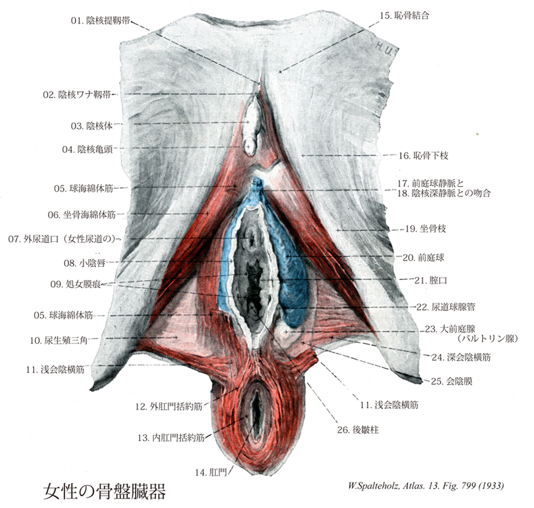
- 799_01【Suspensory ligament of clitoris♀陰核提靱帯 Ligamentum suspensorium clitoridis♀】 Fascial and aponeurotic portion of the superficial investing fascia that attaches the body of the clitoris to the pubic symphysis.
→(浅腹筋膜の正中下端部が靱帯化したもので、陰茎(陰核)ワナ靱帯の深部にあり、恥骨結合前面からおこり、陰茎(陰核)海綿体の基部背面につく。女性では著しく弱い。)
- 799_02Retzius' band【Fundiform ligament of clitoris♀陰核ワナ靱帯;陰核係蹄靱帯(♀) Ligamentum fundiforme clitoridis♀】
→(浅腹筋膜の正中線下端の部は陰茎または陰核海綿体の根元に2条の靱帯を送る。その一つは陰茎または陰核ワナ靱帯で、恥骨結合の上方で白線の前面から起こって、弾性線維を多く含み、下端は2脚に分かれ陰茎または陰核海綿体をすすんだあと、陰嚢または大陰唇の皮下に放散する。ほかの一つは陰茎または陰核提靱帯で、前者の下方で恥骨結合の前面から起こり、陰茎または陰核海綿体の基部の背面につく。いずれも女性では著しく弱い。)
- 799_03【Body of clitoris陰核体 Corpus clitoridis】 Union of the two crura of clitoris below the pubic symphysis.
→(陰核体は左右1対の陰核海綿体が結合性の陰核筋膜で包まれてできる。陰核は陰茎と異なって尿道で貫かれていないので、陰核対には尿道海綿体もない。陰核対には恥骨結合から起こる陰核提靱帯がつく。)
- 799_04【Glans of clitoris陰核亀頭 Glans clitoridis♀】 End of the body of clitoris that ca. swell.
→(陰核亀頭は前庭球と関連している陰核の尖端。部分的に包皮で被われる。)
- 799_05【Bulbospongiosus muscle球海綿体筋 Musculus bulbospongiosus; Musculus bulbocavernosus】 Male: Muscle arising from the perineal body and the inferior aspect of the corpus spongiosum of penis, passing to the perineal membrane and dorsum of penis. It is unpaired. It acts to compress the bulb of penis and transport urethral contents further. ABC Female: Muscle that originates on the ramus of ischium, attaching to and covering the cms of clitoris. It assists in filling the cavernous bodies with blood. I: Pudendal nerve.
→(男性では球海綿体は尿道球の周辺を不体の筋として回るが、会陰の中心腱と尿道海綿体下側の正中縫線から起こる。球海綿体は前方へ放散し、海綿体のまわり下尿生殖隔膜筋膜や尿道海綿体へ向かい、また前筋線維をもって陰茎背部へ付く。この筋は随意的または反射的に尿道球を圧迫し、それにより尿道の内容を駆出する。女性では球海綿体筋は男性のように全長で1つの筋にはなっていない。2つの筋が会陰の中心腱より起こるが、各筋はそれぞれ引き続き前庭球と大前庭腺を被っている。その筋束は前庭球や陰核海綿体に停止し、陰核体後部で反対側からの筋線維と絡み合っている。この筋は大前庭腺を反射的に空にし、血液を前庭球の後方拡大部から送り出し、またオルガスムの際外腟口を収縮させる。)
- 799_06【Ischiocavernosus muscle坐骨海綿体筋 Musculus ischiocavernosus】 Male: Muscle extending from the ramus of ischium over the cms of the penis to the tunica albuginea. Smaller bundles of muscle fibers run over the penis below the pubic symphysis to the contralateral side.
→(坐骨海綿体筋は男性より女性のほうが発達が弱い。この筋は坐骨枝より起こり陰核脚を被い、その腱性線維は外下表面に付く。その筋は陰核海綿体を圧し、血液を押し付け流出を妨げ、それにより陰核の勃起成立を助ける。)
- 799_07【External urethral orifice; External urinary meatus; External urethral opening外尿道口(女性尿道の) Ostium urethrae externum; Orificium urethrae externum】
→(陰核2~3cm下方に位置する。(Feneis))
- 799_08【Labium minus; Lesser lip小陰唇 Labium minus pudendi】 Cutaneous fold that is devoid of fat and hair and contains sebaceous glands. It forms the boundary of the vestibule of vagina.
→(小陰唇は大陰唇の内側にある2つの皮膚とヒダである。皮膚は毛や脂肪を欠き平滑で、粘膜に似ている。陰毛はないが豊富な皮脂腺をもつ扁平なヒダ。成人では小陰唇は大陰唇で被われている。小児では、大陰唇の発達が悪いので、小陰唇の大部分は陰核とともに露出する。後方で左右が連絡する部分に陰唇小体というヒダをつくる。)
- 799_09【Carunculae hymenales; Hymenal caruncle処女膜痕 Carunculae hymenales】 Remains of the hymen on the wall of the vagina following childbirth.
→(処女膜痕は分娩後の裂傷後に、腟口を取り囲む処女膜の残った多数の小膜片。)
- 799_10【Urogenital triangle; Urogenital region尿生殖部;尿生殖三角;尿生殖器部 Regio urogenitalis; Trigonum urogenitale】 Region around the perineum that is located anterior to an imaginary line connecting the two ischial tuberosities.
→(尿生殖三角は左右の坐骨結節をむすぶ線の前方部で泌尿器官を囲む周辺部部域。この領域の浅筋膜のうち脂肪層(キャンパー筋膜)は坐骨直腸窩内の脂肪層に接続するものであり、同時に大腿皮下の浅筋膜にも移行する。陰嚢では脂肪層が平滑筋層(肉様膜)で置き換えられている。肉様膜は寒冷刺激に応じて収縮し、陰嚢皮膚の表面積を減少させる。尿生殖三角線筋膜のうち線維層(コールス筋膜)は後方では尿生殖隔膜後縁に付着し、外側方では恥骨弓の辺縁に付着するほか、前方では前腹壁浅筋膜の線維層(スキャルパ筋膜)へと移行する。このような尿生殖三角浅筋膜の線維層は陰茎あるいは陰核の部位では管状の鞘構造をとり、また陰嚢あるいは大陰唇の部位では著明な1層をなす。浅会陰隙とは、下方を会陰線筋膜の線維層で境され、上方を尿生殖隔膜で境されるような隙間のことである。この隙間は後方では隙間の上壁と下壁がたがいに癒着する形で閉じられ、外側方でも隙間の上壁と下壁が恥骨弓辺縁部に付着する形で閉じられている。しかし浅会陰隙はその前方部で前腹壁浅筋膜と前腹壁筋の間の隙間と自由に交通する。)
- 799_11Thiele's muscle【Superficial transverse perineal muscle浅会陰横筋 Musculus transversus perinei superficialis】 Inconstant expansion of the deep transverse perineal muscle that extends from the ischial tuberosity to the perineal body. I: Pudendal nerve.
→(浅会陰横筋は横走する浅在性の薄い筋である。この筋は坐骨結節や坐骨枝の境界域の起始部ではしばしば坐骨海綿体筋と交通し、そこから分枝する。その線維は会陰体へ連なり、外肛門括約筋と球海綿体筋に放散する。女性ではその筋は多少退化し、わずかに筋膜の被膜のみ同定できる程度である。)
- 799_12【External anal sphincter muscle外肛門括約筋 Musculus sphincter ani externus】 Transversely striated outer sphincter muscle of the anus. It consists of the following three parts. I: Pudendal nerve.
→(外肛門括約筋は肛門挙筋の表層にあり、肛門を囲む横紋筋。内肛門括約筋の衿のように張り付いている。外口門括約筋のほぼ矢状面に位置する筋束が腸間終端を両側から閉鎖する。この筋束は後方では尾骨から張る靱帯(肛門尾骨靱帯)に付着し、前方では会陰中心に付いている。)
- 799_13【Internal anal sphincter muscle; Internal sphincter of anus内肛門括約筋 Musculus sphincter ani internus】 Thickened ring of muscle about 1-2 cm high in the circular muscular layer around the anus.
→(高さ1~2 cmの、輪走する強い肛門輪筋。(Feneis))
- 799_14【Anus肛門 Anus】 Inferior opening of the anal canal, encircled by the subcutaneous and superficial parts of external anal sphincter muscle.
→(肛門は消化下管の末端が外部へ開く部分である。外肛門括約筋の表在部と皮膚部に囲まれた肛門管の開口。 実質的な機能を持つ肛門は、むしろ肛門管と考えてよい。)
- 799_15【Pubic symphysis; Pubis symphysis; Symphysis pubis恥骨結合 Symphysis pubica】 Cartilaginous joint between the rami of the pubis.
→(恥骨結合は骨盤前面で左右の恥骨が正中線上で向かい合ってできる連結。両側の恥骨結合面がうすい硝子軟骨に被われ、その間に線維軟骨でできる恥骨間円板が連結する。その構造は椎間円板の線維輪に似る。女性では妊娠時にこの結合は弱められ、またこのことは分娩時における新生児の頭の産道通過を助ける。モルモットなどでは、女性ホルモンの投与によって、実験的にこの結合を弱めることができる。付属する靱帯に次のものがある。(1)上恥骨靱帯:恥骨結合の上縁で左右の恥骨を結ぶ。(2)恥骨弓靱帯:恥骨結合の下縁で、左右の恥骨下枝を結び、恥骨弓をつくる。下面で尿生殖膜との間隙を陰茎静脈が通る。 Symphysisははsyn(一緒に)physis(生える)、すなわち「自然に癒合したもの」という意味である。解剖学用語としてのsymphysisは線維軟骨結合という一般名詞であるが、慣用的にはpubicaという形容詞なしでも恥骨結合を指すことが多い。正しい読み方はスィンフィスィスである。)
- 799_16【Inferior pubic ramus; Ramus of pubis恥骨下枝;結合部(恥骨枝の) Ramus inferior ossis pubis; Pars symphysica ramus ossis pubis】 The part of the pubis that is anteroinferior to the obturator foramen, between the pubic symphysis and the suture line with the ischium.
→(恥骨下枝は閉鎖溝の下縁の前半をつくる。内外から圧平された骨板で、とくに閉鎖孔に向く上縁は薄く鋭い。下縁はやや肥厚する。下縁の後方部の前面付近は粗に隆起してCrista phalica[INA](陰茎稜)をつくる。)
- 799_17【Veins of bulb of vestibule; Veins of vestibular bulb♀腟前庭球静脈;前庭球静脈(♀) Venae bulbi vestibuli♀】 Vein conveying blood either to the deep dorsal vein of clitoris or internal pudendal vein.
→(前庭球静脈は前庭球動脈に伴行する。前庭球の静脈血を集め、坐骨直腸窩で内陰部静脈へそそぐ。)
- 799_18【Deep veins of clitoris♀陰核深静脈(♀) Venae profundae clitoridis♀】 Veins emptying the clitoris. They correspond to the deep veins of penis.
→(陰核深静脈は陰茎深静脈に相当する陰核の静脈。)
- 799_19【Ramus of ischium坐骨枝;坐骨下枝 Ramus; Ramus inferior (Os ischii)】 The part of the ischium below the obturator foramen. Its anterior end articulates with the inferior pubic ramus.
→(坐骨結節のところから前方に伸びて恥骨下枝に合する部で、閉鎖孔の下縁の後半をつくる。坐骨結節につづく下縁がやや厚く、閉鎖孔に向かう上縁が鋭い三角柱状をなす。)
- 799_20【Bulb of vestibule前庭球 Bulbus vestibuli】 Erectile tissue corresponding to the corpus spongiosum of penis. It is mainly situated at the roots of the labia majora.
→( 前庭球は腟前庭の左右両側にある。男性の尿道海綿体および尿道球にあたるもので、女性では腟によって左右に分かれる。 左右の前庭球は前端で合して陰核亀頭をつくる。 前庭球は海綿体組織ででき、球海綿体筋 で被われる。球海綿体筋は会陰腱中心と前庭球後部から起こり、陰核海綿体に付き、膣口を括約する。 ★前庭球は性的興奮で勃起する。勃起し膨張すると、その後ろにある大前庭腺を圧迫して、その分泌物を腟前庭に排出させる。(解剖学講義))
- 799_21【Vaginal orifice; Vaginal opening腟口 Ostium vaginae】 Opening of the vagina into the vestibule.
→(腟口すなわち腟の開口部は外尿道口の約1cm後方にあり、外尿道口より大きい。大前庭腺の導管は腟口のすぐ外側で、小陰唇との間に開いている。)
- 799_22【Duct of bulbo-urethral gland尿道球腺管;排出管(尿道球腺の) Ductus glandulae bulbourethralis; Ductus excretorius】 The 3-4 cm long excretory duct.
→(尿道球腺管は3~4cm長の腺排出管。)
- 799_23Bartholin's gland; Duverney's gland; Giedemann's gland【Greater vestibular gland; Bartholin's gland大前庭腺;バルトリン腺 Glandula vestibularis major】 Mucous glands on both sides of the posterior end of the bulb of the vestibule. Their long excretory duct opens at the vestibule of vagina between the labia minora and vaginal orifice.
→(バルトリン腺とも呼ばれる。大前庭線は男性の尿道球腺にあたるエンドウ豆大の球状の腺で、腟口の左右両側の深部で浅会陰隙にある。大前庭腺は無色ないし薄い乳白色の粘稠な分泌物を分泌する。分泌は性的興奮によって促進され腟前庭を潤す。バルトリン家はデンマークで三代続いた医学者の家系である。この腺に名をのこすのは孫のKaspar Baritolin II (1655-1738)で、1677年に記載された。このほかにバルトリン管(舌下線管Bartholin's duct: 1685年)にもその名がある。)
- 799_24【Deep transverse perineal muscle♀深会陰横筋(女の) Musculus transversus perinei profundus♀】
→()
- 799_25【Inferior fascia of urogenital diaphragm; Perineal membrane会陰膜;外尿生殖隔膜筋膜;外尿生殖隔膜筋膜 Membrana perinei; Fascia diaphragmatis urogenitalis inferior; Fascia diaphragmatis urogenitalis externa】
→(深会陰横筋の前下方にある筋膜。)
- 799_26【Posterior vaginal column後皺柱;後ヒダ柱;後腟摺柱 Columna rugarum posterior】 Longitudinal ridge in the anterior vaginal wall.
→()