Spalteholz HANDATLAS DER ANATOMIE DES MENSCHEN VON WERNER SPALTEHOLZ
メニューは解剖学(TA)にリンクしてあります。図の番号をクリックすると下記の説明へ、右側の用語をクリックすると解剖学(TA)にジャンプします。
907


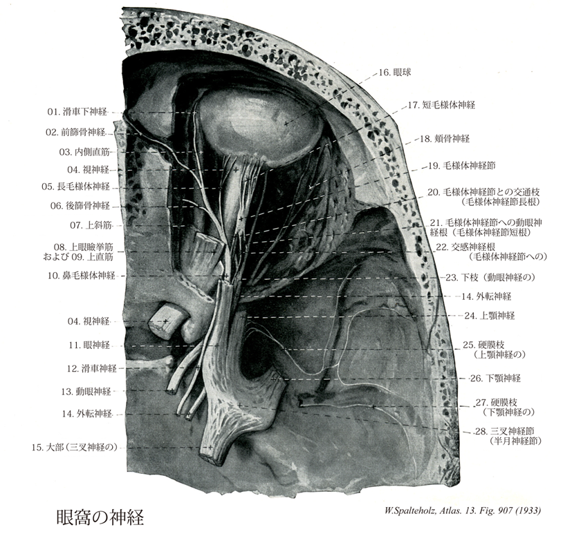
- 907_00【Orbit; Orbital cavity眼窩 Orbita; Cavitas orbitalis】 Orbital cavity that contains the eyeball and its appendages.
→(眼窩は眼球とその付属器とを容れる不規則な四角錐体状の大きなくぼみで、最深部はその後内方にある。錐体底にあたる部はほぼ四辺形の眼窩口で、軽度外下方に傾いており、顔面に開いている。その上縁を眼窩上縁、下縁を眼窩下縁という。眼窩上縁は前頭鱗からなり、その内側半分に2個の切痕または孔があり、その内側のものを前頭切痕(まれに前頭孔)、外側のものを眼窩上孔(まれに眼窩上切痕)とう。眼窩下縁は上顎骨体および頬骨からなり、その下方に眼窩下孔が開口している。眼窩は上・下・内側・外側の4壁を有し、7種類の骨による10部より形成されている。上壁は大部分が前頭骨眼窩面および蝶形骨小翼腹側面よりなり、外側には涙腺窩、小翼内には視神経管があり、ここに視神経および眼動脈を通す。下壁は大部分が上顎骨眼窩面によりなるが、外側の一部が頬骨眼窩面、後方の小部分が口蓋骨眼窩突起により形成されている。また後方から前方へ眼窩下溝その延長部である眼窩下管が走り、これが既述の眼窩下孔に開口する。内側壁は大部分が篩骨眼窩板により形成され、残りの部分のうちの前部は上顎骨前突起および涙骨、後部は蝶形骨体側面最前部によって形成されている。なお篩骨眼窩板上縁と前頭骨眼窩部との間には、前篩骨孔および後篩骨孔があり、前者は鼻腔に行く前篩骨神経および前篩骨動脈を通し、後者は篩骨蜂巣に行く後篩骨神経および後篩骨動脈を通す。また内側壁の前部にある涙嚢窩は、上顎骨の前涙嚢稜と涙骨の孔涙嚢稜との間にあり、稜骨の涙嚢溝が合して形成されたものである。外側壁は前半部は頬骨眼窩面、後半部は蝶形骨大翼眼窩面と上壁の蝶形骨小翼との間には頭蓋腔に通ずる上眼窩裂があり、動眼神経、滑車神経、眼神経、外転神経、上眼静脈などを通す。また外側壁後半部の蝶形骨大翼眼窩面と下壁の上顎骨眼窩面との間には翼口蓋窩および側頭下窩に通ずる下眼窩裂があり、眼窩下神経、頬骨神経、下眼静脈などを通す。)
- 907_01【Infratrochlear nerve滑車下神経 Nervus infratrochlearis】 Nerve passing beneath the muscle sling of the superior oblique to the inner angle of the eye. It supplies the lacrimal sac, Iacrimal caruncle, and surrounding skin.
→(上斜筋の下を通り眼窩内側壁を前進し、滑車上神経内側枝と結合して神経弓を作り、これから眼瞼枝を出し上、下眼瞼および内眼角の皮膚と涙嚢に分布する。)
- 907_02【Anterior ethmoidal nerve前篩骨神経 Nervus ethmoidalis anterior】 Nerve emerging through the anterior ethmoidal foramen into the cranial cavity. It travels outside of the dura mater before passing through the cribriform plate of the ethmoid into the nose
→(前篩骨孔を通って頭蓋腔に入り、篩板の上を前進し、さらに鼻孔に入って内鼻枝と外鼻枝とに分かれる。前者は鼻粘膜の前上部に分布し、後者は鼻骨後面の篩骨神経溝を通り、鼻骨と鼻軟骨の間で鼻背に出て皮膚に分布する。)
- 907_03【Medial rectus muscle内側直筋;内側眼球直筋 Musculus rectus medialis; Musculus rectus bulbi medialis】 o: Common tendinous ring, i: About 5.5 mm from the corneal margin. Action: Adduction of the corneal pole. I: Oculomotor nerve.
→(内側直筋は眼球の鼻側および耳側を走り、その停止腱は角膜縁の後方約6mmの強膜に放射状に停止する。目の動き:視線を内側に向ける。)
- 907_04【Optic nerve [II]視神経;視束[脳神経II] Nervus opticus; Fasciculus opicus [II]】 Nerve emerging medial to the posterior pole of the eyeball and extending to the optic chiasma.
→(視神経は脳神経の1つとして扱われてはいるが、実は前脳胞の延長部である。眼球網膜の第8層である神経細胞層中にある多極神経細胞から出る神経線維が集まって出来る神経である。すなわち杆状体細胞および錐体状細胞よりの興奮は網膜の内顆粒層の双極細胞に伝わり、それがさらに神経細胞層の細胞に連絡し、この神経細胞の出す神経突起である線維はまず眼球の後極よりやや内下方の一ヶ所に集まって、視神経円板を作り、強大な神経幹となり、網膜の続きである視神経鞘に囲まれて後内側に向かう。眼球から約15~20mm隔ったたところで、眼動脈の枝である網膜中心動脈およびこれに伴う静脈が外側から入り込み、その中軸を通って網膜に分布する。左右両側の視神経は眼窩後端の視神経管を通って頭蓋腔に入り、次第に相近づいて蝶形骨体上の視神経溝でほぼ半交叉をして視交叉を作り、そのつづきは視索と名が変わって間脳の外側膝状体および中脳の上丘などの第一次視覚中枢に達して、ここで終わる。網膜が眼胚から発達するので経路に相応する。ヒトの視神経は眼球網膜の神経細胞層中にある多極神経細胞から出る100万本以上の神経線維からなる。すなわち、杆状体細胞および錐体状細胞よりの興奮は網膜の内顆粒層の双極細胞に伝わり、それがさらに神経細胞層の細胞に連絡し、この神経細胞の出す神経突起である線維はまず眼球の後極よりやや内下方の一ヶ所に集まって、視神経円板を作り、強大な神経幹となり、網膜の続きである視神経鞘に囲まれて後内側に向かう。眼球から約15~20mm隔ったたところで、眼動脈の枝である網膜中心動脈およびこれに伴う静脈が外側から入り込み、その中軸を通って網膜に分布する。左右両側の視神経は眼窩後端の視神経管を通って頭蓋腔に入り、次第に相近づいて蝶形骨体上の視神経溝でほぼ半交叉をして視交叉を作り、そのつづきは視索と名が変わって間脳の外側膝状体および中脳の上丘などの第一次視覚中枢に達して、ここで終わる。)
- 907_05【Long ciliary nerves長毛様体神経 Nervi ciliares longi】 Two, long, thin branches that carry sympathetic fibers to the dilator pupillae and afferent fibers from the iris, ciliary body, and cornea.
→(通常2本あって、毛様体神経節から出る短毛様体神経とともに視神経の付近で眼球に入り、強膜を貫いて、これと脈絡膜の間を前にすすみ、毛様体と虹彩にいたる鈍知覚枝である。)
- 907_06【Posterior ethmoidal nerve後篩骨神経 Nervus ethmoidalis posterior】 Nerve emerging at the posterior end of the orbit through the posterior ethmoidal foramen to supply the mucosa of the sphenoidal sinus and posterior ethmoidal cells.
→(鼻毛様体神経の枝で、後篩骨孔を通って、覚枝を後篩骨洞および蝶形骨洞に送る。)
- 907_07【Superior oblique muscle上斜筋;上眼球斜筋 Musculus obliquus superior; Musculus obliquus bulbi superior】 o:Medial to the common tendinous ring on the body of sphenoid, i: After a hook-shaped course, obliquely behind the equator. Its tendon passes through the trochlea. Action: Abduction, intorsion, and depression of the eye. I: Trochlear nerve.
→(上斜筋は眼窩傍結合組織すなわち視神経鞘と(おもに)蝶形骨体の結合組織である総腱輪の内側から起こる。上斜筋は眼窩錐体の内側直近の上を前方に走行する。眼球の縁で上斜筋の丸みのある腱は結合組織性の吊り索(滑車)を通過し鋭角で後方に曲がる。さらに上斜筋の腱は上直筋の下でこれと交差し眼球上後側頭部の強膜に停止する。目の動き:視線を内側かつ下方に向ける。)
- 907_08【Levator palpebrae superioris muscle上眼瞼挙筋 Musculus levator palpebrae superioris】 o: Upper portion of optic canal and dural sheath of optic nerve. Its insertion tendon widens anteriorly and divides into a superior and an inferior layer. I: Oculomotor nerve.
→(上眼瞼挙筋は視神経管の縁の総腱輪の外側で視神経鞘から起こり、眼窩上壁のすぐ下で前頭神経の下を通り上眼瞼にいく。上眼瞼挙筋の腱は分離して上眼瞼挙筋浅板と上眼瞼挙筋深板に分かれる。前者は上眼瞼中を縁に向かって進み、後者は上瞼板筋の平滑筋細胞を伴って上眼瞼の瞼板に付く。下瞼板筋は下眼瞼板と下結膜円蓋の間の下眼瞼に存在する平滑筋層である。)
- 907_09【Superior rectus muscle上直筋;上眼球直筋 Musculus rectus superior; Musculus retus bulbi superior】 o: Common tendinous ring, i: Along an oblique line passing anterior to the equator, 7-8 mm behind the corneal margin. Action: Elevation and intorsion of superior pole. I: Oculomotor nerve.
→(上直筋は、眼球の上部を斜め外側に進んで眼球の周囲に達し、そこで角膜縁の後方約7-8mmの胸膜に停止腱が放射上に胸膜組織と絡まるように停止する。目の動き:視線を外側かつ上方に向ける。)
- 907_10【Nasociliary nerve; *Nasociliary branch of ophthalmic nerve鼻毛様体神経 Nervus nasociliaris】 Furthest medial branch of the ophthalmic nerve. It initially lies beneath the superior rectus and then between the superior oblique and medial rectus muscles.
→(眼球、類の、鼻粘膜の一部および鼻背皮膚に分布する鈍知覚神経で、上眼窩裂を通って眼窩に入り視神経と上直筋との間を通って斜めに前内側にすすみ、前篩骨孔のあたりで滑車下神経と前篩骨神経とに分かれる。その経過中に出す枝はつぎのようである。)
- 907_11【Ophthalmic nerve; Ophthalmic division [Va; V1]眼神経 [三叉神経第1枝] Nervus ophthalmicus [Va; V1]】 First division of the trigeminal nerve, which passes through the superior orbital fissure.
→(眼神経は第五脳神経の第一枝(CN V1)。蝶形骨体上の海綿静脈洞の外側に沿って前方にすすみ、上眼窩裂を通って眼窩に入る。つぎの枝(①涙腺神経、②前頭神経、③鼻毛様体神経)に分かれる。また眼筋にいたる動眼、滑車、外転の3神経および交感神経との間に交通がある。)
- 907_12【Trochlear nerve [IV]滑車神経[脳神経IV] Nervus trochlearis [IV]】 Nerve exiting on the dorsal side, caudal to the tectal plate. It supplies the superior oblique muscle.
→(滑車神経は脳神経中最少のもので、滑車神経核からでて上斜筋を支配する鈍体性運動性神経である。この神経は脳の背側から脳をでる唯一の脳神経で、下丘のすぐ後方で、上小脳脚と上髄帆小帯との間から出て、大脳脚をめぐり、(側頭骨)錐体尖の近くで硬膜を貫いて海綿静脈洞の上壁に達し、動眼神経の外側から上側に向かって前進し、上眼窩裂を通って眼窩内に入り、上直筋、上眼瞼挙筋起始部の上を前内側にすすんで、上斜筋に分布する。)
- 907_13【Oculomotor nerve [III]動眼神経[脳神経III] Nervus oculomotorius [III]】 Nerve containing motor and parasympathetic fibers that exits the oculomotor sulcus and passes through the superior orbital fissure into the orbit.
→(動眼神経の主成分は動眼神経主核から出る体性運動性のもので外側直筋および上斜筋以外の眼筋を支配する。このほかに副交感性の動眼神経副核[Edinger-Westphal核]から出る線維が加わる。以上の2核から出る線維は多数の根をつくって大脳脚内側溝から出て1神経幹となり、滑車神経、外転神経および眼神経とともに、蝶形骨体の両側にある海綿静脈洞の上壁に沿ってすすみ、上眼窩裂を通って眼窩内に入り、上下の2枝に分かれる。上枝は上瞼挙筋および上直筋に、下枝は内側直筋、下直筋および下斜筋に分布する。また下枝からはきわめて短い動眼神経からの根が出て、毛様体神経節に入るが、これは動眼神経副核から出て、下枝を通って毛様体神経節に入る副交感線維にほかならない。)
- 907_14【Abducent nerve[VI]; Abducens nerve [VI]外転神経[脳神経VI] Nervus abducens [VI]】 Cranial nerve emerging from the brain at the angle between the pons and medulla oblongata. It penetrates the dura mater at a point half as high as the clivus, continues laterally in the cavernous sinus, and then passes through the superior orbital fissure into the orbit where it supplies the lateral rectus.
→(外転神経は第六脳神経である。外側直筋に至る鈍体性運動性神経で、その起始核たる外転神経核は橋の中にあり、これから出る神経は橋の後縁で正中線に近く表面に現れ、内頚動脈の外側を通って上眼窩裂から眼窩に入り、外側直筋の内側からそのなかに入る。)
- 907_15【Sensory root of trigeminal nerve感覚根;知覚性根;知覚根;大部(三叉神経の);三叉神経根 Radix sensoria; Portio major; Radix nervi trigemini】 Sensory part of the nerve that emerges caudally from the pons and extends to the trigeminal ganglion.
→(三叉神経の知覚根は体性感覚線維で三叉神経の大部に相当し、橋に入り、三叉神経主感覚核と三叉神経脊髄路核に分布する。)
- 907_16【Eyeball眼球 Bulbus oculi】
→(眼球は名前のように球状(直径約25mm・体積約8cm3)で、視覚器の主要部をなす。眼窩脂肪体、眼筋筋膜、眼球鞘などに包まれて眼窩中にあり、前方からは眼瞼により保護されている。また眼筋の働きにより球関節に似た自由度の高い体軸性運動を行う。眼球の内部には前方に眼房水、後方に硝子体が満ちて、12~22mmHgの内圧が保たれる。眼球の形状を規定するため、前極、後極、赤道、経線、外眼球軸(前・後極を結ぶ)、内眼球軸、視軸などを用いる。眼球軸は角膜と水晶体前・後面の曲率中心を通る軸で、網膜面では中心窩と円板の中間を通る。したがって水晶体後面の屈曲率中心と中心窩を結ぶ視軸とは一致しない。眼球壁は組織発生的に、①眼球線維膜(強膜、角膜)、②眼球血管膜(脈絡膜、毛様体筋、虹彩支質、角膜内皮、胎児期の瞳孔膜)、③眼球内膜(網膜視部、毛様体・虹彩色素上皮層)の3層よりなる。①と②は中胚葉、③は神経外胚葉に由来する。内部の水晶体は体表外胚葉、硝子体は中胚葉由来であり、眼瞼・眼球膜、角膜上皮は皮膚の表皮の続きである。)
- 907_17【Short ciliary nerves短毛様体神経 Nervi ciliares breves】 Up to 20 short nerves that pierce the sclera around CN II. They contain postganglionic parasympathetic fibers from the ciliary ganglion as well as postganglionic sympathetic fibers of the sympathetic root that carry information to the eye. They contain sensory fibers of the nasociliary root that carry impulses away from the eye.
→()
- 907_18【Zygomatic nerve頬骨神経 Nervus zygomaticus】 Nerve that divides in the pterygopalatine fossa. It passes through the inferior orbital fissure to the lateral wall of the orbital cavity and anastomoses with the lacrimal nerve.
→(翼口蓋窩から下眼窩裂を通り眼窩に入り、その外側壁で涙腺神経との交通枝を出し、のちに次の2枝分かれる。涙腺神経との結合により翼口蓋神経節よりの副交感神経線維が涙腺にいくことなる。)
- 907_19【Ciliary ganglion毛様体神経節 Ganglion ciliare】 Ganglion situated on the posterolateral aspect of the optic nerve that contains cells for the postganglionic parasympathetic fibers to the ocular muscles. They constrict the pupils and contract the ciliary muscle for near vision.
→(眼窩内にある自律神経節でそこに含まれれる神経細胞の大多数は副交感神経節後ニューロン(その興奮により眼球内の虹彩にある瞳孔括約筋を収縮させて縮瞳をおこさせると同時に、毛様体筋をも収縮させて毛様小帯を弛緩させることにより水晶体の厚みを増加させる)の細胞体である。この自律神経節には①動眼神経からの枝(副交感性根)、②交感神経枝、③鼻毛様体神経との交通枝(知覚根)、および④数条の短毛様体神経が連絡するが、動眼神経からの枝の中に含まれる副交感神経節前線維のみが上記の神経細胞体とシナプスを形成する。一方、②および③の中にはそれぞれ交感神経節後線維(虹彩の瞳孔散大筋を支配)と眼球の近くをつかさどる神経線維が含まれるが、これらは毛様体神経節内を素通りするだけで同神経節内から新たにおこる副交感性節後線維群とともに④を形成して眼球内にすすむ。)
- 907_20【Communicating branch with ciliary ganglion; Sensory root of ciliary ganglion; Nasociliary root of ciliary ganglion; Communicating branch of nasociliary nerve with ciliary ganglion毛様体神経節との交通枝;毛様体神経節の感覚根;毛様体神経節の鼻毛様体根;毛様体神経節長根 Ramus communicans cum ganglio ciliari; Radix sensoria ganglii ciliaris; Radix nasociliaris ganglii ciliaris; Radix longa(Ganglion ciliare)】 Sensory fibers passing through the ciliary ganglion from the eye to the nasociliary nerve.
→()
- 907_21【Parasympathetic root of ciliary ganglion; Oculomotor root of ciliary ganglion; Branch of oculomotor nerve to ciliary ganglion副交感神経根;動眼神経根;毛様体神経節への動眼神経根;毛様体神経節短根 Radix parasympathica; Radix oculomotoria; Ramus nerve oculomotorii ad ganglion ciliare; Radix brevis(Ganglion ciliare)】 Preganglionic parasympathetic fibers from CN III.
→(毛様体神経節の副交感神経根は毛様体神経節へ副交感神経の節前線維を送っている動眼神経の枝。)
- 907_22【Sympathetic root to ciliary ganglion交感神経根(毛様体神経節への);毛様体神経節交感根 Radix sympathica ganglii ciliaris; Radix sympathica ad ganglii ciliaris】 Postganglionic sympathetic fibers from the internal carotid plexus.
→(毛様体神経節の交感神経根は内頚動脈神経叢から出て毛様体神経節を素通りして眼球に至る節後線維で、その細胞体は上頚神経節にある。)
- 907_23【Inferior branch of oculomotor nerve下枝(動眼神経の) Ramus inferior (Nervus oculomotorius)】 Lower branch supplying the medial rectus, inferior rectus, and inferior oblique muscles.
→(動眼神経の下枝は運動枝を内側直筋・下直筋・下斜筋に送り、副交感神経節前枝をその根部から毛様体神経節に送る。)
- 907_24【Maxillary nerve; Maxillary division [Vb; V2]上顎神経[三叉神経第2枝] Nervus maxillaris [Vb; V2]】 Second division of the trigeminal nerve. It passes though the foramen rotundum to the pterygopalatine fossa and continues through the orbital fissure into the orbit.
→(三叉神経の第2枝で蝶形骨大翼の正円孔を通って頭蓋腔を去り、翼口蓋窩で頬骨神経および翼口蓋神経を出した後、眼窩下神経となって眼窩下裂を経て眼窩に入り、顔面まで達する。知覚枝は下眼瞼の皮膚と結膜、上唇と頬の皮膚と粘膜、口蓋、上顎歯と歯肉、上顎洞、鼻翼および鼻腔の後下部に分布する)
- 907_25【Meningeal branch of maxillary nerve硬膜枝;中硬膜枝(上顎神経の) Ramus meningeus (Nervus maxillaris)】 Branch given off anterior to the foramen rotundum that passes to the dura mater in the frontal region of the middle meningeal artery.
→(上顎神経の硬膜枝は頭蓋内で分かれ、中硬膜動脈とともに分布して脳硬膜にいたる知覚枝で、下顎神経の硬膜枝と交通する。)
- 907_26【Mandibular nerve; Mandibular division of trigeminal nerve [Vc; V3]下顎神経[三叉神経第3枝] Nervus mandibularis [Vc; V3]】 Third division of the trigeminal nerve that passes through the foramen ovale into the infratemporal fossa. It contains sensory fibers and motor fibers for the muscles of mastication.
→(三叉神経節からの感覚線維と運動根が卵円孔で結合してできる三叉神経の第三枝で最も太く、この神経は三叉神経節から出てただちに蝶形骨大翼の卵円孔を通って側頭下窩に出て硬膜、咀嚼筋、頬粘膜、耳介、外耳道付近その他へ枝を与えた後、舌神経、下歯槽神経の2終枝に分かれる。)
- 907_27【Meningeal branch of mandibular nerve; Nervus spinosus硬膜枝(下顎神経の) Ramus meningeus (Nervus mandibularis); Nervus spinosus】 Nerve passing through the foramen spinosum, accompanying the two branches of the middle meningeal artery. It supplies the dura mater, part of the sphenoidal sinus, and the mastoid cells.
→(下顎神経の硬膜枝は頭蓋を出るとすぐ分かれて棘孔を通って再び頭蓋腔に入り、上顎神経の硬膜枝とともに、中硬膜動脈に沿って脳硬膜に分布する知覚枝で、なお蝶形骨大翼乳突蜂巣の内部にも線維を与える。)
- 907_28Gasserian ganglion【Trigeminal ganglion三叉神経節;半月神経節 Ganglion trigeminale; Ganglion semilunare】 Crescent-shaped equivalent of a spinal ganglion of the trigeminal nerve lying in an outpouching in the subarachnoid space (trigeminal cavity) above the foramen lacerum on the medial, anterior surface of the petrous temporal bone.
→(ガッセル神経節またはガッサー神経節とよばれる。三叉神経の大きい扁平な知覚神経節で、中頭蓋窩の正中部分に沿った静脈洞に密接して脳硬膜の三叉神経腔にある。オーストラリアの解剖学者Johann Laurentius Gasser (1723-1765頃)によって報告された。彼自身についてはよく判っていない。)