Spalteholz HANDATLAS DER ANATOMIE DES MENSCHEN VON WERNER SPALTEHOLZ
メニューは解剖学(TA)にリンクしてあります。図の番号をクリックすると下記の説明へ、右側の用語をクリックすると解剖学(TA)にジャンプします。
913


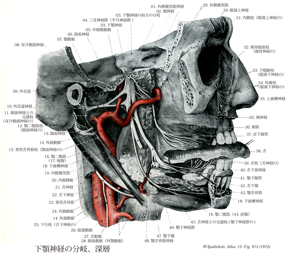
- 913_01【Nerve to lateral pterygoid; Nerve to lateral pterygoid muscle外側翼突筋神経;外翼突筋神経 Nervus pterygoideus lateralis; Nervus pterygoideus externus】 Motor branch supplying the lateral pterygoid. It commonly arises together with the buccal nerve.
→()
- 913_02【Buccal nerve頬神経;頬筋神経 Nervus buccalis; Nervus buccinatorius】 Sensory branch supplying the skin and mucosa of the cheek and buccal gingiva near the first molar tooth.
→(これは他枝とは異なり知覚神経で、外側翼突筋を貫き、またはその下を通り頬筋の外側に出て前にすすみ口角に至る。この間に頬粘膜に分布するとともに、一部は頬の皮膚に至り顔面神経の枝と結合する。)
- 913_03【Mandibular nerve; Mandibular division of trigeminal nerve [Vc; V3]下顎神経[三叉神経第3枝] Nervus mandibularis [Vc; V3]】 Third division of the trigeminal nerve that passes through the foramen ovale into the infratemporal fossa. It contains sensory fibers and motor fibers for the muscles of mastication.
→(三叉神経節からの感覚線維と運動根が卵円孔で結合してできる三叉神経の第三枝で最も太く、この神経は三叉神経節から出てただちに蝶形骨大翼の卵円孔を通って側頭下窩に出て硬膜、咀嚼筋、頬粘膜、耳介、外耳道付近その他へ枝を与えた後、舌神経、下歯槽神経の2終枝に分かれる。)
- 913_04Gasserian ganglion【Trigeminal ganglion三叉神経節;半月神経節 Ganglion trigeminale; Ganglion semilunare】 Crescent-shaped equivalent of a spinal ganglion of the trigeminal nerve lying in an outpouching in the subarachnoid space (trigeminal cavity) above the foramen lacerum on the medial, anterior surface of the petrous temporal bone.
→(ガッセル神経節またはガッサー神経節とよばれる。三叉神経の大きい扁平な知覚神経節で、中頭蓋窩の正中部分に沿った静脈洞に密接して脳硬膜の三叉神経腔にある。オーストラリアの解剖学者Johann Laurentius Gasser (1723-1765頃)によって報告された。彼自身についてはよく判っていない。)
- 913_05【Middle meningeal artery中硬膜動脈 Arteria meningea media】 Artery passing medial to the lateral pterygoid and through the foramen spinosum into the middle cranial fossa, where it distributes vessels between the dura mater and bone.
→(中硬膜動脈は顎動脈より起こり外側翼突筋の内側を通り棘孔から中頭蓋窩に入り、そこで岩様部枝、腹硬膜枝、上鼓室動脈、前頭枝、頭頂枝に分枝する。上記の部位と終末枝を通って前頭蓋窩と中頭蓋窩に分布し、後頭動脈の硬膜枝、上行咽頭動脈、眼動脈、涙腺動脈、茎乳突孔動脈、顎動脈の腹硬膜枝、深側頭動脈と吻合する。)
- 913_06【Chorda tympani; Chorda tympani nerve鼓索神経 Chorda tympani】 Bundle of parasympathetic fibers to the submandibular ganglion and sensory fibers from the taste buds of the anterior two-thirds of the tongue. As a recurrent nerve it traverses the tympanic cavity, running between the malleus and incus, then continues through the petrotympanic fissure (glaserian fissure) or sphenopetrosal fissure to join the lingual nerve.
→(顔面神経管下端の近くで分かれ、鼓索神経小管を通って鼓室に入り、鼓膜の内面でキヌタ骨長脚とツチ骨柄との間を通って前進し、錐体鼓室裂を通って頭蓋底外面に出た後、後耳介神経と中硬膜動脈との内側を前進し、鋭角をなして舌神経に合する。味覚神経線維を舌神経に送り、顎下腺、舌下腺の分泌神経線維を顎下神経節に送るものである。)
- 913_07【Maxillary artery顎動脈;上顎動脈;内顎動脈 Arteria maxillaris; Arteria maxillaris interna】 Thicker terminal branch of the external carotid artery. It lies beneath the temporomandibular joint and behind the ramus of mandible, running laterally or medially from the lateral pterygoid to the pterygopalatine fossa.
→(顎動脈は外頚動脈の最大の終枝である。下顎頚の後で起こり、咀嚼筋を通り、下顎枝の内側(側頭下窩)を前に走って翼口蓋窩に入る。顎動脈は顔面・頭部の深部(脳硬膜・鼓室・咀嚼筋・上顎骨・下顎骨・歯・歯肉・口蓋・鼻腔など)に広く分布する動脈で、その経過中に多くの枝を出している。顎動脈は外側翼突筋の外側(すなわち表層)を走る場合が多いが(94%)、外側翼突筋の内側(すなわち下層)を走る例も6%の頻度で見られる。また、顎動脈が頬神経の下層を通る例も24%にみられる。顎動脈に伴行するべき静脈が、太い単一の血管ではなく、静脈叢の形になっているのは、咀嚼運動の際の咀嚼筋の収縮瘤によって静脈壁が圧迫されて「欝血」congestionを起こすのを防ぐためである。)
- 913_08【Auriculotemporal nerve耳介側頭神経 Nervus auriculotemporalis】 Nerve usually encircling the middle meningeal artery. It sends a small branch to the temporomandibular joint and then ascends between the ear and superficial temporal artery to the skin of the temple.
→(中硬膜動脈を間にはさむ2根をもって始まり、下顎骨の関節突起の内側を通って後に向い、つぎに弓状をえがいて外上方に曲がり、耳下腺の下で浅側頭動脈の後側に達し、つぎに多くの枝に分かれて耳介前側および側頭部の皮膚に分布する。)
- 913_09【External acoustic meatus; External auditory meatus外耳道 Meatus acusticus externus】
→(外耳道は側頭骨の鼓室部を耳介から鼓膜へ至る通路で骨性部分。軟骨性外耳道からなる。)
- 913_10【Nerve to external acoustic meatus外耳道神経 Nervus meatus acustici externi】 Usually two small branches that supply the skin of the external acoustic meatus.
→(外耳道の皮膚に分布し、その鼓膜枝は鼓膜外面に至る。)
- 913_11【Communicating branches with facial nerve; Communicating branches of auriculotemporal nerve with facial nerve顔面神経との交通枝(耳介側頭神経の) Rami communicantes cum nervus faciali; Rami communicantes nervus auriculotemporalis cum nervus faciali】 Branches communicating with the facial nerve. They convery parasympathetic fibers from the otic ganglion via the facial nerve to the paratid gland.
→(耳神経節よりでる副交感性の線維を顔面神経を通り耳下腺へ送る。 (Feneis))
- 913_12【Digastric branch of facial nerve二腹筋枝;顎二腹筋枝(顔面神経の) Ramus digastricus (Nervus facialis); Nervus digastricus】 Branch supplying the posterior belly of digastric.
→(顔面神経の二腹筋枝は顎二腹筋の後腹を支配する。)
- 913_13【Facial nerve [VII]顔面神経[脳神経VII] Nervus facialis [VII]】 Nerve arising from the second pharyngeal arch. It emerges from the brain at the pontocerebellar angle between the pons and inferior olive and passes with the vestibulocochlear nerve to the petrous part of the temporal bone, which it exits via the stylomastoid foramen. It supplies the muscles of facial expression.
→(顔面神経は第七脳神経である。狭義の顔面神経と中間神経とを合わせたもので、混合神経である。その主部をなす狭義の顔面神経は運動神経で、起始核たる顔面神経核は延髄上部から橋背部にかけてあり、これから出る神経は橋の後縁で脳を去り、内耳神経とともに内耳道に入り、その底で内耳神経と分かれ、内耳神経と分かれ、顔面神経管孔を経て顔面神経管に入り、間もなく殆ど直角をなして後外側に曲がる。この曲がるところは鼓室前庭窓の後上で顔面神経膝といい、ここに膝神経節がある。ついで弓状に後下方へ走り、茎乳突孔を通って頭蓋底外面に出て耳下腺中に入り、耳下腺神経叢を作った後、つぎつぎに多くの枝を出して広頸筋およびこれから分化したすべての浅頭筋(表情筋)、茎突舌骨筋、顎二腹筋後腹、アブミ骨筋などに分布する。以上の運動神経線維とは別に、膝神経節中の神経細胞から出る味覚神経線維が集まって、舌下腺および顎下腺に至る副交感性の分泌線維とともに中間神経を作り、広義の顔面神経の一部をなす。膝神経節細胞は偽単極性で、神経細胞より出る一条の突起はただちに分かれて、末梢および中枢の2枝となる。中枢枝は顔面神経に密接しつつ内耳道を経て脳に入って孤束核に終わり、末梢枝は、いわゆる上唾液核から出て舌下腺、顎下腺に至る副交感性の分泌腺にとともにいわゆる鼓索神経を作り、途中で再び分泌線維と分かれて舌神経に入り、舌体に分布して味覚を司る。)
- 913_14【External carotid artery外頚動脈 Arteria carotis externa】 It extends from the carotid bifurcation to its terminal division into the superficial temporal and maxillary arteries posterior to the neck of mandible.
→(外頚動脈は主として前頚部と顔面に分布する動脈で、甲状軟骨上縁の高さで総頚動脈から分かれておこり、顎二腹筋後腹と茎突舌骨筋の内側を通り、耳下腺におおわれて下顎後窩を上行し、下顎頚の高さで顎動脈と浅側頭動脈の2終枝に分かれる。分枝は次のとおりである。①上甲状腺動脈、②上咽頭動脈、③舌動脈、④顔面動脈、⑤後頭動脈、⑥後耳介動脈、⑦浅側頭動脈、⑧顎動脈)
- 913_15【Stylohyoid branch of facial nerve茎突舌骨筋枝(顔面神経の) Ramus stylohyoideus (Nervus facialis)】 Branch supplying the stylohyoid that sometimes arises together with the lingual branch.
→(顔面神経の茎突舌骨筋枝は多くの場合舌枝とともに起こり、茎突舌筋へいたる枝。)
- 913_16【Digastricus muscle; *Digastric muscle顎二腹筋 Musculus digastricus; Musculus biventer mandibulae】 o:Mastoid notch, i: Digastric fossa. It has an intermediate tendon that acts on the lesser horn of the hyoid bone by means of a connective tissue sling. Raises the hyoid bone and opens the mouth.
→(顎二腹筋は舌骨の上方にある細長い筋で中間腱で前腹と後腹との2腹に分かれる。その後腹をもって側頭骨乳突切痕で起始し、斜め前・下方へ走る。舌骨付近で後腹は中間腱に移行し、この腱は二分した茎突舌骨筋によって挟まれ、かつ線維性滑車によって舌骨に固定される。前腹(顎舌骨筋からは皮膚側へ位置しているが)は中間腱から起始し、下顎骨内面で下顎下縁近くの二腹筋窩に停止する。顎二腹筋の前腹(下顎神経の枝である顎舌骨筋神経の支配)と後腹(顔面神経の支配)とは神経支配が異なることは注意を要する。下顎が固定されているときには、舌骨を引き上げる。舌骨が固定されているときは下顎骨を後下方に引く。両者は発生学的にも由来を異にし、前腹は顎舌骨筋・口蓋帆長筋などとともに咀嚼筋と同類(鰓弓のうち顎骨弓mandibular archに属する筋)であり、後腹は茎突舌骨筋・アブミ骨筋などとともに顔面表情筋と同類(鰓弓のうち舌骨弓hyoid archに属する筋)である。ちなみに、咀嚼筋は下顎神経で支配され、顔面表情筋は顔面神経支配である。このように発生学的な由来を知れば、色々な筋の支配を整然と整理することができる。)
- 913_17【Posterior belly of digastric muscle後腹(顎二腹筋の) Venter posterior; Venter mastoideus (Musculus digastricus)】 Portion of the digastric muscle that passes from the mastoid notch to the intermediate tendon. I: Nerve to mylohyoid.
→(顎二腹筋の後腹は乳様突起から中間腱までの部分)
- 913_18【Inferior alveolar nerve下歯槽神経 Nervus alveolaris inferior】 Thickest branch of the mandibular nerve containing sensory and motor fibers. It enters the mandibular canal through the mandibular foramen approximately 1 cm posterior to the lingual nerve.
→(下顎神経の終糸の一つで舌神経の後側に出て、下歯槽動脈に伴って下顎孔を通って下顎管に入るが、その直前に顎舌骨筋神経を出す。下顎管内では数枝に分かれ、これが歯槽下で結合して下歯神経叢を作り、下顎の歯および下顎骨の骨膜や歯肉に分布する。)
- 913_19【Medial pterygoid muscle内側翼突筋 Musculus pterygoideus medialis; Musculus pterygoideus internus】 o: Pterygoid fossa and the maxillary tuberosity. i: Pterygoid tuberosity on inner side of the angle of the mandible, passing obliquely downward and backward. Synergist of the temporal and masseter muscles. I: Mandibular nerve.
→(内側翼突筋は蝶形骨の翼突窩で起始して、下顎角内面に停止する。したがって、この筋は、下顎骨の外面側を走る咬筋浅部と同様な走行方向で下顎骨の内側面を走る。両筋は作用方向は同一であり、したがって協力筋である。)
- 913_20【Internal jugular vein; Jugular vein内頚静脈 Vena jugularis interna】 Main vein of the neck that extends from the jugular foramer to the venous angle.
→(内頚静脈は脳、顔と頚の浅層からの血液を集める。この大きな静脈は、後頭蓋窩の後静脈孔で、S状静脈洞から直接つながって始まり、内頚動脈についで総頚動脈に沿って下行し、鎖骨下静脈と合して腕頭静脈に終わる。上端と下端では肥大しており、それぞれ頚静脈上球ならびに頚静脈下球とよばれる。内頚静脈に注ぐ根として蝸牛小管静脈、咽頭静脈、舌静脈、上甲状腺静脈、顔面静脈、下顎後静脈がある。内頚静脈と鎖骨下静脈とが合流するところを静脈角angulus venosusといい、左側の静脈角には右胸管が開口し、右側の静脈角の近くには右リンパ幹が注いでいる。)
- 913_21【Lingual nerve舌神経 Nervus lingualis】 Branch of the mandibular nerve curving anteriorly between the lateral and medial pterygoid into the floor of the mouth where it lies next to the wisdom tooth immediately beneath the mucosa.
→(下顎神経[CN V3]の終枝の一つで内側翼頭筋と外側翼突筋との間を通って前下方にすすみ、内側翼突筋の前縁に達して弓状に曲がり、つぎに口腔底に沿って顎下腺および顎舌骨筋の上を前に走ってしたの外側縁に至り、下顎骨体中央部の内側で多くの枝に分かれてしたの中に入り、舌の前3分の2と口腔底の粘膜に分布して、その知覚および味覚を司る。舌神経はその基部の近くで顔面神経の枝である鼓索神経と結合して、これから味覚神経線維および顎下腺と舌下腺への分泌線維を受け、また末端で舌下神経の枝と結合する。)
- 913_22【Hypoglossal nerve [XII]舌下神経[脳神経XII] Nervus hypoglossus [XII]】 Motor nerve supplying the tongue. It emerges from the brain between the medulla oblongata and inferior olive with numerous roots. Traveling in the hypoglossal canal, it curves anteriorly between the internal jugular vein and internal carotid artery and continues over the posterior border of the floor of the mouth into the tongue.
→(舌下神経は第12脳神経である。舌筋に分布する鈍運動神経で、その起始核である舌下神経核は延髄の下部にあり、これから出る神経は10~15の線維束に分かれて延髄の前オリーブ溝から出て、後頭骨の舌下神経管内で一幹となってこの管をでる。初めは迷走神経および内頚静脈の後外側にあるが、ついでその後をめぐって迷走神経の外側に現れ、つぎに茎突舌骨筋および顎二腹筋後腹の内側で弓状をなして前下方にすすみ、舌骨舌筋の外側に至って多くの枝、すなわち舌筋枝に分かれて舌筋に分布する。)
- 913_23【Stylohyoid muscle茎突舌骨筋 Musculus stylohyoideus】 o: Styloid process, i: Body of hyoid bone near lesser horn. It accompanies the posterior belly of the digastric muscle and gives it passage through a perforation. It acts to draw the hyoid bone upward and backward during swallowing. I: Facial nerve.
→(茎突舌骨筋は側頭骨の茎状突起の基部後面から起始する。茎突舌骨筋の筋腹は二分して顎二腹筋の中間腱を挟みつける。茎突舌骨筋は舌骨体および大角に停止する。作用として舌骨を挙上する。顔面神経から支配を受ける。)
- 913_24【Internal carotid artery内頚動脈 Arteria carotis interna】 It passes from the carotid bifurcation, without any branches, to the cranial base, continuing in the carotid canal to its terminal division into the middle and anterior cerebral arteries.
→(内頚動脈は、総頚動脈から起こり、頚部では頭蓋底にいたるまでは枝を出さない。ついで頚動脈管をへて中大脳動脈と前大脳動脈に分枝するまでをいう。内頚動脈は頚部、側頭骨錐体部(岩様部)、海綿静脈洞部、大脳部の4つの部分に分けられる。この内頚動脈の海綿静脈洞部と大脳部とは、特別な形態を呈するので、「頚動脈サイフォン」とよばれている。内頚動脈の主な枝として、眼動脈、後交通動脈、前脈絡叢動脈がでる。内頚動脈は、視交叉の外側で小さな前大脳動脈と大きな中大脳動脈とに分岐する。中大脳動脈は内頚動脈の直接の続きで終枝と考えられる。)
- 913_25【Superior root of ansa cervicalis; Superior limb of ansa cervicalis上根(頚神経ワナの);下行枝(舌下神経の) Radix superior (Ansa cervicalis); Ramus descendens nervus hypoglossus】 Root lying for a short distance on the hypoglossal nerve, then descending along the medial side of the internal jugular vein and passing into the inferior root.
→(頚神経ワナの上根は第一・第二頚神経から発する神経線維で、舌下神経と伴行した後、分枝して頚神経ワナの中で下根とつながる。舌骨下筋群を支配する。)
- 913_26【Common carotid artery総頚動脈 Arteria carotis communis】 Artery of the neck without any branches. It runs on both sides of the trachea and larynx and passes deep to the sternocleidomastoid. It arises on the right from the brachiocephalic trunk and on the left from the aortic arch.
→(総頚動脈は頭部に血液を送る血管の主幹。右は腕頭動脈の枝、左は大動脈弓の上行部より出る。そのため左総頚動脈は右のものよりも4~5cm長い。総頚動脈は枝を出さず、気管・喉頭の両側を上行し、甲状軟骨上縁の高さで音叉のような形をなし内・外頚動脈に分かれる。分岐部の後側には頚動脈小体が存在する。また分岐部のないし内頚動脈始部の壁はやや薄く膨隆しており(頚動脈洞)、舌咽神経の枝を介し血圧を感受するという。)
- 913_27【Lingual artery舌動脈 Arteria lingualis】 Second anterior branch of the external carotid artery. It enters the tongue behind the greater horn of hyoid bone, where it is covered by the hyoglossus, and runs near the inferior surface of the tongue to its tip.
→(舌動脈は外頚動脈第二前方枝で、外側は舌骨舌筋に被われて舌面下を走り舌深動脈となる。舌骨上枝、舌背枝、舌下動脈に分枝する。)
- 913_28【Facial artery顔面動脈;外顎動脈 Arteria facialis; Arteria maxillaris externa】 Third anterior branch of the external carotid artery. It lies behind the posterior belly of digastric muscle, stylohyoid, and submandibular gland. It crosses the mandible along the anterior border of the masseter and supplies the muscles of facial expression.
→(顔面動脈は舌動脈のやや上方で、外頚動脈の前側から起こり、下顎角の内側で顎下腺の上面を前方に走り、化学体の下縁をまわって顔面に現れる。顔面に出ると、蛇行しながら口角を経て鼻の側縁に沿って上場し内眼角(メガシラ)に至る。顔面動脈が下顎骨の下縁をまたがって顔面に出るところで体表から脈動を触れる。この部位は咬筋の前縁(歯を強くかみ合わせると触れる)にあたる。)
- 913_29【Lateral pterygoid muscle外側翼突筋 Musculus pterygoideus lateralis; Musculus pterygoideus externus】 o: Lateral surface of lateral plate of pterygoid process and inferior surface of greater wing of sphenoid, i: Two-headed (variant: three-headed) at disco-capsular system of temporomandibular joint and pterygoid fovea. I: Mandibular nerve.
→(外側翼突筋は2頭からなる。上頭は蝶形骨大翼の下面から起こる。下頭は蝶形骨翼状突起外側板に起始する。下頭は側頭下窩を通過して、下顎骨関節突起(翼突筋窩に)停止し、上頭もまた関節円板および関節包に付着する。三叉神経の下顎神経の外側翼突筋神経より支配を受ける。作用として下顎骨を引く。片側が働けば下顎骨前部は対側に働く。)
- 913_30【Supra-orbital nerve眼窩上神経 Nervus supraorbitalis】 Thickest branch of the frontal nerve which supplies the conjunctiva, upper eyelid, frontal sinus, and the skin of the forehead.
→(眼窩上神経は眼窩上縁の眼窩上孔(切痕)を通り前頭部に出て結膜、上眼瞼、前頭洞および前額の皮膚に分布する前頭神経の太い枝である。)
- 913_31【Medial branch of supra-orbital nerve内側枝(眼窩上神経の) Ramus medialis; Ramus frontalis (Nervus supraorbitalis)】 Branch passing medially through the frontal notch.
→(眼窩上神経の内側枝は眼窩上切痕を通り内側方へいたる枝。(Feneis))
- 913_32【Zygomaticofacial branch of zygomatic nerve; Zygomaticofacial nerve頬骨顔面枝(頬骨神経の);頬骨顔面神経 Ramus zygomaticofacialis (Nervus zygomatici)】 Branch passing through the zygomaticofacial foramen above the zygoma.
→(頬骨神経の頬骨顔面枝は頬骨顔面孔を通って顔面に出て頬部の皮膚に分布する。)
- 913_33【Inferior palpebral branches of infra-orbital nerve下眼瞼枝(眼窩下神経の) Rami palpebrales inferiores (Nervus infraorbitalis)】 Branches given off after traversing the infra-orbital foramen to supply the lower lid.
→(眼窩下神経の下眼瞼枝は眼窩下孔の外側で、下眼瞼へ分布する。)
- 913_34【External nasal branches of infra-orbital nerve外鼻枝(眼窩下神経の) Rami nasales (Nervus ethmoidalis anterior nervus infraorbitalis)】 Branches to the lateral aspect of the ala of the nose.
→(眼窩下神経の外鼻枝は鼻翼の外側へ分布する。)
- 913_35【Superior alveolar nerve上歯槽神経;上歯槽枝(眼窩下神経の) Nervi alveolares superiores; Rami alveolares superiores (Nervus infraorbitalis)】 Trunk that gives off the following three branches.
→(中上歯槽枝および前上歯槽枝の2枝は後上歯槽枝とともに上歯槽神経と総称され、これらは合して歯槽管の中で上歯槽神経叢を作り、これから出る上歯枝は上顎歯の歯根尖孔から歯髄中に分布し、上歯肉枝は歯槽の槽間中隔を通じて歯肉、歯根膜に分布する。)
- 913_36【Buccinator muscle頬筋 Musculus buccinator】 Muscle arising from the pterygomandibular raphe and adjacent areas of the maxilla and mandible to the height of the first molar teeth, and inserting into the orbicularis oris at the angle of the mouth. It forms the cheek, moves food from the oral vestibule between the dental arcades during mastication, prevents entrapment of the mucous membrane of the mouth, and is active during laughing and crying. I: Facial nerve.
→(頬筋は頬の筋性土台に該当し、口角部で口輪筋に付着する。頬筋は弓状に上顎骨歯槽突起の臼歯部、かつ下顎骨歯槽突起から起こる。上および下顎間は腱性の翼突下顎放線によって橋渡しされ、この放線もまた頬筋の起始である。上咽頭収縮筋の一部がこの放線の後部で起始する。口角付近で、線維索が交叉するので、頬の上方に位置する部分は下唇に広範囲わたって達することもあるし、達しないこともある頬筋は上顎の第2大臼歯のレベルで耳下腺管によって貫通され、しかも本筋は脂肪体からこれを隔てる浅筋膜(頬咽頭筋膜)を有する唯一の顔面筋である。頬筋は上・下歯列弓および頬粘膜間に入り込んだ植物片を再度歯列弓間に押し戻し、咀嚼および植物片のかたちづくりに重要な役割を果たしている。本筋は口腔前庭を圧縮して、空気あるいは液体を口裂を通してふき出す(泡をふき出す、口笛をふく、吐き出す:“トランペット吹きの筋”)。両側の頬筋の収縮はは口角の外側部をくぼませる。参考:この筋は頬粘膜に密に結合しているが、皮膚との間は脂肪組織で隔てられている。上顎第2大臼歯の高さで耳下腺管に貫かれる。)
- 913_37Stensen's (Stenon) duct【Parotid duct耳下腺管 Ductus parotideus】 Excretory duct that extends around the anterior border of the masseter, usually over the buccal fat pad, and opens opposite to the upper second molar tooth.
→(耳下腺管はステンセン管ともよばれる。または、ステノン管ともよばれ、日本ではステノ氏孔などともいう。耳下腺管は頬骨弓の下方約2cmの部を水平に走り、頬筋を貫いて上顎第2大臼歯対側の口腔粘膜に開口する。デンマークの解剖学者Niels Steno [Nicholas Stensen] (1638-1686)によって、1661年頃に発見された。後年、ステンセンはローマカトリックの司教となっている。)
- 913_38【Tongue舌;シタ Lingua】
→(舌は筋がよく発達した器官で、舌の前方の大部分は舌体、舌の前端部を舌尖、舌の後部を舌根という。また舌の上面を舌背といい、その正中線に舌正中溝があり、舌体と舌根との境界にはV字形の分界溝がある。分界溝の中央には舌背孔とよばれる陥凹があり、これは胎生期に、ここから甲状腺の原基が陥入したため、甲状腺と連なっていた甲状舌管のなごりである。舌の外側縁を舌縁といい、舌の下面正中線には口腔粘膜との間に舌小帯とよばれる粘膜ヒダがあり、舌の下面で、舌根両側から舌尖に向かう軟らかい鋸状の釆状ヒダとよばれる粘膜ヒダがる。舌の表面は舌粘膜でおおわれ、その深層にある舌筋と固く結合している。舌体の粘膜は舌乳頭とよばれる乳頭が非常に発達しており、舌乳頭は糸状乳頭、円錐乳頭、茸状乳頭、葉状乳頭、有郭乳頭に区別されている。舌根には舌乳頭がなく、多数の舌小胞とよばれる小丘状の高まりがみられる。舌小胞はリンパ小節の集団によって構成されており、これらの舌小胞を総称して舌扁桃とよばれている。舌体では舌粘膜が強靭な舌腱膜とよばれる密な結合組織で粘膜下の筋と固く結合しており、舌の正中面では舌腱膜に連続して密な結合組織が中隔をなしており、これを舌中隔とよんでいる。味覚器官をもち、咀嚼、燕下、および構音を助ける。)
- 913_39【Lingual branches of lingual nerve舌枝(舌神経の) Rami linguales (Nervus lingualis)】 Numerous branches to the anterior two-thirds of the mucosa of the tongue containing sensory fibers and taste fibers.
→(舌神経の舌枝は味覚および味覚線維を有する舌前2/3へ分布する多数の枝。)
- 913_40【Sublingual nerve舌下部神経;舌下枝(舌神経の) Nervus sublingualis】 Branch passing lateral to the sublingual gland into the mucosa of the floor of the mouth and into the gingiva of the anterior mandibular teeth.
→(舌神経が舌に入る際に出て舌下腺およびその周囲に分布し舌下腺中では舌下神経節を形成する。)
- 913_41Wharton's duct【Submandibular duct顎下腺管 Ductus submandibularis; Ductus submaxillaris [Whartoni]】 Excretory duct that drains the submandibular gland. It loops around the posterior border of the mylohyoid, accompanied by glandular tissue and opens at the sublingual caruncle.
→(ワルトン管、ウォルトン管ともよばれる。顎下腺の導管。腺質を伴って顎舌骨筋の後縁をまわり、舌下小丘に開口する。イギリスの解剖学者Thomas Wharton (1614-1673)による。Wharton's jelly(胎児期に出現する膠様組織)にもその名を残している。)
- 913_42Rivinus' gland【Sublingual gland舌下腺 Glandula sublingualis】 Predominantly mucous gland contained lying on the floor of the mouth in the mylohyoid muscle with numerous excretory ducts.
→(舌下腺は大口腔線のうちででは最も小さい腺で、口腔底の舌下ヒダ内にある細長い扁平な腺。導管の一部は顎下腺と同じく、舌下乳頭に開口し、他の一部は舌下ヒダに開口している。混合腺であるが、粘液性が漿液性より圧倒的に優勢である(これが他の2つの唾液腺との区別しやすい点である)。半月も認められる。介在部および線条部の発達が悪く、なかなかこれらをみとめにくいのも他の唾液腺との違いである。)
- 913_43【Mylohyoid muscle顎舌骨筋;口底隔膜 Musculus mylohyoideus; Diaphragma oris】 o: Mylohyoid line, i: Median fibrous raphe and body of hyoid bone. It forms the muscular floor of the mouth; supports the tongue. Raises the floor of the mouth and the hyoid bone. Draws the mandible inferiorly. I: Nerve to mylohyoid.
→(顎舌骨筋は両側性に下顎骨の内面側から舌骨筋線の部分で起始する。左右両筋部は後方で収斂して、正中縫線で合一して筋板を形成し、この筋板は舌骨体に付着し、かつ両側下顎骨半を連結する。両側顎舌骨筋は口底隔膜を形成する。)
- 913_44【Anterior belly of digastric muscle前腹(顎二腹筋の) Venter anterior; Venter mandibularis (Musculus digastricus)】 Portion of the digastric muscle that extends from the mandible to the intermediate tendon. I: Nerve to mylohyoid.
→(顎二腹筋の前腹は下顎骨から中間腱までの部分。顎舌骨筋神経から支配を受ける。)
- 913_45【Communicating branches with lingual nerve舌神経との交通枝(顎下神経節の) Ramus communicans cum nervo linguali】
→()
- 913_46【Submandibular ganglion顎下神経節 Ganglion submandibulare; Ganglion submaxillare】 Ganglion that varies in shape and lies along the lingual nerve, usually above the submandibular gland. It contains cells for the postganglionic parasympathetic fibers to the submandibular and sublingual glands.
→(舌神経が顎下腺の上を通るところでその下側にあり、直径3~3.5mm、舌神経と交通して(舌神経との交通枝)、直接これから知覚根を、また間接にはこれを介して鼓索神経から副交感根を受け、さらに顔面動脈を包む交感神経叢からは交感根(顎下神経節への交感枝)を受け手、顎下腺、舌下腺などに腺枝を与える。)
- 913_47【Submandibular gland顎下腺 Glandula submandibularis; Glandula submaxillaris】 Predominantly serous gland that is situated almost entirely beneath the mylohyoid muscle.
→(顎下線は顎舌骨筋の下で、下顎骨と顎二腹筋の間の三角形の窩(顎下三角)の中にある長さ2.5~3.5cm、厚さ約1.5cm、成人平均重量(一側)3.5~9.0gのやや扁平な楕円体。複合管状胞状線で、腺房は漿液細胞が大部分を占める混合性である。導管系は介在導管と線条導管が耳下腺、舌下腺に比べてはるかによく発達し、これらの導管上皮細胞には、管腔側に多少とも分泌顆粒様構造をもつことが多い。とくに齧歯目の顎下腺では、腺房は漿粘液性の分泌顆粒をもったただ1種類の細胞からなり、介在導管と線条導管の間には多数の分泌顆粒をいれた上皮細胞の一群がみられる。これを顆粒性膨大部(Granular convoluted tubes)または線条導管分泌部(secretory protion of striated duct)とよぶ。その発達は性ホルモン依存性で雌より雄がよく発達し(性的二形、sexual dimorphism)マウスやラットではこの部の総体積は終末部のそれを凌駕する。主としてマウスの顎下腺で証明された神経成長因子、上皮成長因子、レニン、カリクレインなどの特蛋白は、この部分で産生放出されると考えられている。顎下腺管(Ductus submandibularis) (Wharton's ductともいう)は大舌下腺管とともに舌下小丘に開く。血管は顔面、舌動脈の枝が、神経は鼓索神経が顎下神経を経て、また血管を介して交感性線維が分布する。)
- 913_48【Nerve to mylohyoid; Mylohyoid nerve顎舌骨筋神経;顎舌骨筋枝(下歯槽神経の) Nervus mylohyoideus】 Motor nerve traversing the mylohyoid sulcus and then passing beneath the mylohyoid. It supplies the mylohyoid and anterior belly of the digastric.
→(顎舌骨筋と顎二腹筋前腹に運動神経を与えた後、オトガイおよび顎下部に皮神経を送る。)