Spalteholz HANDATLAS DER ANATOMIE DES MENSCHEN VON WERNER SPALTEHOLZ
メニューは解剖学(TA)にリンクしてあります。図の番号をクリックすると下記の説明へ、右側の用語をクリックすると解剖学(TA)にジャンプします。
1015


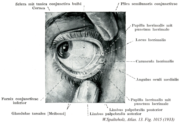
- 1015_01【Sclera強膜 Sclera】 Membrane of the eyeball composed of interwoven collagen fibers. It has a bluishwhite appearance and is visible through the conjunctiva.
→(眼球の形状を保つ強靱な膠原線維組織層。角膜となっている前部6分の1を除いた部分。前方では隔膜固有質に、後方では篩板から視神経外鞘を経て脳硬膜に、それぞれつづいている。強膜と角膜を合わせて眼球線維膜という。強膜の厚さは眼球後極で~1.0mm、前部で~0.6mm、赤道で~0.4mmである。視神経線維束を通す篩板は後極の内側3.5mm、視神経乳頭の直後方にあたる。視神経は~数十本の掌側としてこれを通る。渦静脈、長・短毛様体動脈および神経が強膜を貫く。強膜はは外から内へ、①強膜上皮、②強膜固有質、③強膜褐色板の3沿うよりなる。虹彩角膜角に沿って強膜固有質が内方へ皮厚し(強膜距)毛様体筋腱により貫かれる。この部の直前に輪状に走る強膜静脈洞(Schlemmn管)があり、眼房水は虹彩角膜間隙(Fontana腔)からこれを通って渦静脈に排出される。角膜縁をとり膜浅い強膜溝の深層にこれらの構造がある。眼球前部の強膜上板毛細血管網に富み、その炎症性変化を臨床的に「網膜充血」という。強膜前部は眼球結膜、後部は眼球鞘(Tenon鞘)によりおおわれる。内面は脈絡外隙を間に脈絡外板に接する。)
- 1015_02【Bulbar conjunctiva眼球結膜 Tunica conjunctiva bulbi】 Portion of the conjunctiva covering the eyeball. It consists of stratified, nonkeratinized squamous epithelium with only a small number of goblet cells and a lamina propria of loosely organized structures, containing few cells and permeated by elastic fibers.
→(眼球結膜は結膜のうち眼球を被う部分。杯細胞に乏しい角化していない重層扁平上皮である。固有層は疎で細胞に乏しく弾性線維を含む。)
- 1015_03【Cornea角膜 Cornea】 Transparent, anterior part (1/6) of the eyeball that is anteriorly convex and posteriorly concave. It is 0.9 mm thick in the middle and 1.2 mm thick at the margin.
→(角膜は眼球前方部の透明部分。厚さ約1mm、直径10~12mmの前弯した楕円形の膜で、角膜頂、角膜縁、前および後面を区別する。弯曲度は前面(曲率半径約7.8mm)よりも後面の方が強い。前面は光学的に縦径線が横径線に比してやや強く弯曲し、正視眼ではこの差は水晶体弯曲度の逆の関係により補正されるている。角膜の特徴として、角膜血管が入る辺縁部以外ではまったく血管が存在しない。組織学的に5層が区別される。①角膜上皮、②前境界板(Bowman膜)、③角膜固有質、④後境界板(Descement膜)、⑤角膜内皮)
- 1015_04【Inferior conjunctival fornix下結膜円蓋 Fornix conjunctivae inferior】 Reflection of bulbar conjunctiva onto the palpebral conjunctiva behind the inferior eyelid.
→(眼球結膜が下眼瞼後方で、眼瞼結膜へと折り変えるところ。(Feneis))
- 1015_05Meibomian glands【Tarsal glands瞼板腺;マイボーム腺 Glandulae tarsales】 Elongated string of holocrine glands in the superior and inferior tarsi with openings near the posterior palpebral margin. They produce a sebaceous discharge that lubricates the margins of the eyelids.
→(マイボーム腺ともよばれる。上・下瞼板に埋没している皮脂腺で、結膜の間に、上眼瞼に30~40、下眼瞼に20~30個の多房状腺があり、後眼瞼縁に導管口が開く。特殊化した皮脂腺で眼瞼縁を保護し、結膜面をうるおす涙の露出を防ぐ。眼瞼を反転すると平行に並んだ真珠首飾り状の腺体を結膜を透かしてみることができる。瞼板腺の急性化膿性炎症を内麦粒腫、慢性化膿性炎症を霰粒種という。ドイツの解剖学者Heinrich Meibom (1638-1700)によって、1666年に記載された。が、第一発見者は、Casserius (1609)であるという。)
- 1015_06【Plica semilunaris conjunctivae結膜半月ヒダ Plica semilunaris conjunctivae】 Fold at the medial angle of eye that connects the superior and inferior conjunctival fornices.
→(結膜半月ヒダは眼瞼結膜によってつくられる内眼角における上および下結膜円蓋の間の結合ヒダ。 多くの動物にみられる結膜粘膜にひだ。通常急速時には部分的に目の背部に隠れるが、鳥類にみられるように、角膜を清掃するためのウインク様の動作をすると、広がって角膜の一部または全体を覆う。)
- 1015_07【Lacrimal papilla涙乳頭 Papilla lacrimalis】 Single small, medial, conical elevation on each eye at the inner margin of the upper and lower eyelids on top of which the lacrimal punctum sits.
→(上および下眼瞼の内縁の内側にある小さな円錐状の高まり。尖端に涙点がある。(Feneis))
- 1015_08【Lacrimal punctum涙点 Punctum lacrimale; Punctum lacrimale inferius et superius】 Punctate beginning of the lacrimal drainage system situated on the lacrimal papilla.
→(涙点は上および下眼瞼縁の内側交連の近くにある涙管の細く丸い涙排出系の始まりの開口部。)
- 1015_09【Lacus lacrimalis; Lacrimal lake涙湖 Lacus lacrimalis】 Space in the medial angle of eye around the lacrimal caruncle.
→(涙湖は内眼角における涙丘周囲の間隙。)
- 1015_10【Lacrimal caruncle涙丘 Caruncula lacrimalis】 Mucosal protuberance at the medial angle of eye with stratified squamous or columnar epithelium.
→(涙丘はめがしらにある米粒大の隆起。その周辺の陥凹部を涙湖という。組織学的には皮膚に似ている。)
- 1015_11【Medial angle of eye内眼角;メガシラ Angulus oculi medialis; Angulus oculi nasalis】 Medial end of the palpebral fissure that has a convex, rounded shape for the lacrimal lake.
→(内眼角は眼裂の内側端でもある。まるく弯出し涙湖をなす。)
- 1015_12【Posterior palpebral margin後眼瞼縁 Limbus posterior palpebrae】 Inner margin of the eyelids facing the conjunctiva.
→(結膜に対する眼瞼縁。(Feneis))
- 1015_13【Anterior palpebral margin前眼瞼縁 Limbus anterior palpebrae】 Margin of the eyelids facing the outer skin of the eyelid.
→(前眼瞼縁は眼瞼皮膚に対する眼瞼縁。)