Spalteholz HANDATLAS DER ANATOMIE DES MENSCHEN VON WERNER SPALTEHOLZ
メニューは解剖学(TA)にリンクしてあります。図の番号をクリックすると下記の説明へ、右側の用語をクリックすると解剖学(TA)にジャンプします。
526 


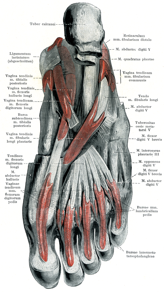
- 526_01【Calcaneal tuberosity踵骨隆起 Tuber calcanei】 Tuberosity on the posterior aspect of the calcaneus.
→(踵骨の後半部は大きな骨塊となって後方に飛び出している。この部分は踵骨隆起と呼ばれ、いわゆるかかとの主要部を成している。その後面には表面にギザギザした稜線が横に走っているが、ここはアキレス腱がつく場所である。)
- 526_02【Flexor retinaculum of foot屈筋支帯[足の];破裂靱帯 Retinaculum musculorum flexorum pedis; Ligamentum laciniatum】 Multilayered band situated over the long flexor tendons passing from the medial malleolus to the calcaneus. Its superficial portion invests the tibial nerve and posterior tibial artery and veins. Its deep portion forms an osteofascial canal with compartments containing the posterior tibial flexor muscles, flexor digitorum longus, and flexor hallucis longus.
→(足の屈筋支帯は下腿筋膜の厚くなったもので、内果の下部から扇状に広がって、前部は舟状骨に後部は踵骨につき中間部は足底腱膜に移行する。屈筋支帯は後脛骨筋と長趾屈筋の腱を被い、またその間の隔壁を骨に送ったのち、載距突起についてさらに長母指屈筋腱溝を被う深葉と、脛骨神経および後脛骨動静脈を被う浅葉とに分かれる。)
- 526_03【Tendinous sheath of tibialis posterior後脛骨筋の腱鞘 Vagina tendinis musculi tibialis posterioris】 Tendon sheath surrounding the tibialis posterior beneath the flexor retinaculum. It begins where it is crossed by the flexor digitorum longus.
→(後脛骨筋の腱鞘は屈筋支帯に被われて、後脛骨筋の腱の周囲をかこみ、さらに遠位側にのびる。)
- 526_04【Tendinous sheath of flexor hallucis longus長母趾屈筋の腱鞘;長母指屈筋の腱鞘(足の) Vagina tendinum musculi flexoris hallucis longi】 Tendon sheath surrounding the long flexor tendon of the great toe, extending to the proximal end of the sole of the foot, where it crosses under the tendon of the flexor digitorum longus.
→(長母趾屈筋の腱鞘は下伸筋支帯の下にあって、そこからさらに下方にのびる。)
- 526_05【Tendinous sheath of flexor digitorum longus長指屈筋の腱鞘(足の);長趾屈筋の腱鞘 Vagina tendinum musculi flexoris digitorum longi】 Tendon sheath surrounding the long flexors of the toes, located posterior and inferior to the medial malleolus and covered by the flexor retinaculum.
→(長趾屈筋の腱鞘は足根部内側で長趾屈筋の腱をおおい、屈筋支帯の深部で足に達する滑膜性腱鞘。)
- 526_06【Subtendinous bursa of tibialis posterior後脛骨筋の腱下包;後脛骨筋腱包 Bursa subtendinea musculi tibialis posterioris】
→(")
- 526_07【Plantar tendinous sheath of fibularis longus; Plantar tendinous sheath of peroneus longus長腓骨筋の足底腱鞘;足底長腓骨筋腱鞘 Vagina plantaris tendinis musculi fibularis longi; Vagina plantaris tendinis musculi peronei longi】 Tendon sheath on the sole of the foot for passage of the long tendon of the peroneus.
→(長腓骨筋の足底腱鞘は足底部における長腓骨筋腱の周りにある。)
- 526_08【Flexor digitorum longus tendon長趾屈筋腱 Tendo musculus flexor digitorum longus】
→()
- 526_09【Abductor hallucis muscle母趾外転筋;母指外転筋(足の) Musculus abductor hallucis】 o: Calcaneal tuberosity. i: Medial sesamoid bone and proximal phalanx of great toe. Medial abduction, supports longitudinal arch of foot. I: Medial plantar nerve.
→(母趾外転筋は踵骨隆起の内側突起、屈筋支帯および足底腱膜から起始する。腱となり内側種子骨を介して母趾の基節骨底内側面および短母趾屈筋の内側腱に停止する。内側足底神経の支配を受ける。この筋の収縮は母趾の屈筋と外転とをもたらす(体重を支えていない下肢の場合)。また、体重を支えている下肢においては、この筋の収縮が内側縦足弓の維持に役立つ。 )
- 526_10【Tendinous sheaths of toes趾の腱鞘;趾の屈筋腱鞘;腱鞘(足の指の) Vaginae tendinum digitorum pedis】
→(趾の腱鞘は指の近くに見られる長および短趾屈筋腱の周囲にある。)
- 526_11【Inferior fibular retinaculum; Inferior peroneal retinaculum下腓骨筋支帯;遠位腓骨筋支帯 Retinaculum musculorum fibularium inferius; Retinaculum musculorum peroneorum inferius; Retinaculum musculorum fibularium distale】 Lower retinaculum that holds the peroneus muscles in place. It passes from the extensor retinaculum to the lateral surface of the calcaneus. One band passes to the fibular trochlea, dividing the peroneus brevis and peroneus longus muscles overlying it. It strengthens the dorsal fascia of the foot.
→(下腓骨筋支帯は下伸筋支帯の外側脚につづいて踵骨外側面から踵骨隆起外側面下部に至る。)
- 526_12【Abductor diditi minimi muscle of foot小趾外転筋;小指外転筋(足の) Musculus abductor digiti minimi pedis】 o:Pisiform, flexor retinaculum. i: Proximal phalanx of little finger. Abduction. I: Ulnar nerve.
→(小趾外転筋は踵骨の足底面、特に踵骨隆起の外側突起、足底腱膜および第5中足骨粗面から起こる。その停止は第5の基節骨底に停止する。外側足底神経の支配を受ける。この筋は体重を支えない下肢においては第5趾を屈曲、外転させる作用を示し、足に体重がかかる場合には外側縦足弓を上方に引き、外側縦足弓を維持するのに役立つ。)
- 526_13【Quadratus plantae muscle; Flexor accessorius muscle足底方形筋;副趾屈筋 Musculus quadratus plantae; Musculus flexor accessorius】 o: Calcaneus. i: Lateral border of tendon of flexor digitorum longus. Toe flexion and support of longitudinal arch of foot. I: Lateral plantar nerve.
→(足底方形筋は踵骨底側面に起こり、長趾屈筋の腱に停止する。同筋は副趾屈筋とも呼ばれるが、それは長趾屈筋の停止腱が趾を引く方向を矯正するからである(趾の底屈時)。外側部の趾へ行く腱は、線維性の腱鞘によって長軸方向に固定される前に、中足骨上を斜走する。この腱の斜走は足底方形筋の索引によって中足骨長軸に沿った方向となる。外側足底神経の支配を受ける。この筋の収縮により長趾屈筋腱は後方へ引っ張られるために、第2~5趾の屈曲が得られる。 踵骨からおこって長母趾屈筋腱に停止し、その補助に働く。神経支配:外側足底神経。(イラスト解剖学))
- 526_14【Common tendinous sheath of fibulares; Common tendinous sheath of peronei腓骨筋の総腱鞘;総腓骨筋腱鞘 Vagina communis tendinum musculorum fibularium; Vagina communis tendinum musculorum peroneorum】 Tendon sheath that extends from beneath the fibular retinacula to the cuboid.
→(腓骨筋の総腱鞘は上、下腓骨筋支帯の下にあって、立方骨までのび、長、短腓骨筋の腱を囲む。)
- 526_15【Fibularis (peroneus) longus tendon長腓骨筋腱 Tendo musculus peroneus longus; Tendo musculus fibularis longus】
→()
- 526_16【Tuberosity of fifth metatarsal bone [V]第5中足骨粗面 Tuberositas ossis metatarsi quinti [V]】 Bony protuberance on the lateral aspect of the base of the fifth metatarsal. Attachment site of the fibularis brevis muscle.
→(第5中足骨底の外側には第5中足骨粗面(短腓骨筋の着くところ)があり、体表から骨の突起として触れる。これが独立した小骨(Vesaliusの骨)となることがある。)
- 526_17【Flexor digiti minimi brevis muscle of foot短小趾屈筋;短小指屈筋(足の) Musculus flexor digiti minimi brevis pedis】 o: Base of fifth metatarsal and long plantar ligament, i: Proximal phalanx of little toe. Flexion and abduction of little toe. 1: Lateral plantar nerve.
→(短小趾屈筋と小趾対立筋は第5中足骨底、長足底靱帯および長腓骨筋腱鞘から共通腱をもって起こる。小趾の短屈筋は第5趾の基節骨底に、小趾の対立筋は第5中足骨外側面に停止する。人では対立筋は短小趾屈筋の弱い分束としてしか出現せず、その停止部でしか同定できない。外側足底神経の浅枝による支配を受け、中足趾節関節で第5趾を屈曲させる作用を示す。)
- 526_18【Third plantar interosseous第3底側骨間筋 Musculus interosseus plantaris III】
→()
- 526_19【Plantar interosseous muscles底側骨間筋 Musculi interossei plantares】 o: Muscle arising from a single head on third through fifth metatarsals. i: Bases of proximal phalanges. Adduction and flexion at metatarsophalangeal joints. I: Lateral plantar nerve.
→(3つの底側骨間筋は第3~5中足骨底側面と長足底靱帯から起こり、第3~5趾基節骨底内側面へ付着する。普通、指背腱膜には達しない。外側足底神経の支配をうけるこれらの筋の収縮により、足の第2趾に向かうような各趾の内転、各趾の中足趾節関節の屈曲、および趾節間関節の伸展が得られる。)
- 526_20【Opponens digiti minimi of foot小趾対立筋;小指対立筋(足の) Musculus opponens digiti minimi pedis】 Part of the flexor digiti minimi brevis that is occasionally present.o:Distal half of fifth metatarsal.
→(短小趾屈筋と小趾対立筋は第5中足骨底、長足底靱帯および長腓骨筋腱鞘から共通腱をもって起こる。小趾の短屈筋は第5趾の基節骨底に、小趾の対立筋は第5中足骨外側面に停止する。人では対立筋は短小趾屈筋の弱い分束としてしか出現せず、その停止部でしか同定できない。)
- 526_21【Lumbrical bursaes of foot虫様筋包;虫様筋嚢(足の) Bursae musculi lumbricalium pedis】
→(")
- 526_22【Intermetatarsophalangeal bursa中足趾節間包;中足趾節間嚢 Bursae intermetatarsophalangeae; Bursae intermetatarsophalangicae】
→()