Spalteholz HANDATLAS DER ANATOMIE DES MENSCHEN VON WERNER SPALTEHOLZ
メニューは解剖学(TA)にリンクしてあります。図の番号をクリックすると下記の説明へ、右側の用語をクリックすると解剖学(TA)にジャンプします。
601 


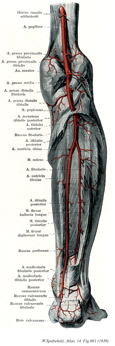
- 601_01【Adductor hiatus内転筋腱裂孔;腱裂孔;内転筋間裂孔 Hiatus adductorius; Hiatus canalis adductorii】 Slitlike opening between the insertions of the adductor magnus on the femur. It opens in the popliteal fossa.
→(内転筋管の上方は腸恥窩につづき、下方は大内転筋の骨幹停止部と内側上顆停止腱との間、すなわち[内転筋]腱裂孔によって膝窩に開く。)
- 601_02【Popliteal artery膝窩動脈 Arteria poplitea】 Artery extending from the end of the adductor canal to its division at the inferior border of the popliteus.
→(膝窩動脈は内転筋管裂孔で大腿動脈よりつづいておこり、膝窩のほぼ中央の深層を下行して、膝窩筋の下縁付近で前および後脛骨動脈に分岐して終わる。)
- 601_03【Superior lateral genicular artery外側上膝動脈;腓側近位膝動脈 Arteria superior lateralis genus; Arteria genus proximalis fibularis】 Artery passing above the lateral femoral condyle and beneath the tendon of the biceps femoris anteriorly to the genicular anastomosis.
→(外側上膝動脈は腓腹筋の外側頭の上縁を通り、外側広筋に分布した後、膝関節動脈網へ。)
- 601_04【Superior medial genicular artery内側上膝動脈;脛側近位膝動脈 Arteria superior medialis genus; Arteria genus proximalis tibialis】 Artery passing beneath the tendon for the adductor magnus anteriorly to the genicular anastomosis.
→(内側上膝動脈は腓腹筋内側頭の上縁を通って前方へ向かい、大内転筋腱の深層に出て膝関節動脈網へ。一部は下行膝動脈と吻合枝、また筋枝を内側広筋に与える。しばしば弱小となり、このときは下行膝動脈関節枝によって代償される。)
- 601_05【Sural arteries腓腹動脈;腓腹枝(膝窩動脈の) Arteriae surales】 Branches supplying the calf muscles, and the fascia and skin of the leg.
→(腓腹動脈は内外の二つの太い枝よりなり、腓腹筋、ヒラメ筋、足底筋へ。一部の枝は、腓腹筋両頭の間を下行して下腿後面の皮膚へ。)
- 601_06【Middle genicular artery中膝動脈 Arteria media genus; Arteria genus media】 It passes inferiorly and posteriorly to the cruciate ligaments and synovial folds.
→(中膝動脈は細い。膝関節後面のほぼ中央で分岐し、斜膝窩靱帯および関節包を貫いて膝関節の内部へ入り、交叉靱帯と滑膜へ。しばしば外側上膝動脈より分岐する。)
- 601_07【Inferior lateral genicular artery外側下膝動脈;腓側遠位膝動脈 Arteria inferior lateralis genus; Arteria genus distalis fibularis】 Artery passing beneath the lateral head of the gastrocnemius and beneath the fibular collateral ligament to the genicular anastomosis.
→(外側下膝動脈は腓腹筋外側頭および膝関節の外側側副靱帯の深層を通って膝関節の前面に出て、膝関節動脈網へ。)
- 601_08【Inferior medial genicular artery内側下膝動脈;脛側遠位膝動脈 Arteria inferior medialis genus; Arteria genus distalis tibialis】 Artery passing beneath the medial head of the gastrocnemius and tibial collateral ligament to the genicular anastomosis.
→(内側下膝動脈は、はじめ腓腹筋内側角深層を膝窩筋の上縁に沿って下内方へ走り、次いで脛骨内側顆の下方で内側側副靱帯の深層を通って前方にまわり、膝関節動脈網へ。)
- 601_09【Popliteus muscle膝窩筋 Musculus popliteus】 o: Lateral femoral epicondyle. i: Posterior surface of tibia. Medial rotation of the leg with flexed knee joint. I: Tibial nerve.
→(膝窩筋は本来は下腿の深屈筋群に属する。哺乳類以外では同筋は脛骨と腓骨をつなぐ。哺乳類では、人でも、膝窩筋は膝関節と密接に関係しながら発達し、屈曲した下腿が内旋するのを助ける。膝窩筋は膝関節外側側副靱帯下方の、大腿骨外側顆外表面から起こる。その停止はヒラメ筋線より近位の、脛骨後面である。膝窩筋は膝窩の底をなし、腓腹筋頭および血管、神経におおわれている。起始腱は、その上を弓状膝窩靱帯が通り、膝窩筋下陥没の上方にある。この陥没は成人では常に膝関節腔と交通している。半膜様筋の一部は膝窩筋の筋膜へ放散する。この筋は脛骨神経の支配を受ける。この筋の収縮により大腿骨に対する脛骨の内旋が得られる。立位で体重を支えている下肢においては、脛骨に対する大腿骨の外旋をもたらす、といってもより、後者の場合は、体重を支えて伸展している膝関節を屈曲し始める膝窩筋作用であり、これよより緊張した膝関節靱帯がゆるめられる(膝関節の固定解除unlocking)。膝窩筋の一部が外側半月に付着している関係上、この筋の収縮は外側半月を膝関節屈曲初期に後方へ引く役割も果たす。)
- 601_10【Posterior tibial recurrent artery後脛骨反回動脈 Arteria recurrens tibialis posterior】 Inconstant artery that passes beneath the popliteus to the knee joint.
→(後脛骨反回動脈は不定の動脈で、本幹が下腿骨間膜を貫いて下腿の前面に出る直前で分岐する。膝窩筋の前面を上行して膝窩動脈の枝の内側下膝動脈と吻合して膝関節動脈網へ。)
- 601_11【Anterior tibial artery前脛骨動脈 Arteria tibialis anterior】 It extends from its origin at the inferior border of the popliteus to the inferior border of the inferior extensor retinaculum. After penetrating the interosseous membrane, it lies between the tibialis anterior and extensor digitorum longus, then between the tibialis anterior and extensor hallucis longus.
→(前脛骨動脈は、腋窩の遠位部、すなわち膝窩筋の下縁の高さで、膝窩動脈が二分して生ずる枝の一つ。分岐後、下腿骨間膜の上部を越え、骨間膜の前面に出て下行する。経過中に下腿の前外側にある筋とその付近に枝を送る。動脈はとくに上部と下部で膝関節と足関節との周囲の膝蓋動脈網に小枝を送り吻合する。前脛骨動脈は、足関節のすぐ上方で表層に現れ、足関節の前側で前脛骨筋の腱の外側に沿って走り、足背で足背動脈となる。)
- 601_12【Circumflex fibular branch of posterior tibial artery; Circumflex peroneal branch of posterior tibial artery腓骨回旋枝;腓骨枝(後脛骨動脈の) Ramus circumflexus fibularis; Ramus circumflexus peronealis (Arteria tibialis posterioris)】 Branch arising at the very beginning of the posterior tibial artery and traveling anteriorly around the fibula to the genicular anastomosis.
→(後脛骨動脈の腓骨回旋枝は多くは後脛骨動脈の起始の直下でおこるが、ときに膝窩動脈の下端または前脛骨動脈よりおこり、外下方に走って腓骨頭の下でその外面をまわって下腿の前面に出て膝関節網と周囲の筋へ。)
- 601_13【Posterior tibial artery後脛骨動脈 Arteria tibialis posterior】 It passes beneath the tendinous arch of soleus under the superficial flexor group and passes from posterior to the medial malleolus.
→(後脛骨動脈は腋窩の近位部で腋窩動脈からつづいてはじまり、ヒラメ筋腱弓の深層を通ってヒラメ筋と後脛骨筋の間を下行し、距腿関節を後面を経て、屈筋下におおわれて内果と踵骨隆起の間を通り、母趾外転筋の深層で内側および外側足底動脈に分かれて終わる。脛骨神経が伴行する。)
- 601_14【Tibial nutrient artery脛骨栄養動脈 Arteria nutricia tibiae; Arteria nutriens tibiae】 Artery entering the nutrient foramen below the soleal line.
→(脛骨栄養動脈は後脛骨動脈上部より起こり、頸骨の後面にある栄養孔に入る。)
- 601_15【Soleus muscleヒラメ筋 Musculus soleus】 o: Head of fibula, upper one-third of the posterior surface of tibia, i: Achilles tendon. Plantar flexion and supination.
→(ヒラメ筋の下腿後区浅層の筋の1つ。大半は腓腹筋におおわれ、腓骨頭から腓骨後面、腱性弓(ヒラメ筋腱弓)および脛骨(ヒラメ筋線と内側縁)に起始する。ヒラメ筋腱弓は腓骨から脛骨へ至り、神経血管索をまたぐ。ヒラメ筋はアキレス腱を介して踵骨隆起に停止する。ヒラメ筋の筋腹は腓腹筋の筋腹よりも遠位に伸び、微細な腱様層によっておおわれている。その上を腓腹筋が滑走する。ヒラメ筋は複合羽状筋で、前頭平面に位置する腱様膜によって浅層の筋層に分けることができる。脛骨神経の支配を受ける。この筋は腓腹筋、足底筋とともに距腿関節における足の底屈を生じさせる。すなわち、これらの3筋は歩行、歩行時に、地面を後方に蹴るための力を出すのである。)
- 601_16【Fibular artery; Peroneal artery腓骨動脈 Arteria fibularis; Arteria peronea】 Artery that extends to the calcaneus and is covered for most of its length by the flexor hallucis longus.
→(腓骨動脈は個体発生的には下臀動脈(坐骨神経伴行動脈)からつづいた下肢の動脈の本幹で、前および後脛骨動脈はその分子にすぎない。したがって腓骨動脈の欠損例はない。膝窩筋の下縁の下方で後脛骨小津脈より分岐して、腓骨の内側縁に沿って後脛骨筋と長母趾屈筋の筋注を下行、脛腓靱帯結合うしろで踵骨枝を分岐して終わる。この動脈はしばしば強大化して、後脛骨動脈の欠損を代償したり(約5%)、また貫通枝によって足背動脈の主流となったりする(約7%)。この枝に貫通枝、交通枝、外果枝、踵骨枝がある。)
- 601_17【Fibular nutrient artery腓骨栄養動脈 Arteria nutricia fibulae】 Branch passing to the fibula.
→(腓骨栄養動脈は腓骨動脈から起こり腓骨に分布する。)
- 601_18【Flexor hallucis longus muscle長母趾屈筋;長母指屈筋(足の) Musculus flexor hallucis longus】 o: Fibula, i: Distal phalanx of great toe. Plantar flexion, supination, flexion of great toe. I: Tibial nerve.
→(長母趾屈筋はずっと内側に(おもな腱は第1末節骨底に)停止する。その起始は下腿の外側、つまり腓骨後面の遠位2/3,骨間膜の狭い紐状部分および後筋間中隔である。その腱は長趾屈筋腱の下を横切り(足底腱交叉)、足底で第2および第3(まれに第4)趾末節骨に腱性停止を送る。距骨後面と載距突起下面において長母趾屈筋の腱は溝の中を走る。同筋は腱鞘に包まれる。腱鞘は内果先端のレベルにはじまり、遠位へ伸び、第1中足骨底に至る。停止腱は第1中足骨頭から末節骨に至るまで腱鞘に包まれている。腱鞘は線維性のおおいによって母趾の各分節に付く。)
- 601_19【Tibialis posterior muscle後脛骨筋 Musculus tibialis posterior】 o:Tibia, fibula, interosseous membrane, i: Navicular, cuneiform bones I-III, and metatarsals II-IV. Plantar flexion and supination. I: Tibial nerve.
→(後脛骨筋は骨間膜の広い領域から起こる。狭い辺縁部は腓骨と脛骨の近位部に起こり、浅い線維束は浅深の屈筋間にある結合組織から起こる。後脛骨筋の腱は内果上方で長趾屈筋の腱の下を横切り(下腿腱交叉)、その主束は舟状骨粗面につき、その外側束は(しばしば)すべての遠位足根骨と第2~4中足骨底の足底面に付く。内果溝で後脛骨筋の腱は腱鞘に包まれ、内果下方では屈筋支帯におおわれている。深屈筋群へ脛骨神経の筋枝の中で後脛骨筋への枝はほかの枝よりもずっと近位、ヒラメ筋腱弓のレベルで出る。母趾とそのほかの趾への長屈筋群に対する近位の枝は下腿中位1/3へうつるレベルで脛骨神経から分かれる。脛骨神経からの筋枝は普通下腿遠位1/2からも分かれる。後脛骨筋は脛骨神経の支配を受ける。この筋の収縮により距腿関節における足の底屈、距骨下関節および横足根関節による足の内反が得られる。この筋は内層の縦足弓を維持するうえにも重要である。この筋の収縮が足底で数個の骨を互いに引き寄せるうえに役立つことにも注意すべきである。)
- 601_20【Flexor digitorum longus muscle長趾屈筋;長指屈筋(足の) Musculus flexor digitorum longus】 o: Tibia, i: Distal phalanges of the second through fifth toes. Plantar flexion, supination, flexion of toes. I: Tibial nerve.
→(長趾屈筋はヒラメ筋線より遠位の脛骨後面およびヒラメ筋腱弓の一部から起こる。その停止腱は基節骨の領域で短趾屈筋の腱を貫通し(第2~5)趾の末節骨に停止する。長趾屈筋の腱は腱鞘に包まれて、内果溝を後脛骨筋腱の背外側に走り、屈筋死体の下を載距突起内側縁に沿って足底に至る。舟状骨粗面のレベルでは長母趾屈筋腱の浅層を通る。この際、長母趾屈筋の健束が長趾屈筋の腱に混じる。この腱交叉位遠では足底方形筋が長趾屈筋の腱に付く。この付加的な屈筋は長趾屈筋停止腱の牽引方向を趾放線の長軸方向と関連させる。同一趾へ向かう長趾屈筋(「貫通筋」と短趾屈筋「被貫通筋」)の停止腱は腱鞘(滑液鞘に包まれる。腱鞘は第1中足骨頭の上方から始まり、末節骨までのびている。これらの滑液鞘は線維鞘に包まれる。線維鞘は手指におけると同じように横走線維と交織する線維(輪状および十字部)からなる。)
- 601_21【Perforating branch of fibular artery貫通枝;穿通枝(腓骨動脈の) Ramus perforans (Arteria fibularis)】 It pierces the interosseous membrane immediately above the malleolus and passes to the lateral malleolar network and dorsum of foot.
→(腓骨動脈の貫通枝は外果の上方約5cmの付近で分岐し、ただちに下腿骨間膜を貫いて下腿の前面に出て、前外果動脈と吻合する。)
- 601_22【Lateral malleolar branches of fibular artery外果枝;後腓側踝動脈(腓骨動脈の) Rami malleolares laterales arteria fibularis; Arteria malleolaris fibularis posterior】 Branches that often arise from the communicating branch and pass to the lateral malleolus.
→(腓骨動脈の外果枝は二、三の小枝。しばしば交通枝から出ることがある。外顆動脈網へ)
- 601_23【Medial malleolar branches of posterior tibial artery内果枝;後脛側踝動脈(後脛骨動脈の) Rami malleolares mediales ; Arteria malleolaris tibialis posterior】 Branches passing behind the medial malleolus to the medial malleolar network.
→(後脛骨動脈の内果枝は本幹の下端に近く、内果の後方でおこり、内果動脈網へ。)
- 601_24【Communicating branch; Communicating branch of fibular artery交通枝(腓骨動脈の) Ramus communicans】 Transverse branch that anastomoses with the posterior tibial artery.
→(腓骨動脈の交通枝は前方より下方で分岐し、長母趾屈筋の深層を通って後脛骨動脈に吻合する。)
- 601_25【Calcaneal branches of posterior tibial artery踵骨枝;内側踵骨枝;脛側踵骨枝(後脛骨動脈の) Rami calcanei; Rami calcaneares mediales; Rami calcaneares tibiales (Arteria tibialis posterior)】 Branches passing to the surface of the calcaneus.
→(後脛骨動脈の踵骨枝は屈筋支帯の深層で分岐する数本の枝で、踵骨隆起の後面の皮下へ。)
- 601_26【Calcaneal branches of lateral malleolar branch of fibular artery踵骨枝;外側踵骨枝;腓側踵骨枝(腓骨動脈の外果枝の) Rami calcanei; Rami calcaneares fibulares (Arteria peronea)】 Branches that mainly pass to the lateral side of the calcaneus.
→(腓骨動脈の外果枝の踵骨枝は腓骨動脈の終枝で、踵骨隆起の外側面へ。)
- 601_27【Calcaneal anastomosis踵骨動脈網;踵骨網 Rete calcaneum】 Arterial network on the posterior calcaneus.
→(踵骨動脈網は踵骨隆起の後面とその内外両側面にある皮下組織のなかにつくられる動脈網で、後脛骨動脈からの内側踵骨枝と、腓骨動脈の外側踵骨枝によってつくられそのほか外顆動脈網と内果動脈網よりも枝を受ける。)