Spalteholz HANDATLAS DER ANATOMIE DES MENSCHEN VON WERNER SPALTEHOLZ
メニューは解剖学(TA)にリンクしてあります。図の番号をクリックすると下記の説明へ、右側の用語をクリックすると解剖学(TA)にジャンプします。
063


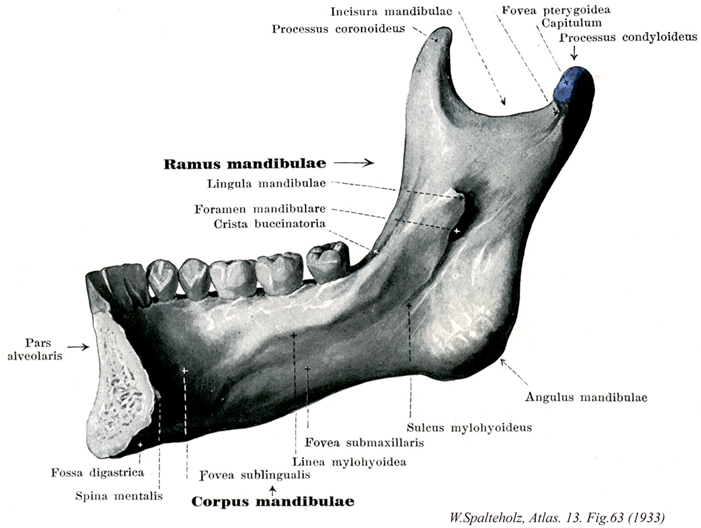
- 063_00【Mandible下顎骨 Mandibula】
→(下顎を形成する。下顎を支え、頭蓋と顎関節をつくる骨で、水平な馬蹄形の部(下顎体)と、その後端から上方に向かう部(下顎枝)に分けられる。本来有対の骨として生じ、生後1年目で下顎底の前端で癒合して一つの骨となる。下顎体の上縁は歯槽部で、下縁は下顎底という。歯槽部には各側8本の歯をいれる八つのへこみ(歯槽)があり、全体として歯槽弓をつくる。各歯槽を境する骨壁を槽間中隔といい、大臼歯の歯槽はさらにその歯根の間を隔てる低い根管中隔で分けられている。体の正中線上前面で左右の骨が癒合した部分は高まり、その下縁は三角形をなして突出(オトガイ隆起)し、ヒトの特徴であるオトガイをつくる。その外側、下縁に接する小突出部をオトガイ結節という。外面ではオトガイ結節から斜線が下顎枝の前縁に向かう。また第2小臼歯の下方にオトガイ孔がある。下顎体の内面には前方正中部に四つの隆起からなるオトガイ棘があり、上二つはオトガイ舌筋、下二つはオトガイ舌骨筋がつく。その下外側で下縁に切歯て卵形のへこみ(二腹筋窩)がある。そこから斜めに下顎枝の前縁に向かう線(顎舌骨筋線)があり、左右のこの線の間をはる顎舌骨筋が口底をつくる。この線の上前はへこみ(舌下腺窩)、またこの線の下方、第2~3大臼歯の所もへこむ(顎下線窩)。下顎底が下顎枝にうつる所は下顎角といわれ、小児で鈍角であるが成長とともに直角に近づく。下顎枝の上縁は深い切れ込み(下顎切痕)によって二つの突起に分かれ、前のもの(筋突起)には側頭筋がつき、後のもの(関節突起)の先に横楕円形の下顎頭があて、側頭骨鱗部にある関節窩と顎関節を作る。下顎頭の下はすこしくびれ(下顎頚)、その前面に外側翼突筋のつく翼突筋窩がある。下顎枝外面は平らで下顎角に近く咬筋のつく咬筋粗面、内面には内側翼突筋のつく翼突筋粗面がある。下顎枝内面中央には下顎孔があり、その前縁は上内方に尖り(下顎小舌)口腔から触れるので、下歯槽神経の伝達麻酔の際、針をさす指標となる。下顎孔の後下から溝(顎舌骨筋神経溝)が出て前下方に斜めに向かう、この上の高まりが顎舌骨筋線である。下顎管は下顎孔からはじまり下顎体の中央で二分し、外側管はオトガイ孔で外側にひらき、内側管は切歯のそばに終わるが、その経過中に各歯槽に向かって小管を出している。有顎魚の下顎を支配する骨格は本来下顎軟骨(Meckel軟骨)で、上顎を支配する支持する軟骨は(口蓋方形軟骨)と顎関節をつくる。ともに鰓弓軟骨の変化したものである。硬骨魚類では下顎軟骨のまわりに若干の皮骨が生じて下顎を支え、そのうち前外面にあり、顎縁の歯をつけた大きい歯を歯骨という。顎関節は下顎軟骨と口蓋方形軟骨それぞれの後部の化骨物(関節骨と方骨)の間につくられる。両棲類、爬虫類も同じ状態であるが、哺乳類では歯骨のみが大きくなって下顎骨となり、顎関節は歯骨と燐骨(側頭骨鱗部に相当する骨)の間に新生されたものである。そして関節骨と方骨はツチ骨、キヌタ骨になっている。多くの哺乳動物では下顎骨は生体でも対をなした状態にとどまっている。Mandibulaはmandere(噛む)という動詞に由来し、語尾のbulaは「道具」を意味する接尾辞である。下顎骨にはすべての咀嚼筋が付。)
- 063_01【Ramus of mandible下顎枝 Ramus mandibulae】 Projection that forms the ascending ramus of the mandible.
→(下顎体の後端から上(やや)後方に延びた、扁平な板状部で矢状位に立つ。)
- 063_02【Body of mandible下顎体 Corpus mandibulae】 Horizontal part of the mandible to which the rami of the mandible are attached.
→(下顎体は後方に向かって開いたL字形の左右両半からなり、ほぼ垂直に立つ厚い骨板である。一般に前方が高く後方が引く。下顎体を内外両面に分けるほか、上縁とその周囲を歯槽部、下縁を下顎底という。下顎底は歯槽部より広く、そのため側面はやや傾斜し、とくに前部が前下方に突出して顔のオトガイ(頤)をるくる。この下顎底が広いことが人類の下顎骨の著しい特徴である。)
- 063_03【Mandibular notch下顎切痕 Incisura mandibulae】 Notch between the condylar and coronoid processes. The masseteric nerve and vessels pass over it to supply the masseter muscle.
→(下顎枝の上縁は深くくぼんだ下顎切痕により前後の突起に分かれる。)
- 063_04【Coronoid process of mandible筋突起(下顎骨の) Processus coronoideus; Processus muscularis (Mandibulae)】 Muscular process that is separated by the mandibular notch from the condylar process to posterior. Attachment site of the temporal muscle.
→(下顎切痕の前方の筋突起は三角形の薄い骨板である。側頭筋の停止部。)
- 063_05【Lingula of mandible下顎小舌 Lingula mandibulae】 Thin bony projection in front of the mandibular foramen that gives attachment to the sphenomandibular ligament.
→(下顎孔の前縁に上内方に尖った板状の下顎小舌が直立する。蝶下顎靱帯が着く。)
- 063_06【Mandibular foramen下顎孔 Foramen mandibulae】 Opening on the internal aspect of the ramus of the mandible. Beginning of the mandibular canal which lies about 1 cm above the ocdusal plane.
→(下顎枝の中央には下顎孔がある。下顎管のはじまるところ。)
- 063_07【Buccinator crest of mandible頬筋稜(下顎骨の) Crista buccinatoria; Crista musculus buccinatoria mandibula】 Rounded bony ridge extending from the coronoid process to the medial, distal side of the third molar tooth. It forms the medial boundary of the retromolar triangle.
→(下顎の筋突起基部から第三大臼歯の辺りに走る隆線で、頬筋の下顎骨起始部が付着する。)
- 063_08【Alveolar part of mandible歯槽部(下顎骨の) Pars alveolaris (Mandibulae)】 Pectinate process on the base of mandible that houses the roots of the teeth.
→(歯槽部は上顎骨の歯槽突起に対応する。この部の弯曲度は下顎体の中部および下縁のそれより強く、そのためにこの上にならぶ歯槽(成人では左右合わせて16)の後端に近いものは下顎底に比較して内側に寄る。歯槽の列が歯槽弓をつくること、槽間中隔、根間中隔、歯槽隆起(切歯の歯槽隆起から口輪筋の一部、第2,第3大臼歯のそれから頬筋の一部が起こる)などをみることは上顎骨の歯槽部と同じである。ただし、窩gかうの大臼歯は2根であるから、根管中隔は歯槽内を前後に2分する1枚の骨板として認められる。歯槽部は前方の切歯部では幅が狭く、後方の歯槽部では幅が広い。第2大臼歯歯槽より後方は、下顎枝の内面につづく傾斜した三角形の骨面(臼後三角)がのこされるが、ここに第3大臼歯(智歯)が生ずれば狭くなる。)
- 063_09【Digastric fossa二腹筋窩 Fossa digastrica】 Paired pea-sized or beansized depressions near the mental protuberance just above the inferior margin of the mandible for attachment of the anterior belly of the digastric muscle.
→(オトガイ棘の下外側下縁に接して楕円形の二腹筋窩がある。顎二腹筋前腹が着く)
- 063_10【Mental spines; Genial spines; Genial tuberclesオトガイ棘 Spinae mentalis】
→(下顎体の内面には正中線の下端に近く、密接して上下にならぶ2対の小突起、オトガイ棘がある。上方の小さな棘は舌筋の付くところで下オトガイ棘といい、JNAではオトガイ舌筋棘と呼ばれていた。下方の小さな棘はオトガイ舌骨筋の着くところである。JNAではオトガイ舌骨筋棘と呼ばれていた。)
- 063_11【Pterygoid fovea翼突筋窩 Fovea pterygoidea】 Anteromedial depression below the head of the mandible for attachment of the lateral pterygoid muscle.
→(下顎頚の前内側面に翼突筋窩がある。外側翼突筋がつく。)
- 063_12【Head of mandible; *Head of condylar process of mandible下顎頭;下顎顆;下顎小頭 Caput mandibulae; Condylus mandibulae; Capitulum mandibulae】 Articular head of the mandible.
→(関節突起の尖端には横方向にふくれた下顎頭があり、顎関節の関節頭となっている。)
- 063_13【Condylar process of mandible関節突起;顆突起(下顎骨の) Processus condylaris (Mandibulae)】 Articular process.
→(下顎切痕の後方の関節突起は上端に長楕円形の下顎頭を作る。)
- 063_14【Angle of mandible; *Angle of jaw下顎角 Angulus mandibulae】 Angle formed between the body and the ramus of the mandible. It is sharpest in adults and wider in newborns and elderly, edentulous individuals (ca.140°).
→(下顎枝の後縁と下顎体の下縁との合する角は下顎角といわれ、少し外方に曲がる。成人で最も角度が強く、新生児や、無歯の老齢顎ではとくに平坦である。)
- 063_15【Mylohyoid groove顎舌骨筋神経溝;顎舌骨神経溝 Sulcus mylohyoideus; Sulcus nervi mylohyoidei】 Groove that begins at the mandibular foramen and extends anteroinferiorly, transmitting the mylohyoid nerve and the mylohyoid branch of the inferior alveolar artery.
→(下顎孔の前縁からは浅い顎舌骨神経溝が下顎体の方に走っている。この溝は前方に行くにしたがって不明瞭になる。この溝の上方にはこれとほぼ平行する顎舌骨筋線(顎舌骨筋がつく)という隆起がオトガイの方へ向かって走っている。)
- 063_16【Submandibular fossa顎下腺窩 Fovea submandibularis】 Depression below the mylohyoid line on the posterior half of the body of mandible.
→(顎舌骨筋線前部の下外側には顎下腺窩がある。顎下腺のためにできた凹み。)
- 063_17【Mylohyoid line顎舌骨筋線 Linea mylohyoidea】 Oblique ridge extending on the body of the mandible from posterosuperior to anteroinferior, giving origin to the mylohyoid muscle. At its posterior end the mylopharyngeal part of the superior constrictor muscle of the pharynx takes origin. The lingual nerve enters the oral cavity between the two muscles.
→(二腹筋窩の上外側から起こって斜めに上後方に向、歯槽部後端の下を通り下顎枝内面の前部に至る純な隆起線を顎舌骨筋線という。この線の大部分から口腔底を閉ざさず顎舌骨筋が広く起こり、後方小部分から上咽頭収縮筋の一部が起こる。顎舌骨筋線の下にこれと平行して走り、下顎枝内面の下顎孔に達する顎舌骨筋神経溝は同名の神経および頚静脈の通路を示し、下顎孔の近くではとくに明瞭な溝をつくる。)
- 063_18【Sublingual fossa舌下腺窩 Fovea sublingualis】 Concavity that lodges the sublingual gland on the anterior part of the body of the mandible above the mylohyoid line.
→(顎舌骨筋線前部の上内側には舌下腺窩がある。舌下腺のためにできた凹み。)