Spalteholz HANDATLAS DER ANATOMIE DES MENSCHEN VON WERNER SPALTEHOLZ
メニューは解剖学(TA)にリンクしてあります。図の番号をクリックすると下記の説明へ、右側の用語をクリックすると解剖学(TA)にジャンプします。
541 


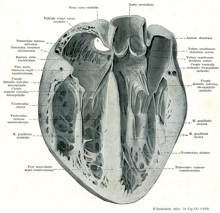
- 541_01【Inferior vena cava下大静脈 Vena cava inferior; Vena cava caudalis】 It arises at the union of the right and left common iliac veins, lies on the right side of the aorta, and opens into the right atrium of the heart.
→(下大静脈は下肢および骨盤と腹部の器官の大部分から血液を受ける本幹で、第5腰椎体の右側で左右の総腸骨静脈の合流として始まり、このあと脊柱に沿って大動脈の右側を上行、肝臓の後面をこれに接して通過し、第八胸椎の高さで横隔膜の大静脈孔を貫いて胸腔に入り、ただちに右心房にそそぐ。下大静脈に流入する枝には総腸骨静脈、下横隔静脈、第3・第4腰静脈、肝静脈、腎静脈、右副腎静脈、右精巣静脈、右卵巣静脈、蔓状静脈叢などがある)
- 541_02Eustachian valve【Valve of inferior vena cava下大静脈弁 Valvula venae cavae inferioris】 Semilunar fold in front of the opening of inferior vena cava. During fetal development it directs blood toward the foramen ovale.
→(オイスタヒ弁とも呼ばれる。右心房内の下大静脈開口部前縁にみられる弁で、冠状静脈弁(テベシウス弁valve of Thebesius)とともに胎生期の静脈弁に由来するという。)
- 541_03【Trabeculae carneae of atrium肉柱(心房の) Trabeculae carneae atriorum cordis】
→(")
- 541_04【Openings of smallest cardiac veins細小静脈孔 Foramina venarum minimarum】 Openings into the right atrium.
→(細小静脈孔は最小心静脈の多数の開口。心臓組織からの血液を直接、右心房その他に注ぐ。)
- 541_05【Atrioventricular septum房室中隔 Septum atrioventriculare】 Portion of the membranous part of interventricular septum between the right atrium and left ventricle above the root of the septal cusp.
→(房室中隔は左心房と左心室の間の膜性部のなかで、房室弁起始より上方にある部分。)
- 541_06【Membranous part of interventricular septum膜性部;膜部(心室中隔の) Pars membranacea septi interventricularis】 Short, thin, fibrous portion of the upper part of the interventricular septum near the exit of the aorta. It arises from endocardium.
→(心室中隔の膜性部は上方は大動脈流出路にある中隔の部分で、薄く、線維性。心内膜からつくられる。)
- 541_07【Posterior cusp of tricuspid valve後尖(三尖弁の) Cuspis posterior; Cuspis dorsalis (Valva tricuspidalis)】
→(三尖弁の後尖は三尖弁のうち後側にある。)
- 541_08【Septal cusp of tricuspid valve中隔尖(三尖弁の) Cuspis septalis (Valva tricuspidalis)】 Cusp or leaflet arising from the interventricular septum.
→(三尖弁の中隔尖は三尖弁の内部にあり、中隔からおこる弁尖端。)
- 541_09【Right ventricle右心室 Ventriculus dexter】
→(右心室は心臓の最下位部を占め、後上方にある右房室口で右心房と交通し、前上方にある肺動脈口で肺静脈に連なる。)
- 541_10【Trabeculae carneae of right ventricle肉柱(右心室の) Trabeculae carneae (Ventriculus dexter)】
→()
- 541_11【Anterior papillary muscle of right ventricle前乳頭筋(右心室の) Musculus papillaris anterior (Ventriculus dexter)】 Largest papillary muscle, located anteriorly and often overlying the septomarginal trabecula. It is connected with the anterior and posterior cusps.
→(右心室の前乳頭筋は前方にあり、大きい。)
- 541_12【Muscular part of interventricular septum筋性部;筋部(心室中隔の) Pars muscularis (Septum interventriculare)】
→(心室中隔の筋性部は中隔のうち大部分を占める厚い筋性の部分は左右両心室の筋層で作られる。)
- 541_13【Ascending aorta上行大動脈;大動脈上行部 Pars ascendens aortae; Aorta ascendens】 Ascending part of the aorta up to its exit from the pericardium.
→(左心室からおこり、肺動脈幹の後ろを上行して大動脈弓にいたる5cmほどの部。基部の内腔は膨らんで大動脈洞(バルサルバ洞)をなし、ここから左右の冠状動脈が出る。(イラスト解剖学))
- 541_14【Left atrium左心房 Atrium cordis sinistrum; Atrium sinistrum】
→(左心房は心臓の後上部にあって、後面をつくっている。左心房は右心房よりもやや小さいが、壁はやや厚い。左心房の後壁の上部に、左右両肺からそれぞれ2本ずつ、前部で4本の肺静脈が開口している。左心房は前下方で房室口によって左心室に通じる。)
- 541_15【Left semilunar cusp of aortic valve; Left coronary cusp of aortic valve左半月弁;左冠尖;左半月帆(大動脈弁の) Valvula semilunaris sinistra; Valvula coronaria sinistra (Valva aortae)】
→()
- 541_16【Right semilunar cusp of aortic valeve; Right coronary cusp of aortic valve右半月弁;右冠尖;前半月帆(大動脈弁の) Valvula semilunaris dextra; Valvula coronaria dextra (Valva aortae)】
→()
- 541_17【Anterior cusp of mitral valve; Anterior cusp of left atrioventricular valve; Anterior cusp of bicuspid valve前尖(僧帽弁の;左房室弁の) Cuspis anterior (Valva mitralis)】 Anterior leaflet situated near the septum.
→(僧帽弁の前尖は前方で、心室中隔側にある。)
- 541_18【Posterior cusp of mitral valve; Posterior cusp of left atrioventricular valve後尖(僧帽弁の) Cuspis posterior (Valva mitralis)】 Posterior leaflet situated near the lateral wall. Its free margin is more deeply grooved than that of the anterior cusp.AB
→(僧帽弁の後尖は後方で、側壁にある。)
- 541_19【Anterior papillary muscle of left ventricle前乳頭筋;左乳頭筋(左心室の) Musculus papillaris anterior; Musculus papillaris sinister (Ventriculus sinister)】 Larger, anterior papillary muscle arising from the lateral wall of the left ventricle.
→()
- 541_20【Posterior papillary muscle of left ventricle後乳頭筋;右乳頭筋(左心室の) Musculus papillaris posterior; Musculus papillaris dexter (Ventriculus sinister)】 It arises from between the interventricular septum and the lateral wall.
→()
- 541_21【Left ventricle左心室 Ventriculus sinister】
→(左心室は心臓の左下部を占め、後上方にある左房室口で左心房と交通し、右上隅にある大動脈口によって大動脈につらなる。左心室の壁は右心室に比べ2~3倍厚い。心室中隔は、右心室に向かって膨隆しているので、心室を横断面でみると、左心室の内腔は円いのに対して、右心室の内腔は半月状である)
- 541_22【Trabeculae carneae of left ventricle肉柱(左心室の) Trabeculae carneae ventriculi sinistri】
→(左心室の内面には、右心室と同様に、多数の発達した肉柱がみられる。)