Spalteholz HANDATLAS DER ANATOMIE DES MENSCHEN VON WERNER SPALTEHOLZ
メニューは解剖学(TA)にリンクしてあります。図の番号をクリックすると下記の説明へ、右側の用語をクリックすると解剖学(TA)にジャンプします。
021


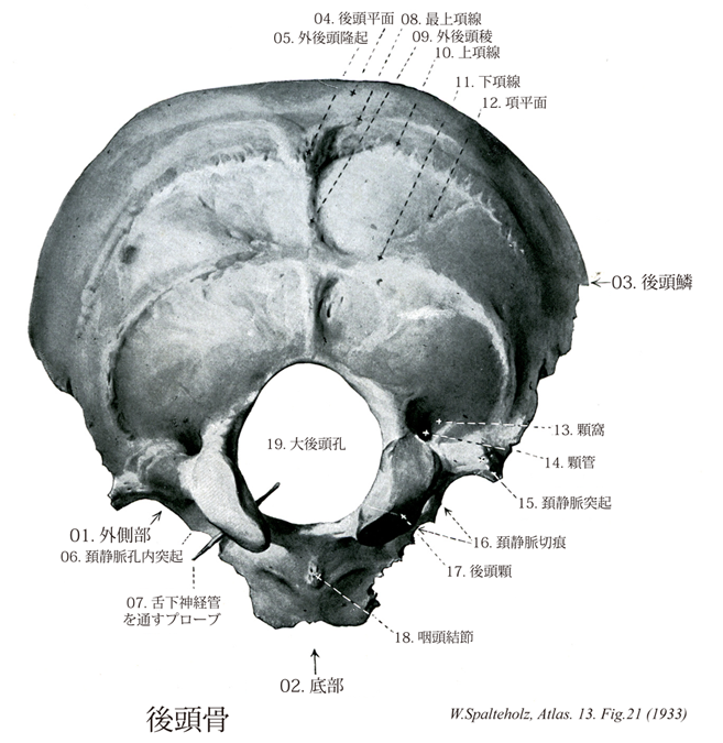
- 021_00【Occipital bone後頭骨 Os occipitale】 Bone located between the sphenoidal, temporal, and parietal bones.
→(脳頭蓋の後下部にある単一の骨で、頭蓋の脊柱上端に連なる部をつくる。前端に近く大きな大後頭孔があって、それより前方の底部、両側の外側部、後方の後頭鱗の3部に分けられる。前方は蝶形骨体、外方は側頭骨の岩様部、上方は頭頂骨と接するその形はほぼ舟状で、内面はくぼみ、外面はふくれる。後頭骨は胎生期後半には4つの部分に分離している。これらの4部が癒合して単一の骨になるのは生後3~4年たってからであるが、各部の名前だけは成人の骨にも残されている。)
- 021_01【Lateral part of occipital bone外側部(後頭骨の) Pars lateralis ossis occipitalis】 The part that is lateral to the foramen magnum.
→(後頭骨の外側部は大後頭孔の両側にある部分で、下面に後頭顆がある。そのやや外側の前縁には頚静脈切痕という切れ込みがあり、これは頚静脈孔の壁の下半を作る(壁の上半は側頭骨の同名の切痕)。この頚静脈孔は後頭骨および側頭骨から出る小さい[頚静脈]孔内突起によって前後の2部に分かれる。前部は小さく、舌咽神経、迷走神経、副神経、および下錐体静脈洞がここを通る。後部は大きく、内頚静脈の通路である。頚静脈切痕の後方の骨部は外側方に突出して頚静脈突起となり、その肥厚した外側縁は側頭骨の岩様部と軟骨性に結合する。外側部の下面には大後頭孔の前半の両側にあたり、後頭顆が突出する。その表面は滑らかで、少し外方に傾斜した楕円形の凸面で、その長軸は前内側から後外側に向かう。後頭窩は環椎の上関節窩に対する関節頭をつくる。横溝(外側部と底部が別個に形成され、癒着した部にあたる)によって前後に2分されることもある。また、正中面に対する角度には個人差が大きい。後頭窩の基底部を後内方から前外方に斜に舌下神経管が貫く。後頭顆の後には顆窩があり、顆導出静脈が通る顆管がここに開く。顆管の太さは個人差がはなはだしく、同一個体でも両側のものの差が著しいことが少なくない。外側部の上面には後頭窩の上に相当して頚静脈結節があり、それが大後頭孔に向かう面の基部に舌下神経管の内口が開く。頚静脈突起は上面ではとくに突隆し、それを後から内前方へS状洞溝の末端がめぐって頚静脈切痕の後縁に達している。顆管の内口が明瞭に見られるときは、この屈曲点よりやや前方に位置することが被い。)
- 021_02【Basilar part of occipital bone底部(後頭骨の) Pars basilaris (Os occipitale)】 The part that ascends from the foramen magnum to the spheno-occipital synchondrosis.
→(後頭骨の底部は大後頭孔前縁の前方にある長方形の板状部で、内頭蓋底と外頭蓋底の斜台の下半分を作る。両側縁は側頭骨の錐体と軟骨結合している。)
- 021_03【Squamous part of occipital bone後頭鱗(後頭骨の) Squama occipitalis】 The part that is posterior to the foramen magnum.
→(後頭鱗は大後頭孔の後方にある広い扁平な骨部で、頭蓋冠の後頭の部分と頭蓋底の後部を作る。その縁は不正三角形の広大な鱗状部である。その鋸歯状で大部分はラムダ縫合をもって頭頂骨と接するが、下方では側頭骨とも接する。後頭骨はその大半が軟骨性骨化によって生ずるが、後頭鱗のうち下項線から上方の部分だけは線維性骨窩によって生ずる膜性骨である。しかも後者は数個との骨化中心から出来るので、それら相互の癒合の様子次第で小さい2~4個の、または大きい1個の頭頂間骨(インカ骨)が独立する変異が生ずる。)
- 021_04【Occipital plane後頭平面 Planum occipitale】 Surface above the external occipital protuberance.
→(後頭平面は上項線より上の後頭骨の後極の外面。)
- 021_05【External occipital protuberance外後頭隆起;後頭結節;外後頭結節 Protuberantia occipitalis externa】 Easily palpable bony projection at the border between the occipital and nuchal planes.
→(外後頭隆起は凸面をなす後頭鱗の外面のほぼ中央に外後頭隆起がある。)
- 021_06【Intrajugular process頚静脈孔内突起;孔内突起 Processus intrajugularis】 Process that occasionally divides the jugular foramen into a lateral compartment for the passage of the internal jugular vein and a medial compartment for the transmission of nerves.
→(頚静脈孔内突起は後頭骨および側頭骨の頚静脈切痕中央から出る小さな、先のとがった骨性突起。この2つの骨は靱帯により結合し頚静脈孔を前後の2部に分けており、前部は小さく、ここを通るものは、舌咽神経、迷走神経、副神経、下錐体静脈洞で、後部は大きく内頚静脈が通る。)
- 021_07【Hypoglossal canal舌下神経管 Canalis nervi hypoglossi; Canalis hypoglossi】 Passageway that begins superolateral to the foramen magnum and ends anterolateral to the occipital condyle. It transmits CN XII and a venous plexus.
→(後頭顆の上方には後内方から前外方に舌下神経の通路である舌下神経管が走る。)
- 021_08【Highest nuchal line最上項線;界上項線 Linea nuchalis suprema; Linea nuchalis supraterminalis; Linea nuchae suprema】 Curved line that extends laterally from the superior margin of the external occipital protuberance. It gives origin to the occipital belly of the epicranius muscle.
→(最上項線は後頭骨の外面上を上項線の上方で平行に走る線。帽状腱膜および後頭筋が付着する。)
- 021_09【External occipital crest外後頭稜 Crista occipitalis externa】 Bony ridge that is occasionally present between the external occipital protuberance and the foramen magnum.
→(外後頭隆起から下方へのびて大後頭孔までいたる隆起線を外後頭稜という。)
- 021_10【Superior nuchal line上項線;分界項線 Linea nuchalis superior; Linea nuchalis terminalis; Linea nuchae superior】 Transverse ridge at the level of the external occipital protuberance. The area between it and the highest nuchal line gives origin to the trapezius muscle.
→(上項線は後頭骨の外後頭隆起から後頭側角へ側方にのびる隆線。僧帽筋、胸鎖乳突筋、および頭板状筋が付着する。上項線は外後頭隆起の直下で外後頭稜に達するが、この到達した点がイニオンである。)
- 021_11【Inferior nuchal line下項線;項平面線 Linea nuchalis inferior; Linea plani nuchalis】 Transverse ridge that extends from the superior nuchal line to the foramen magnum. The area between the inferior and superior nuchal lines gives attachment to the semispinalis capitis muscle.
→(下項線は上項線と大後頭孔の間にあり上項線よりかなり下方にある横走する隆起線である。)
- 021_12【Nuchal plane項平面(後頭骨の) Planum nuchale】 Surface below the external occipital protuberance. Attachment site for muscles of the neck.
→()
- 021_13【Condylar fossa顆窩 Fossa condylaris】 Depression located posterior to the occipital condyle into which the condylar canal opens.
→(後頭顆の後方に顆窩という凹みがある。)
- 021_14【Condylar canal顆管 Canalis condylaris】 Passageway for transmission of a vein that begins at the sigmoid sinus and ends posterior to the occipital condyle.
→(顆窩には顆導出静脈を通す顆管が開口する。導出静脈とは、頭蓋腔の内部と外部の静脈を連絡する静脈で、頭蓋骨の骨組織中を走るもの。)
- 021_15【Jugular process of occipital bone頚静脈突起 Processus jugularis】 Projection located lateral to the jugular foramen that is visible internally and externally. It corresponds to the transverse process of a vertebra.
→(後頭骨顆の後部、頚静脈孔の外側にある突起、脊椎の横突起に相当する。)
- 021_16【Jugular notch of occipital bone頚静脈切痕(後頭骨の) Incisura jugularis ossis occipitalis】 Recess that, together with the petrous part of temporal bone, forms the jugular foramen.
→(後頭骨の外側部の前部に頚静脈切痕があり、側頭骨岩様部の頚静脈切痕と合して頚静脈孔をつくる。)
- 021_17【Occipital condyle; *Condyle後頭顆 Condylus occipitalis】 Spherical eminence for articulation with the atlas.
→(後頭骨下面にある2つの細長い卵形をした関節面を有する高まりが後頭顆である。これは第1頚椎の上関節窩と関節する。)
- 021_18【Pharyngeal tubercle; Pharngeal tubercle of occipital bone咽頭結節;後頭骨の咽頭結節 Tuberculum pharyngeum】 Small protuberance on the inferior aspect of the basilar part of the occipital bone that provides attachment to the pharyngeal raphe.
→(後頭骨の底部の下面は筋の付着部となるため全般に粗で、その中央に小さい咽頭結節がある。咽頭結節は咽頭後壁の咽頭縫線が着く所である。その両側には上咽頭収縮筋、そのさらに側方には頭長筋、前頭直筋が着く。)
- 021_19【Foramen magnum大後頭孔;大孔 Foramen magnum; Foramen occipitale magnum】 Large opening in the occipital bone for the medulla oblongata. vessels, and nerves.
→(大後頭孔(大孔)は大きさや形に変化があるが、一般に前後に長い卵円形である。大後頭孔は頭蓋腔と脊柱管とを結ぶ孔で、したがって脳の脊髄につづく部である延髄下部が副神経脊髄根、椎骨動脈、静脈叢などとともにこれを通る。)