Spalteholz HANDATLAS DER ANATOMIE DES MENSCHEN VON WERNER SPALTEHOLZ
メニューは解剖学(TA)にリンクしてあります。図の番号をクリックすると下記の説明へ、右側の用語をクリックすると解剖学(TA)にジャンプします。
610 


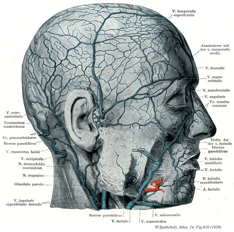
- 610_01【Posterior auricular vein後耳介静脈;耳介後静脈 Vena auricularis posterior; Vena retroauricularis】 Superficial vein lying behind the ear.
→(後耳介静脈は耳の後部表面にある静脈で耳下腺の直下部で下顎後静脈の後部と合流し、外頚静脈を形成する。)
- 610_02【Mastoid emissary vein乳突導出静脈 Vena emissaria mastoidea】 Vein connecting the sigmoid sinus with the occipital vein via the mastoid foramen.
→(乳突導出静脈はS状静脈洞を乳突孔を通って、上矢状静脈洞と浅側頭静脈とを結ぶ静脈。乳突孔の頭蓋内の開口部は、通常はS状静脈洞の下行脚descending limbに下行する。)
- 610_03【Anterior auricular veins前耳介静脈;耳介前静脈 Venae auriculaes anteriores; Venae praeauriculares】 Branches draining the external acoustic meatus and auricle.
→(前耳介静脈は外耳道、耳介からくる枝。)
- 610_04【Parotid veins耳下腺静脈;耳下腺枝(下顎後静脈の) Venae parotideae; Rami parotideae】 Branches draining the parotid gland.
→(耳下腺静脈は耳下腺からくる枝。)
- 610_05【Transverse facial vein顔面横静脈 Vena transversa faciei】 Vein accompanying the transverse facial artery caudal to the zygomatic arch.
→(顔面横静脈は頬骨弓の下方を同名動脈に伴う。)
- 610_06【Occipital vein後頭静脈 Vena occipitalis】 Vein beginning in the venous plexus of the scalp. It frequently opens into the vertebral vein or also into the internal or external jugular vein.
→(後頭静脈は頭皮の静脈網から起こり、多くは椎骨静脈に、ときにはまた内頚静脈や外頚静脈にもひらく。)
- 610_07【Sternocleidomastoid muscle胸鎖乳突筋 Musculus sternocleidomastoideus】 o: Two-headed muscle arising from the sternum and clavicle, i: Mastoid process; superior nuchal line. Rotates the face to the contralateral side and bends the head to the ipsilateral side. Bilateral contraction elevates the face. I: Accessory nerve, cervical plexus (C1-C2).
→(胸鎖乳突筋は側頚部にある強大な斜めに縦走する浅層の筋。胸骨柄前面と鎖骨の胸骨端から2頭をもっておこり、両頭は合して強い筋腹をつくって後上方に走り、乳様突起および後頭骨の上項線につく。作用は複雑で、両側が同時に働くとオトガイを上げて後頭部を片側が働けば頭を対側にまわすが、その浅オトガイが対側に向かって上り、頭は逆に同側に傾く。支配神経は副神経外枝と頚神経叢筋枝(C2, C3)であり、したがって僧帽筋と同系の筋である。また、第6咽頭弓に発生する鰓弓筋で、鎖骨上窩を囲む2頭(胸骨頭と鎖骨頭)をもって始まる。胸骨頭は胸骨柄の上縁から、鎖骨頭は鎖骨の胸骨端から起こる。筋膜は頚筋膜浅葉に鞘状に包まれており、斜め上方に向かって幾分螺旋状に回転しながら頚部外側面を横切り、よく発達した腱となって乳様突起と上項線に停止する。筋の表面は、起始部で腹側に、停止部で外側に向く。参考:副神経外枝の僧帽筋枝は、外枝がこの筋に入る前に分かれることと、筋内で分かれて再び外に現れることがある。胸鎖乳突筋はドイツ語ではKopfnicker(頭をこっくりとうなずかせる筋)と呼ばれるが、これは作用の点からは正しくない。この筋が片側だけ収縮すると、頭はその側へ傾き反対側を振り向いて、あたかも「首をかしげる」状態になる。また両側の物が同時に収縮すると、頭を胴体にめり込ませるように働くのえある。Musculus sternocleidomastoideusというラテン名はあまりにも長たらしいので、米英では多少簡略化してsternomastoid muscleともよぶ。片側の胸鎖乳突筋が先天的に短い場合、または出産時の外傷などによって瘢痕化して短縮すると、この筋の作用を考えればすぐわかるように、頭は病側へ傾くと共に健側にねじれたままの状態になるこれを斜径torticolis, wryneck(性格には筋性斜径)といい、かなり頻度の高いものである。略語(SCM))
- 610_08【Trapezius muscle僧帽筋 Musculus trapezius】 Muscle that consists of three parts that act together to position the scapula and clavicle, draw both toward the vertebral column, and brace the shoulder girdle. I: Accessory nerve; brachial plexus C2-C4.
→(背部第1層にみられる扁平な菱形の筋で背部上半部を占める。僧帽筋は上肢の運動の時に肩甲骨を動かす重要な筋である。とくに上腕の外転のときに、肩甲骨を後内側に引くと同時に下角を外側に回し、関節窩が上外側を向くようにする。僧帽筋は下行部、横走部、上行部に分けられる。[臨床]僧帽筋の完全麻痺(副神経と上部腕神経の同時の傷害)の場合、肩は健側よりも深く位置するようになる項肩線は弓状を呈さず、乱れる。肩甲骨は正中線より、はるかに離され、関節窩は前下方を向く。肩は(肩甲挙筋の)弱いエネルギーにより持ち上げることが出来るにすぎず、わずかに(菱形筋により)後方にもたらされるにすぎない。腕の外側への挙上は大きく減少する。腕は通常水平面まで外転され得ない。腕の前方への挙上は(前鋸筋による肩甲骨の回転により)ほとんど制限さされないが、矢状面での挙上は強く妨げられる。副神経のみが傷害された場合、僧帽筋の下行部の機能は(上頚神経の付随的支配により)種々の程度に保存される。肩甲骨の位置の変化はそれほど著明ではない。しかし、腕を横または後へ挙上することは、ちょうどその程度に応じて制限される。)
- 610_09【Parotid gland耳下腺 Glandula parotidea; Glandula parotis】 It occupies the retromandibular fossa, extending to the temporomandibular joint and the ramus of mandible.
→(耳下腺はヒト最大の唾液腺で、左右の耳の前下方にあり、下は下顎角まで、上は頬骨弓まで、後方は胸鎖乳突筋まで、内側は側頭下窩の下顎骨下顎枝まで広がっている。その分泌管の耳下腺管によって上顎第2大臼歯の頬粘膜に開口する。終末部(線房)は純漿液性の分泌物からなる(これは他の大唾液腺との大きな違いである)。介在部および線条部もよく発達している。小葉内(腺の実質内)に多数の脂肪細胞が散在するもの、大きな特徴の一つで他の唾液腺と容易に区別できる点である。Parotisという語は、para(傍)とotis(耳)との複合語で、耳の傍らにあるものという意味である。17世紀のフランスの解剖学者リオランの命名である。)
- 610_10【External jugular vein外頚静脈;外側浅頚静脈 Vena jugularis externa; Vena jugularis superficialis dorsalis】 Vein lying between the platysma and supetficial layer of cervical fascia and usually emptying into the subclavian vein. It is fed by the following veins.
→(外頚静脈は側頚部の皮下静脈であり、頚部のみならず頭部の表在性静脈血を集める。後耳介静脈と下顎後静脈が合して下顎角の後方ではじまり、広頚筋におおわれて胸鎖乳突筋の表面を斜めに下行し、大鎖骨上窩で鎖骨下静脈にそそぐ。下顎後静脈前枝を介して内頚静脈と連絡しているので、これら2静脈ならびに鎖骨下静脈とともに胸鎖乳突筋を斜めに取り囲む動脈輪を形成している。受け入れる静脈根は後頭静脈、後外頚静脈、頚横静脈と肩甲上静脈、前頚静脈である。)
- 610_11【Parotid vens; Parotid branches of deep facial vein耳下腺枝(顔面静脈の) Venae parotideae; Rami parotidei vena facialis】 Branches draining the parotid gland.
→(顔面静脈の耳下腺枝は耳下腺からそそぐ。)
- 610_12【Facial vein顔面静脈 Vena facialis】 Vein beginning at the medial angle of eye that lies behind the facial artery and then beneath the submandibular gland.
→(顔面静脈は顔面動脈の分布域である顔面浅部からの静脈を集める。顔面静脈は内眼角から始まり(眼角静脈)、顔面動脈の後ろに沿って斜めに下方に走り、内・外頚動脈、舌下神経との浅側を後下方に向かい、舌骨の高さで内頚静脈または外頚静脈にそそぐ。顔面静脈は吻合に富み、また顔面の深部の静脈や頭蓋内の静脈(硬膜静脈洞)とも連絡している。たとえば、顔面静脈は内眼角の付近で、眼窩内の上眼静脈の根もと吻合し、さらに頭蓋腔内の顔面静脈洞とも連絡する。また、鼻や上唇の近くでも深部の静脈と連絡する。)
- 610_13【Superficial temporal veins浅側頭静脈 Venae temporales superficiales】 Veins accompanying the superficial temporal artery.
→(浅側頭静脈は浅側頭動脈に伴行し、耳下腺内で顎静脈と合流し、下顎後静脈をつくる。)
- 610_14【Middle temporal vein中側頭静脈 Vena temporalis media】 Vein from the temporalis that empties into the superficial temporal veins.
→(中側頭静脈は側頭筋から起こり浅側頭静脈にそそぐ。)
- 610_15【Supratrochlear veins滑車上静脈;前頭静脈 Venae supratrochleares; Venae frontales】 Vein beginning at the coronal suture that drains the medial half of the forehead. It joins the angular vein.
→(冠状縫合に始まる前額の内半部の静脈。眼角静脈と合一する。)
- 610_16【Supra-orbital vein眼窩上静脈 Vena supraorbitalis】 Vein from the lateral part of the forehead that joins the supratrochlear veins.
→(眼窩上静脈は外側の額部からきて滑車上静脈と合一する。)
- 610_17【Nasofrontal vein鼻前頭静脈 Vena nasofrontalis】 Connection between the ophthalmic vein and the union of the supratrochlear vein with the angular vein.
→(鼻前頭静脈は滑車上静脈と眼角静脈の合一部を上眼静脈とむすぶ。)
- 610_18【Angular vein眼角静脈 Vena angularis】 Beginning of the facial vein at the angle of eye formed by the union of the supratrochlear and supra-orbital veins. It anastomoses with the ophthalmic vein and communicates via the nasofrontal vein with the superior ophthalmic vein. Similar to the latter, it does not have any valves. Potential infection pathway from the face to the orbits and cranial cavity.
→(眼角静脈は眼窩上静脈と滑車上静脈によって形成され、眼角での顔面静脈起始に相当し、滑車上静脈と眼窩上静脈の合一でつくられる。眼静脈と吻合。鼻前頭静脈を介して上眼静脈とむすばれ、弁を欠く。)
- 610_19【External nasal veins外鼻静脈 Venae nasales externae】 Veins from the lateral side of the nose.
→(外鼻静脈は鼻の外側面からくる静脈。)
- 610_20Stensen's (Stenon) duct【Parotid duct耳下腺管 Ductus parotideus】 Excretory duct that extends around the anterior border of the masseter, usually over the buccal fat pad, and opens opposite to the upper second molar tooth.
→(耳下腺管はステンセン管ともよばれる。または、ステノン管ともよばれ、日本ではステノ氏孔などともいう。耳下腺管は頬骨弓の下方約2cmの部を水平に走り、頬筋を貫いて上顎第2大臼歯対側の口腔粘膜に開口する。デンマークの解剖学者Niels Steno [Nicholas Stensen] (1638-1686)によって、1661年頃に発見された。後年、ステンセンはローマカトリックの司教となっている。)
- 610_21【Superior labial vein上唇静脈 Vena labialis superior; Vena labialis maxillaris】 Veins draining the upper lip.A
→(上唇の静脈。)
- 610_22【Inferior labial veins下唇静脈 Venae labiales inferiores; Venae labiales mandibularis】 Usually multiple veins draining the lower lip.A
→(下唇静脈は多くの場合たくさんある。)
- 610_23【Facial artery顔面動脈 Arteria facialis】 Third anterior branch of the external carotid artery. It lies behind the posterior belly of digastric muscle, stylohyoid, and submandibular gland. It crosses the mandible along the anterior border of the masseter and supplies the muscles of facial expression.
→(顔面動脈は舌動脈のやや上方で、外頚動脈の前側から起こり、下顎角の内側で顎下腺の上面を前方に走り、化学体の下縁をまわって顔面に現れる。顔面に出ると、蛇行しながら口角を経て鼻の側縁に沿って上場し内眼角(メガシラ)に至る。顔面動脈が下顎骨の下縁をまたがって顔面に出るところで体表から脈動を触れる。この部位は咬筋の前縁(歯を強くかみ合わせると触れる)にあたる。)
- 610_24【Submental veinオトガイ下静脈 Vena submentalis】 Companion vein of the submental artery. It anastomoses with the sublingual and anterior jugular veins.
→(オトガイ下静脈はオトガイ下動脈に伴行する。舌下静脈および前頚静脈と吻合。)
- 610_25【Masseteric veins; Masseter veins咬筋静脈 Venae massetericae】
→()