Spalteholz HANDATLAS DER ANATOMIE DES MENSCHEN VON WERNER SPALTEHOLZ
メニューは解剖学(TA)にリンクしてあります。図の番号をクリックすると下記の説明へ、右側の用語をクリックすると解剖学(TA)にジャンプします。
1014


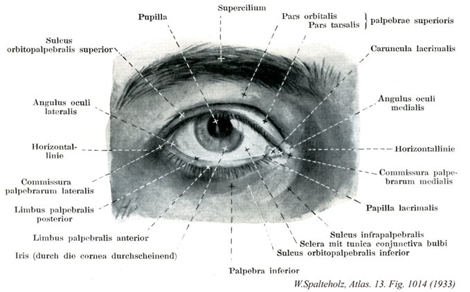
- 1014_01【Pupil瞳孔 Pupilla】 Opening in the iris surrounded by the pupillary margin. Its diameter ca. change in response to light entering the eye and the distance to an object being viewed.
→(瞳孔は虹彩の中心にある瞳孔縁に囲まれた孔。ほぼ円形の開口。角膜、前眼房を通過した光が、ここを経て水晶体、硝子体、そして網膜に達する。瞳孔の直径は明るさにより両側性に変化し(瞳孔反射)、明所では~1.0mm(縮瞳)、暗所では~8.0mm(散瞳)ぐらいである。入射光の強さおよびみえるものまでの距離に応じてその径を変えることが出来る。不交感神経遮断剤(アトロピンなど)や交感神経刺激剤(アドレナリンなど)の点眼により持続的散大が可能で、眼科学的に広く応用されている。瞳孔は胎生期には虹彩内皮のつづきである瞳孔膜によりとじられているが、妊娠末期にこれが消失する。眼胞から眼杯が形成される過程で脈絡裂閉鎖が不完全な場合、いろいろの程度に虹彩瞳孔縁の欠損が残り、これを虹彩披裂(Coloboma iridis)という。虹彩筋は輪走する瞳孔括約筋と、放射状の瞳孔散大筋がある。虹彩筋はすべて色素上皮細胞より分化する神経外胚葉由来の筋上皮組織である。)
- 1014_02【Suprapalpebral sulcus; Superior orbitopalpebral sulcus上眼瞼溝;上眼溝;前頭眼瞼溝;上眼窩眼瞼溝 Sulcus suprapalpebralis; Sulcus frontopalpebralis; Sulcus orbitopalpebralis superior】 Groove located above the upper eyelid.
→()
- 1014_03【Lateral angle of eye外眼角;メジリ Angulus oculi lateralis; Angulus oculi temporalis】 Sharp lateral angle of the eye that simultaneously forms the lateral end of the palpebral fissure.
→(眼裂の外側端でもある。(Feneis))
- 1014_04【Lateral palpebral commissure外側眼瞼交連 Commissura lateralis palpebrarum】 Junction of the upper and lower eyelids at the lateral angle of eye.
→(外側眼瞼交連は外眼角部での上眼瞼の下限瞼への移行。)
- 1014_05【Posterior palpebral margin後眼瞼縁 Limbus posterior palpebrae】 Inner margin of the eyelids facing the conjunctiva.
→(結膜に対する眼瞼縁。(Feneis))
- 1014_06【Anterior palpebral margin前眼瞼縁 Limbus anterior palpebrae】 Margin of the eyelids facing the outer skin of the eyelid.
→(前眼瞼縁は眼瞼皮膚に対する眼瞼縁。)
- 1014_07【Iris虹彩 Iris】 Round disc with a central opening (pupil) situated in the frontal plane that varies in color in different individuals. It forms the posterior end of the anterior chamber and becomes continuous at its margin with the ciliary body. It has a diameter of 10-12 mm.
→(虹彩は、前頭面に位置し、眼の血管層の前方部分をつくる隔膜で、色に個人差のある円板。中央に開口部(瞳孔)があり、直径は約10~12mm。前眼房の後境界で、その縁は毛様体へ移行する。周囲辺縁は強膜岬角に付着している。瞳孔をかたちづくるあたかもカメラの絞りのような器官で、虹彩内皮、虹彩支質、虹彩筋、虹彩色素上皮層より構成され、血管に富む。虹彩の脈管と神経はは虹彩の動脈としては毛様体縁に沿う大虹彩動脈輪、瞳孔縁に沿う大虹彩動脈輪、瞳孔縁に沿う小虹彩動脈輪、両者を放射状につなぐ小動脈があり、長後毛様体動脈、前毛様体動脈、脈絡膜毛細血管叢より供給される。静脈血はこれらに伴う静脈のほか、渦静脈に流入する。虹彩の神経支配として長毛様体神経(三叉神経由来の体知覚性神経)と短毛様体神経(毛様体神経節由来の自律神経)があり、後者には動眼神経副核由来の節前線維から興奮を受けて伝達する節細胞の軸索すなわち副交感神経節後線維と、内頚動脈神経叢を経て毛様体神経節に達し、節内でそれに合流する胸部交感神経核由来の交感神経節後線維が含まれる。瞳孔括約筋は副交感神経、散大筋は交感神経の支配を受ける。)
- 1014_08【Eyebrow眉 Supercilium】 Eyebrow with its thicker, brushlike hair.
→(眉はほぼ眼窩上縁に沿う弓状の皮膚の高まりを眉といい、その表面の毛を眉毛という。しかし「眉」を両方に用いることが多い。)
- 1014_09【Superior eyelid; Upper eyelid上眼瞼;ウワマブタ Palpebra superior】 Larger, upper eyelid.
→()
- 1014_10【Orbital part of superior eyelid上瞼板部;眼窩部(上眼瞼の) Pars supratarsalis; Pars orbitalis (Palpebra superior)】
→()
- 1014_11【Tarsus part of superior eyelid瞼板部(上眼瞼の) Pars tarsalis (Palpebra superior)】
→()
- 1014_12【Lacrimal caruncle涙丘 Caruncula lacrimalis】 Mucosal protuberance at the medial angle of eye with stratified squamous or columnar epithelium.
→(涙丘はめがしらにある米粒大の隆起。その周辺の陥凹部を涙湖という。組織学的には皮膚に似ている。)
- 1014_13【Medial angle of eye内眼角;メガシラ Angulus oculi medialis; Angulus oculi nasalis】 Medial end of the palpebral fissure that has a convex, rounded shape for the lacrimal lake.
→(内眼角は眼裂の内側端でもある。まるく弯出し涙湖をなす。)
- 1014_14【Medial palpebral commissure内側眼瞼交連 Commissura medialis palpebrarum】 Junction of the upper and lower eyelids at the medial angle of eye.
→(内側眼瞼交連は内眼角部での上眼瞼の下眼瞼への移行。)
- 1014_15【Lacrimal papilla涙乳頭 Papilla lacrimalis】 Single small, medial, conical elevation on each eye at the inner margin of the upper and lower eyelids on top of which the lacrimal punctum sits.
→(上および下眼瞼の内縁の内側にある小さな円錐状の高まり。尖端に涙点がある。(Feneis))
- 1014_16【Palpebromal sulcus瞼頬溝;瞼鼻溝 Sulcus palpebromalaris; Sulcus palpebronalis】
→()
- 1014_17【Sclera強膜 Sclera】 Membrane of the eyeball composed of interwoven collagen fibers. It has a bluishwhite appearance and is visible through the conjunctiva.
→(眼球の形状を保つ強靱な膠原線維組織層。角膜となっている前部6分の1を除いた部分。前方では隔膜固有質に、後方では篩板から視神経外鞘を経て脳硬膜に、それぞれつづいている。強膜と角膜を合わせて眼球線維膜という。強膜の厚さは眼球後極で~1.0mm、前部で~0.6mm、赤道で~0.4mmである。視神経線維束を通す篩板は後極の内側3.5mm、視神経乳頭の直後方にあたる。視神経は~数十本の掌側としてこれを通る。渦静脈、長・短毛様体動脈および神経が強膜を貫く。強膜はは外から内へ、①強膜上皮、②強膜固有質、③強膜褐色板の3沿うよりなる。虹彩角膜角に沿って強膜固有質が内方へ皮厚し(強膜距)毛様体筋腱により貫かれる。この部の直前に輪状に走る強膜静脈洞(Schlemmn管)があり、眼房水は虹彩角膜間隙(Fontana腔)からこれを通って渦静脈に排出される。角膜縁をとり膜浅い強膜溝の深層にこれらの構造がある。眼球前部の強膜上板毛細血管網に富み、その炎症性変化を臨床的に「網膜充血」という。強膜前部は眼球結膜、後部は眼球鞘(Tenon鞘)によりおおわれる。内面は脈絡外隙を間に脈絡外板に接する。)
- 1014_18【Bulbar conjunctiva眼球結膜 Tunica conjunctiva bulbi】 Portion of the conjunctiva covering the eyeball. It consists of stratified, nonkeratinized squamous epithelium with only a small number of goblet cells and a lamina propria of loosely organized structures, containing few cells and permeated by elastic fibers.
→(眼球結膜は結膜のうち眼球を被う部分。杯細胞に乏しい角化していない重層扁平上皮である。固有層は疎で細胞に乏しく弾性線維を含む。)
- 1014_19【Infrapalpebral sulcus; Inferior orbitopalpebral sulcus下眼瞼溝;下眼溝;眼瞼下溝;下眼窩眼瞼溝 Sulcus infrapalpebralis; Sulcus palpebralis inferior; Sulcus orbitopalpebralis inferior】 Groove located below the lower eyelid.
→(下眼瞼下にある溝。)
- 1014_20【Inferior eyelid; Lower eyelid下眼瞼;シタマブタ Palpebra inferior】 Smaller, lower eyelid.
→()