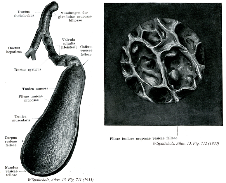
Spalteholz HANDATLAS DER ANATOMIE DES MENSCHEN VON WERNER SPALTEHOLZ
メニューは解剖学(TA)にリンクしてあります。図の番号をクリックすると下記の説明へ、右側の用語をクリックすると解剖学(TA)にジャンプします。
711


- 711_00【Gallbladder胆嚢;タンノウ Vesica biliaris; Vesica fellea】 Pear-shaped organ measuring 8-12 cm in length.
→(胆嚢はナスビの形のふくろ(長さ約9cm、太さ約4cm)で、胆汁を貯える。肝臓の下面の下面にあって、胆嚢窩に浅くはまりこんでいるので、肝臓の下面の被膜と共通の結合組織でおおわれ、下面と底は腹膜におおわれる。胆嚢の底はふくろの底の部分、体はふくらみの部分、頚は細くなった部分である。底が前方に向き、肝臓の下縁から少し前に突出している。頚がうしろ向き、胆嚢管につながる。胆嚢の内面には網状のひだが突出し、丈の高い単層円柱上皮でおおわれる。上皮細胞は粘液分泌を行う。よく発達した筋層がある。胆嚢管は長さ約3cmのやや迂曲する管で、内腔にらせん状に突出するひだがあり、らせんひだとよばれる。胆管と合流して総胆管となる。肝管を流れてくる胆汁は、通常、胆嚢管にはいって胆嚢に貯えられ、必要に応じて胆嚢管から総胆管を経て必要に応じて胆嚢管から総胆管を経て十二指腸に放出される。とくに食事が十二指腸に達すると、十二指腸壁から血中にコエシストキニンが放出され、このホルモンの左葉で胆嚢が収縮し、胆汁が排出される。)
- 711_01【Bile duct; Common bile duct総胆管;胆管 Ductus choledochus; Ductus biliaris】 Duct draining the gallbladder that is formed by the union of the common hepatic and cystic ducts and passes to the major duodenal papilla.
→(総胆管は肝管と胆嚢管の合流点から十二指腸下行部の内側面に下行する6~8cmの管で、肝十二指腸間膜の中を、肝固有動脈、門脈と伴行する。十二指腸に終わる手前で膵頭を貫き、膵管と合流する。膵頭癌に際して総胆管が圧迫されて黄疸を起こすことは、この局所解剖学的関係による。総胆管は膵管と合流するところ、あるいはその直後に胆膵管膨大部をつくったのち、大十二指腸乳頭において十二指腸に開口する。総胆管の内面は単層円柱上皮で覆われ、固有層には小さい胆管粘液腺がある。筋層はおよそ内輪外斜の走行を示すが、とくに総胆管の下部では輪走筋が発達して、総胆管括約筋とよばれる。また胆膵管膨大部には(胆嚢管)膨大部括約筋が発達している。オッディーの括約筋の名で親しまれるこの筋は、消化管ホルモンや神経の作用を受けながら、胆汁と膵液の放出の肝門をなしている。)
- 711_02【Common hepatic duct; Hepatic duct総肝管;肝管 Ductus hepaticus communis; Ductus hepaticus】 Part of the bile duct between the junction of the right and left hepatic ducts and the cystic duct.
→(総肝管は肝門から右肝管と左肝管が出て、肝門の直下で合流して総肝管となる。総肝管は太さ約4mm、長さ3.5~5cmの管で、肝十二指腸間膜の中を右下方へ走り、胆嚢からくる胆嚢管と鋭角に合流する総胆管となる。)
- 711_03【Cystic duct胆嚢管 Ductus cysticus】 Duct that drains the gallbladder. It joins the common hepatic duct to form the bile duct.
→(胆嚢管は直径が3mmほどの細い管で弓状をなして走り、長さは3~4cmある。その起始には粘膜面にラセンヒダがあるが、総胆管に近い部分では内面は比較的平坦である。胆嚢管は基本的には胆嚢と同じ組織構成を有し、胆嚢管に連なる。胆嚢管の粘膜にはハイステルのラセン襞が形成されて一種のバルブの働きと管の内圧が変化しても胆嚢管の変形を防ぐ働きをしている。)
- 711_04【Gallbladder mucosa; Mucous membrane of gallbladder粘膜(胆嚢の) Tunica mucosa vesicae biliaris】 Mucous membrane of gallbladder composed of simple columnar epithelium.
→(胆嚢の粘膜は単層円柱上皮。)
- 711_05【Mucosal folds; Rugae of gallbladder粘膜ヒダ;ヒダ(胆嚢の) Plicae mucosae vesicae biliaris; Rugae vesicae biliaris】 Mucosal folds that project into the lumen, producing a locular relief.
→(胆嚢の粘膜ヒダは内腔へ突出する粘膜のヒダで、多数の小房をもつレリーフ状を呈する。)
- 711_06【Muscular layer of gallbladder筋層(胆嚢の) Tunica muscularis vesicae biliaris】 Muscular coat in the wall of the gallbladder.
→(胆嚢の筋層は線維筋層として知られる膠原線維を伴った縦、横、斜走する疎らな平滑筋層がある。)
- 711_07Hartmann's pouch【Body of gallbladder; Corpus of gallbladder胆嚢体 Corpus vesicae biliaris; Infundibulum vesicae felleae】 Floor of the gallbladder, which is directed caudally.
→(胆嚢体は胆嚢底と頚の間の部分。)
- 711_08【Fundus of gallbladder胆嚢底 Fundus vesicae biliaris; Fundus vesicae felleae】 The body of gallbladder is attached to the liver.
→(胆嚢底は下方に向いたまるい胆嚢端部。)
- 711_09【Glands of bile duct; Bile duct gland胆管粘液腺;総胆管腺;胆管腺 Glandulae ductus choledochi; Glandulae ductus biliaris; Glandulae mucosae biliosae】 Mucous glands situated in the bile duct.
→(胆管粘液腺は総胆管壁にある粘液腺。)
- 711_10Heister's valve【Spiral fold; Heister's valve; Spiral valve of Heisterラセンヒダ;ハイスターのラセンヒダ;ラセン弁 Plica spiralis; Valvula spiralis】 Spiral-shaped fold in the neck of gallbladder and cystic duct.
→(胆嚢管の起始には粘膜面にラセンヒダがある。胆嚢管のラセンヒダの作用には不明の点が多い。一般に体の中にある管は一方通行の路であるが、胆嚢管だけは二方通行になっている。すなわち肝臓から胆嚢に送られてくる胆汁の通る方向と、胆嚢で濃縮された胆汁が逆に総胆管へ送られる方向の二つがあるわけで、これらの両胆汁の交通整理にラセンヒダが役立っているという説もある。)
- 711_11【Neck of gallbladder胆嚢頚 Collum vesicae biliaris; Collum vesicae felleae】 Part of the gallbladder that is separated from the infundibulum by a bend to the right.
→(胆嚢頚は胆嚢管への移行する部分。)
