Spalteholz HANDATLAS DER ANATOMIE DES MENSCHEN VON WERNER SPALTEHOLZ
メニューは解剖学(TA)にリンクしてあります。図の番号をクリックすると下記の説明へ、右側の用語をクリックすると解剖学(TA)にジャンプします。
960


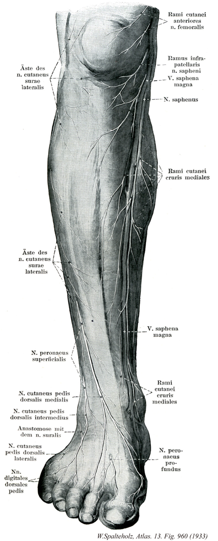
- 960_00【Leg下腿;スネ Crus】
→(膝と踝の間をいう。)
- 960_01【Lateral sural cutaneous nerve外側腓腹皮神経;腓側腓腹皮神経 Nervus cutaneus surae lateralis; Nervus cutaneus surae fibularis】 Nerve given off in the popliteal fossa that supplies the skin of the lateral side of the leg as well as the upper two-thirds of its posterior side.
→(外側腓腹皮神経は膝窩よりあらわれ、たいていは下腿の後外側面の上2/3の皮膚へ分布する。)
- 960_02【Superficial fibular nerve; Superficial peroneal nerve浅腓骨神経 Nervus fibularis superficialis; Nervus peroneus superficialis】 Terminal branch of the common fibular nerve. It descends between the peroneus muscles and extensor digitorum longus.
→(浅腓骨神経は総腓骨神経の終枝の一つ、腓骨筋と長趾伸筋の間を下行する。 (Netter)浅腓骨神経は、長趾伸筋と腓骨筋の間を下行し、長腓骨筋と短腓骨筋に筋枝を出した後、下腿の虫部から下部1/3に移る高さで下腿筋膜を貫く。この高さで、浅腓骨神経は、内側足背皮神経と中間足背神経とに分かれる。内側足背皮神経は足根の前面を走行して足背に至り、下部下腿前面と足背の皮膚と筋膜に枝を送る。下伸筋支帯の下縁近くで、この神経は2本の足背趾神経に分岐する。このうち1本は、足背および母趾の内側面と背側面を支配し、他の1本は第2,第3趾の背側面と側面とを支配する。中間足背皮神経は、足背外側部に沿って走行し、近傍の皮膚や筋膜に枝を出し、第3と第4趾および第4と第5趾に行く2本の足背趾神経に分かれる。また、中間足背皮神経は、外側足背皮神経と交通する。)
- 960_03【Medial dorsal cutaneous nerve内側足背皮神経;脛側足背皮神経 Nervus cutaneus dorsalis medialis; Nervus cutaneus dorsi pedis tibialis】 Branch traveling over the extensor retinaculum and supplying the skin of the dorsum of foot, the medial side of the great toe, and the adjacent halves of the second and third toes.
→(伸筋の支帯の上を通り、母趾の背内側面、第二、第三趾の対向縁の皮膚へ分布。 (Feneis))
- 960_04【Intermediate dorsal cutaneous nerve中間足背皮神経;中足背皮神経 Nervus cutaneus dorsalis intermedius; Nervus cutaneus dorsi pedis medius】 Lateral cutaneous branch of the superficial fibular nerve lying in the middle portion of the dorsum of foot.
→(中間の足背へいたる浅腓骨神経の外側枝。 (Feneis))
- 960_05【Sural nerve腓腹神経 Nervus suralis】 Continuation of the medial sural cutaneous nerve after it joins the sural communicating branch.
→(腓腹神経は脛骨神経の皮枝で腓腹筋の2頭のあいだを下行するが、その途中で総腓骨神経からの交通枝を受けるのがふつうである。腓腹神経からの小枝が下腿後面の皮膚に分布する。外顆より遠位では腓腹神経は小伏在静脈と伴行しながら、足の外側縁から小指外側縁に至る皮膚に枝を起こる。 (Netter)腓腹神経は皮神経であり、膝窩の中部あるいは下部で脛骨神経より起こる(図13)。腓腹筋の両頭の間を下行しながら、腓腹神経は下腿筋膜を貫通して小さな内側腓腹皮神経(内側腓腹皮神経は下腿筋膜貫通して小さな内側腓腹皮神経(内側腓腹皮神経はもっとも大きく、脛骨神経より直接起こることもある)を出し、外側腓腹皮神経から腓腹神経との交通枝を受ける。腓腹神経は、小伏在静脈近傍を通ってアキレス腱の外側へと下行を続けながら、下腿背側面および外側面を覆う皮膚と筋膜に枝を与える。外果と踵骨腱(アキレス腱)の間に達すると腓腹神経は外側踵骨枝を出し、足根と踵の外側面の皮膚と筋膜枝を出す(これに対応する内側踵骨枝は脛骨神経から起こる)。腓腹神経の終末枝は、外側足背皮神経として足および小趾の外側に沿って前方へ走る。)
- 960_06【Lateral dorsal cutaneous nerve外側足背皮神経;腓側足背皮神経 Nervus cutaneus dorsalis lateralis; Nervus cutaneus dorsi pedis fibularis】 Branch to the lateral portion of the dorsum of foot. It anastomoses with the intermediate dorsal cutaneous nerve.
→(外側足背皮神経は腓腹神経の延長で、足背の外側縁とその付近を支配する部分である。)
- 960_07【Dorsal digital nerves of deep fibular nerve背側趾神経;背側指神経(深腓骨神経の) Nervi digitales dorsales pedis; Nervi digitales dorsales hallucis fibularis et digiti secundi tibialis】 Sensory branches to the adjacent sides of the great toe and second toes.
→(母趾および第2趾の対向縁への枝。 (Feneis))
- 960_08【Anterior cutaneous branches of femoral nerve前皮枝(大腿神経の) Rami cutanei anteriores (Nervus femoralis)】 Cutaneous nerves supplying the distal three-fourths of the anterior side of the thigh as far as the patella.
→(大腿神経の前皮枝は大腿の前面と内側面に分枝し、大腿前面を膝蓋にいたる下部3/4の皮膚へ分布し、感覚を伝える。)
- 960_09【Infrapatellar branch of saphenous nerve膝蓋下枝(伏在神経の) Ramus infrapatellaris (Nervus saphenus)】 Branch that penetrates the sartorius and reaches the skin below the patella.
→(伏在神経の膝蓋下枝は縫工筋を貫き膝蓋骨上下の皮膚に分布する。)
- 960_10【Great saphenous vein; Long saphenous vein大伏在静脈 Vena saphena magna】 Vein possessing valves that arises on the medial side of the foot and ascends medially, collecting most of the medial superficial cutaneous veins. It passes through the saphenous opening to empty into the femoral vein.
→(大伏在静脈は古くは薔薇静脈とよんだこともある。ギリシャ語のsaphisは「見える」という意味であるというが、他方アラビア語では「かくれた」という意味を表すという。このように語原的にはギリシャ語とアラビア語では反対の意味に解されているのは興味深いが、いずれにしてもVena saphenaの語原ははっきりしていない。大伏在静脈は下肢最大の皮静脈で、下肢の内側に沿って皮下組織の中を上行する。足背の内側縁にはじまり、内果の前を通るが、この部分で皮膚のうえからその走行をみることができる。伏在神経と伴行して下腿の内側を通り、膝関節の後内側を経て大腿内側面を上行し、鼡径靱帯の下方で深く入り、伏在裂孔で大腿静脈にそそぐ。この経過の途中、周辺より多くの皮静脈がこれに合流するが、とくに大腿の内側と後面よりの皮静脈は1本に合して伏在裂孔のやや下方で大伏在静脈にそそぐことがある。これを副伏在静脈という。また大腿の前面や外側面よりの皮静脈が合しこれにそそぐときは、とくに外側伏在静脈とよぶことがある。このときは大内々側面よりのものは内側伏在静脈という。)
- 960_11【Saphenous nerve伏在神経 Nervus saphenus】 Longest, purely sensory branch of the femoral nerve. It begins in the femoral triangle, passes beneath the 「vastoadductor membrane,」 which it pierces, continues between the sartorius and gracilis to beneath the skin, and then travels with the great saphenous vein as far as the medial side of the foot.
→(大腿三角から足に至る大腿神経の枝。伏在神経は大腿動脈の外側を沿って走り、動脈とともに内転筋管内を下降する。膝関節の内側で皮下にでて大伏在静脈に沿って下行し、下腿と足背との内側面の皮膚に分布する。)
- 960_12【Medial cutaneous nerve of leg; Medial crural cutaneous nerve内側下腿皮枝;脛側下腿皮枝(伏在神経の) Rami cutanei cruris mediales (Nervus saphenus)】 Branches of the saphenous nerve that extend to the skin of the leg and foot.
→(伏在神経の主幹より起こり、下腿および足の内側面の皮膚へ分布。 (Feneis))
- 960_13【Deep fibular nerve; Deep peroneal nerve深腓骨神経 Nervus fibularis profundus; Nervus peroneus profundus】 Nerve passing deep to the peroneus longus and then lateral to the tibialis anterior to the dorsum of foot.
→((Netter)深腓骨神経は、長腓骨筋と長趾伸筋の間で腓骨頚前下方へ斜めに回り、下腿骨間膜の前方へ達する。この神経は前脛骨筋の外側を下行し、始めは長趾伸筋の内側に位置し、ついで長母趾伸筋の内側を通る。長母趾伸筋の腱は、足根の上方でこの神経の上を斜めに横切る。深腓骨神経は下行する経過において、始めは前脛骨動静脈の外方、ついでこれら血管の前方に位置し、最後には脛骨下端と足根の前面においてこれらの血管の外側に再び位置することとなる。ここでこの神経は、内側終末枝と外側終末枝の2小枝に分岐する。固いでは、深腓骨神経は前脛骨筋、長趾伸筋、長母趾伸筋および第3腓骨筋に筋枝を出し、足根に関節枝を送り、さらに前脛骨動静脈に小枝を出す。内側終末は1本の背側趾神経を派生し、この神経は二分して第1、2趾の隣接面を支配する。内側終末枝は足背動脈、近傍の中足指節関節と指節間関節にも枝を出し、時として第1腓側骨間筋に小枝を送る。外側終末枝は、短趾伸筋の下を外方に曲がり、やや広がって数本の細い枝を出し、短趾伸筋、その内側部(短母趾伸筋)、近傍の足根関節、足根中足関節、そして時として第2および第3の背側骨間筋を支配する。)