Spalteholz HANDATLAS DER ANATOMIE DES MENSCHEN VON WERNER SPALTEHOLZ
メニューは解剖学(TA)にリンクしてあります。図の番号をクリックすると下記の説明へ、右側の用語をクリックすると解剖学(TA)にジャンプします。
089


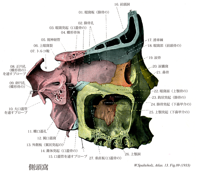
- 089_01【Orbital plate of ethmoid; Orbital plate of ethmoid bone眼窩板;紙様板(篩骨の) Lamina orbitalis; Lamina papyracea】 Especially thin plate of bone that forms part of the medial wall of the orbit.
→(篩骨迷路の外側壁の長方形の平滑な面は眼窩内側壁の主要部をつくる眼窩板である。この上縁の前・後篩骨孔がある。眼窩板より下方は口蓋骨と上顎体に結合する面で、ここにも篩骨蜂巣の一部が開放している。)
- 089_02【Ethmoidal foramina篩骨孔(篩骨の) Foramina ethmoidalia】
→()
- 089_02a【Ethmoidal foramina篩骨孔(前頭骨の) Foramina ethmoidalia】
→()
- 089_02b【Anterior ethmoidal foramen前篩骨孔 Foramen ethmoidale anterius】 Anterior opening in the medial wall of the orbit between the frontal bone and the ethmoid. It transmits the anterior ethmoidal nerve and the anterior ethmoidal vessels from the anterior cranial fossa.
→(篩骨眼窩板の上縁と前頭骨眼窩部との間にある前側の前篩骨孔(前篩骨神経ならびに前篩骨孔動静脈の通路)がある。前篩骨孔は鼻腔に通ずる。)
- 089_02c【Posterior ethmoidal foramen後篩骨孔 Foramen ethmoidale posterius】 Posterior opening in the medial wall of the orbit between the frontal bone and the ethmoid for the passage of the posterior ethmoidal vessels and the posterior ethmoidal nerve.
→(篩骨眼窩板の上縁と前頭骨眼窩部との間にある後側の後篩骨孔(後篩骨神経ならびに後篩骨動静脈の通路)がある。後篩骨孔は篩骨蜂巣に通ずる。)
- 089_03【Orbital process of palatine bone眼窩突起(口蓋骨の) Processus orbitalis (Os palatinum)】 Anterosuperiorly projecting process located between the maxilla, ethmoid, and sphenoid.
→(垂直板の上縁では前部から眼窩突起が上方に起こる。)
- 089_04【Body of sphenoid bone体(蝶形骨の);蝶形骨体 Corpus (Ossis sphenoidalis)】 The part of the sphenoid between the wings of the sphenoid and their processes.
→(蝶形骨体は蝶形骨の中央部にあり立方体をなしている。上面中央部には鞍状を呈したトルコ鞍があり、その中央に横位楕円形の下垂体窩がある。トルコ鞍の後方には鞍背という上方に突出した骨板があり、その両側外側端の突起を後床突起という。鞍背の後部は台形をなして後頭骨の底部とともに斜台を形成する。下垂体窩の前には体の前部との境界線である鞍結節とよべる横走する稜があり、その両側端にある中床突起は発育が弱く明瞭なものは少ない。鞍結節の前には細い横走する[視神経]交叉溝があり、その両外側は視神経管につづく。交叉溝の前部は蝶形骨隆起とよばれているが、これは隆起ではなく滑らかな平面である。体の前部は小翼と後部は大翼と結合している。下錐体窩の外側と大翼の根部との間には、内側頚動脈溝という前後に走る溝があり、外側に蝶形骨小舌という突起状の骨板がある。体の下面は鼻腔、咽頭腔の上壁をなし、中央に蝶形骨吻が前下方に突出し鋤骨翼にはさまれる。体の前面中央部には蝶形骨稜という上下に走る稜線があり、篩骨の垂直板と相接する。蝶形骨稜の両側でがいおうに蝶形骨甲介が認められる。これはバルタン小骨ともよばれ、発生学的には篩骨の一部であったものが8~12歳に蝶形骨体と癒合したものでとくに若年頭蓋で著明である。体の内面は空洞状をなし蝶形骨洞とよばれ、その正中部には蝶形骨洞中隔があり、洞を左右に分けている。その前面には蝶形骨洞口という開口部が両側にあり蝶篩陥凹に通じている。)
- 089_05【Optic canal; *Optic foramen視神経管;視神経孔 Canalis opticus; Foramen opticum; Canalis fasciculi optici】 Canal for transmission of the optic nerves and the ophthalmic artery.
→(視神経孔Optic foramenともよばれる。眼窩の上壁の最も深部で蝶形骨の小翼の蝶形骨体よりの根部は視神経管が貫通し、この管の外側には前床突起が延びだしている。視神経および眼動脈が通る。)
- 089_06【Superior orbital fissure上眼窩裂 Fissura orbitalis superior】 Opening in the upper part of the orbit between the greater and lesser wings of the sphenoid that connects the cranial and orbital cavities. It transmits the ophthalmic, oculomotor, trochlear, and abducens nerves and the superior ophthalmic vein.
→(眼窩の外側壁の後端には、上壁との間に上眼窩裂(蝶形骨の大翼および小翼の間にある上部裂隙)がある。上眼窩裂は頭蓋腔(中頭蓋窩)に通じ、眼筋の支配神経(動眼神経・滑車神経・外転神経)・眼神経・上眼静脈が通る。)
- 089_07【Sella turcicaトルコ鞍 Sella turcica】 It is located above the sphenoidal sinus and houses the pituitary gland.
→(トルコ鞍は蝶形骨体上面には、トルコ風の馬の鞍に似ている骨隆起で中頭蓋窩の中央部にある。この骨のくぼみには、重要な内分泌腺の一つである下垂体が入る。)
- 089_08【Foramen rotundum of sphenoid bone正円孔;正円管(蝶形骨の) Foramen rotundum (Ossis sphenoidalis)】 Foramen that opens anteriorly into the pterygopalatine fossa. It transmits the maxillary nerve.
→(蝶形骨大翼が蝶形骨体から出る根部を貫く3孔が前内方から後外方にならぶ。最前のものは正円孔で、前方に向かって翼口蓋窩に開く。中の最も大きい卵円孔と最後の棘孔はともに頭蓋底下面に開く。正円孔は上顎神経が通る。)
- 089_09【Foramen ovale of sphenoid bone卵円孔(蝶形骨の) Foramen ovale】 Opening for the passage of the mandibular nerve anteromedial to the foramen spinosum.
→(卵円孔は大翼の後内側端に位置し、三叉神経の下顎神経の通路の開口で、棘孔の内前方にある。海綿静脈洞と翼突静脈叢を連絡することがある。)
- 089_10【Greater palatine canal大口蓋管;翼口蓋管 Canalis palatinus major; Canalis pterygopalatinus】 Canal formed by the palatine bone and the maxilla for transmission of the descending palatine artery and the greater palatine nerve.
→(大口蓋孔は上方に向かい大口蓋管となって翼口蓋窩に通じる。大口蓋管は上顎骨と口蓋骨との大口蓋溝が合わさってできる管で、大口蓋動・静脈と大口蓋神経が通る。)
- 089_11【Sphenopalatine foramen蝶口蓋孔;蝶口蓋口 Foramen sphenopalatinum】 Opening in the superior part of the pterygopalatine fossa that connects it with the nasal cavity. The palatine bone contributes the greater portion and the sphenoid the lesser portion.
→(蝶口蓋孔は上鼻道の後端で、蝶形骨と口蓋骨との間にある孔で、鼻腔と翼口蓋窩とを連絡する。蝶口蓋孔は鼻腔の後半部に分布する血管・神経の通路として重要である。鼻腔の外側壁の後部にある口蓋骨の垂直板の上端に蝶口蓋孔がみられる。)
- 089_12【Pterygopalatine fossa翼口蓋窩 Fossa pterygopalatina】 Continuation of the infratemporal fossa to medial between the maxillary tuberosity, the perpendicular plate of the palatine bone, and the pterygoid process. It narrows inferiorly to continue as the greater palatine canal.
→(翼口蓋窩は側頭下窩の内側にある縦に細長い窩で、翼状突起の外側板と上顎骨体との間にある裂隙。上方から下方へ行くにしたがって前後幅は狭くなる。内側は口蓋骨垂直板、上壁は蝶形骨体よりなり、外側は遊離面をなし、また下壁は上顎骨体、蝶形骨翼状突起、口蓋骨錐体突起により閉ざされている。この窩は内・外・前・後・上・下のすべての方向と交通しており、内方では蝶口蓋孔(上後鼻神経が通る)を通じて鼻腔と、外方では翼上顎裂を通じて側頭下窩と、前方では眼窩列を通じて眼窩と、後方では翼突管(翼突管神経が通る)を通じて外頭蓋底と、上方では正円孔(上顎神経)を通じて内頭蓋底と、下方では大口蓋管(大口蓋神経および下行口蓋動・静脈)を通じて口腔とそれぞれ交通している。)
- 089_13【Lateral plate of sphenoid process; Lateral pterygoid plate外側板(翼状突起の);外側翼状板 Lamina lateralis (Processi pterygoideus ossis sphenoidalis)】
→(蝶形骨の翼状突起の外側板は内側板にくらべて広く、やや短く、矢状面に対して斜めに位置する(その外面からは外側翼突筋が起こる)。外側翼突筋の下頭が起始する。内外両側板は前縁で連結し、その前面を縦に走る浅い翼口蓋溝は翼状突起が上顎体および口蓋骨垂直板と合してつくる翼口蓋窩の後壁をつくる。また、内外両側板は後方に開いた翼突窩をつくる(ここから内側翼突筋が起こる)。しかし、内側板と外側板とはその下部では離れて翼突切痕をはさむ。ここには口蓋骨の錐体突起がはまりこんでこれを補う。外側板の後縁は鋭く、その上部から小さい翼棘突起を出すことが多い。)
- 089_14【Pyramidal process of palatine bone錐体突起(口蓋骨の) Processus pyramidalis (Os palatinum)】 The inferoposterior end of the perpendicular plate of the palatine bone, which is inserted in the pterygoid notch.
→(垂直板の下部は水平板より矢状径が広くなり、水平板より後に大きく突出する錐体突起となって蝶形骨翼状突起の翼突切痕にはまる。)
- 089_15【Lesser palatine canals小口蓋管;口蓋管 Canales palatini minores; Canales palatini】 Canals in the pyramidal process for passage of the lesser palatine nerves and arteries.
→(大口蓋孔の下部から後下方に通常1~2本の小さい小口蓋管(小口蓋神経および同名の血管の通路)がわかれ、その下端は錐体突起の下面に開いて小口蓋孔を作る。)
- 089_16【Frontal sinus前頭洞 Sinus frontalis】 It can extend beyond the squamous part of frontal bone into the orbital part of frontal bone. It opens below the middle nasal concha above the sphenoidal sinus.
→(前頭洞は眉間の辺りにある副鼻腔をなす空洞。篩骨漏斗により同側の中鼻道に連なる。)
- 089_17【Trochlear spine滑車棘 Spina trochlearis】 Small bony spicule that is occasionally present in the anterosuperior part of the medial angle of the eye. It gives attachment to the superior oblique muscle.
→(前頭骨の滑車窩に小さい滑車棘をまれに見る(上斜筋の腱の方向を転ずる滑車が着く)。)
- 089_18【Orbital part of frontal bone眼窩部(前頭骨の) Pars orbitalis (Os frontale)】 The part that forms the roof of the orbit.
→(前頭骨の眼窩部は左右にあって眼窩上壁の大部分をつくる薄い骨板である。両側の間に馬蹄形の大きい篩骨切痕があって、ここには篩骨の篩板がはまる。眼窩部の上面は前頭鱗内面のつづきで大脳をのせるが、全体としてふくれ上がり、脳隆起および指圧痕がとくに著しい。)
- 089_19【Lacrimal bone涙骨 Os lacrimale】 Bone located in the orbit in front of the orbital plate of the ethmoid.
→(涙骨は左右1対の不正長方形の薄い骨で、上顎骨の前頭突起後方の眼窩の内壁の一部をなす。これに続く鼻涙管の骨壁の一部もつくる。この骨も結合組織性骨化によって生ずる。涙骨の全体の形は手指の爪に似ているが、厚さは爪よりも薄い。下鼻甲介、篩骨、前頭骨、上顎骨と連結する。外面は眼窩に向かい、中央を縦走する稜縁の前方にある溝状のくぼみが涙嚢窩の構成に加わる部分である。外側面は眼窩の内側壁の前部を形成し、内側面は鼻腔(中鼻道)の外側壁の一部を作る。上縁は前頭骨眼窩部と、下縁は上顎骨眼窩面と、前縁は上顎骨前頭突起と、後縁は篩骨眼窩板とそれぞれ接している。外側面の前半部には縦に走る涙骨溝があり、これは上顎骨の前頭突起の同名溝と合して涙嚢窩を形成する。涙嚢孔の後方の境界を後涙嚢稜といい、下方へ延びて涙嚢鈎となり、上顎骨前頭突起の涙嚢溝および下鼻甲介の涙骨突起とともに鼻涙管壁の一部を形成する。Lacrimaleはlacrima(涙)の形容詞である。)
- 089_20【Fossa for lacrimal sac涙嚢窩 Fossa sacci lacrimalis】 Widened depression at the beginning of the nasolacrimal canal for the lacrimal sac.
→(眼窩の内側壁の前端部には浅い溝状の凹みがある。この窩みは涙嚢窩といわれ、下方に向かって鼻涙管となり鼻腔に通じる。涙嚢窩と鼻涙管は、それぞれ涙嚢と鼻涙管をいれる。)
- 089_21【Nasal bone鼻骨 Os nasale】 Bone located between the right and left frontal processes of the maxilla. Its superior end articulates with the frontal bone.
→(鼻骨は三角形に近い長方形の薄い骨で、左右のものが正中で接合して鼻背の骨格を作る。骨化様式は結合組織性骨化である。鼻腔を前上方からおおう台形の骨である。上方は狭く、下方は広い。上縁は前頭骨鼻部の鼻棘に接し、下縁は遊離縁で骨鼻孔の梨状口の上縁をなす。外側縁は上顎骨の前頭突起と結合し、内側縁は他側の鼻骨と結合し、両者間に鼻骨間結合をなす。鼻骨の前面は軽度膨隆し、後面は軽度陥凹している。前面のほぼ中央に鼻骨孔があり、この孔は後面で篩骨孔につづき、ここを前篩骨神経の外鼻枝が通る。)
- 089_22【Orbital surface of maxilla眼窩面(上顎骨の) Facies orbitalis (Maxilla)】 The part of the body that forms the largest surface of the floor of the orbit.
→(上顎骨の眼窩面は眼窩の底となる滑らかな三角形の面で、やや外下りに傾く。)
- 089_23【Uncinate process of ethmoid鈎状突起(篩骨の) Processus uncinatus (Ossis ethomoidalis)】 Hooked process directed posteroinferiorly. It is almost completely concealed by the middle nasal concha. It projects across the wide opening to the maxillary sinus.
→(篩骨胞と下鼻甲介の間を、中鼻甲介の起部前端から起こる長い薄い板状の鈎状突起が前上方から後下方に斜めに走り、その下端は下鼻甲介に達する。)
- 089_24【Ethmoidal process of inferior concha; Ethmoidal process of inferior nasal concha篩骨突起(下鼻甲介の) Processus ethmoidalis (Concha nasalis inferior)】 Process that is united with the undnate process of the ethmoid.
→(篩骨突起は上縁の後部から上に向かい、篩骨の鈎状突起の下端と連接し、前者と共に上顎洞裂孔の後下部を閉じる。篩骨突起より後方の上縁は口蓋骨の鼻甲介稜に着く。下鼻甲介の弯曲には個人差が多い。)
- 089_25【Maxillary process of inferior concha上顎突起(下鼻甲介の) Processus maxillaris (Concha nasalis inferior)】 Lateral process that forms part of the medial wall of the maxillary sinus.
→(上顎突起は上縁の中央部で外下側に折れ曲がる三角形の骨片で、上顎洞裂孔の下半をふさいでその下縁につく。)
- 089_26Highmore, Antrum of【Maxillary sinus上顎洞;ハイモア腔 Sinus maxillaris】 It measures over 3 cm vertically and sagittally and 2.5 cm in the frontal plane. Its floor is usually at least 1 cm below the floor of the nose and its lowest point is usually at the level of the first molar.
→(上顎洞は上顎体中にある最大の副鼻腔で、その形はだいたいにおいて上顎体の形に一致するが、尖端を外上方、すなわち頬骨突起の方に出しているので錐体状に近く、その底は鼻腔面にむく。ここにはなはだ大きい上顎洞裂孔があるが、完全な頭蓋ではこの裂孔は口蓋骨の垂直板、篩骨の鈎状突起および下鼻甲介の上顎、篩骨稜突起によりその一部がふさがれて著しく小さくなる。(生体では、さらに鈎状突起まで鼻粘膜に被われるため、中鼻甲介の下の半月裂孔に開く小さな開口を残すのみとなる。)上顎洞はその前壁が最も厚く、つぎは後壁、上壁の順で内側壁が最も薄い。下壁は歯槽突起に入り、場所によってその厚さが異なるが、大臼歯および小臼歯の歯根をおおう部、とくに第1、第2臼歯の付近で最も薄く、それらの歯根はしばしば洞に達する。また、下壁には歯槽中隔の為に多くの骨の高まりやくぼみを見るのを常とする。なお、上顎洞の前後稜壁には多くの細い歯槽溝または歯槽管および歯槽孔が見られる。『ハイモア洞』:イギリスの自然科学者Nathaniel Highmore (1613-1685)の名を冠するが、レオナルド・ダ・ビンチがすでに観察している。ハイモアは、この他にも精巣縦隔(Highmore's body)に名を残している。)
- 089_27【Perpendicular plate of palatine bone垂直板;鉛直板;上顎板;鉛直部(口蓋骨の) Lamina perpendicularis; Lamina maxillaris; Pars perpendicularis (Os palatinum)】 Vertical bony plate contributing to the walls of the nasal and maxillary sinuses.
→(垂直板の内側面は鼻腔外側壁の後部をつくり、前後に走る上下二つの稜があり、上のもの(篩骨稜)には中鼻甲介後端がつき、下のもの(鼻甲介稜)は発達よく、下鼻甲介をつける垂直板の上縁は深く切れ込むが(蝶口蓋切痕)、上方に蝶形骨体があるので孔(翼口蓋孔)となり、鼻腔と翼口蓋窩を連絡する。蝶口蓋切痕の前の突起(眼窩突起)はうしろの突起(蝶形骨突起)より大きく、上前方に向かって5面あり、内側の3面は他骨との接触面で、前は上顎骨、中のは篩骨(この部分は篩骨洞をおおうためへこむ)、うしろのは蝶形骨体につく。外側面に2面あり、ともに自由面で、上の面は眼窩底の一番後ろをつくり、下の面は翼口蓋窩に面する。蝶形骨突起は上内方に向かい、下面は内面で鼻腔外側壁をつくり、上(外)面は翼状突起につき、内方にのびて鋤骨翼に達し、これと静脈のとおる管(咽頭管)をかこむ。垂直板の外側面(上顎面)は上顎骨体内面をおおう部のうしろに、縦の前後の二つの粗面があり、前のは上顎骨内面に、うしろのは蝶形骨翼状突起につく。2面の間には蝶口蓋切痕から下る第3の面があって、上は翼口蓋窩の底をつくり、下方は垂直な溝(大口蓋溝)となり、上顎骨の同名溝と合して大口蓋管をつくり、大口蓋孔で口蓋にひらく。大口蓋神経、下行口蓋動脈が通る。この管から通常2本の小管(小口蓋管)が分かれて、錐体突起の基部をつらぬき、その下面下、内側に小孔(小口蓋孔)でひらく。)