Spalteholz HANDATLAS DER ANATOMIE DES MENSCHEN VON WERNER SPALTEHOLZ
メニューは解剖学(TA)にリンクしてあります。図の番号をクリックすると下記の説明へ、右側の用語をクリックすると解剖学(TA)にジャンプします。
1016


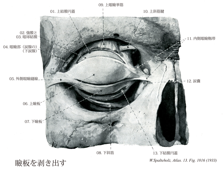
- 1016_01【Superior conjunctival fornix上結膜円蓋 Fornix conjunctivae superior】 Reflection of bulbar conjunctiva onto the palpebral conjunctiva located high up behind the superior eyelid.
→(眼球結膜が上眼瞼の上後方で、眼瞼結膜へと折り変えるところ。(Feneis))
- 1016_02【Sclera強膜 Sclera】 Membrane of the eyeball composed of interwoven collagen fibers. It has a bluishwhite appearance and is visible through the conjunctiva.
→(眼球の形状を保つ強靱な膠原線維組織層。角膜となっている前部6分の1を除いた部分。前方では隔膜固有質に、後方では篩板から視神経外鞘を経て脳硬膜に、それぞれつづいている。強膜と角膜を合わせて眼球線維膜という。強膜の厚さは眼球後極で~1.0mm、前部で~0.6mm、赤道で~0.4mmである。視神経線維束を通す篩板は後極の内側3.5mm、視神経乳頭の直後方にあたる。視神経は~数十本の掌側としてこれを通る。渦静脈、長・短毛様体動脈および神経が強膜を貫く。強膜はは外から内へ、①強膜上皮、②強膜固有質、③強膜褐色板の3沿うよりなる。虹彩角膜角に沿って強膜固有質が内方へ皮厚し(強膜距)毛様体筋腱により貫かれる。この部の直前に輪状に走る強膜静脈洞(Schlemmn管)があり、眼房水は虹彩角膜間隙(Fontana腔)からこれを通って渦静脈に排出される。角膜縁をとり膜浅い強膜溝の深層にこれらの構造がある。眼球前部の強膜上板毛細血管網に富み、その炎症性変化を臨床的に「網膜充血」という。強膜前部は眼球結膜、後部は眼球鞘(Tenon鞘)によりおおわれる。内面は脈絡外隙を間に脈絡外板に接する。)
- 1016_03【Bulbar conjunctiva眼球結膜 Tunica conjunctiva bulbi】 Portion of the conjunctiva covering the eyeball. It consists of stratified, nonkeratinized squamous epithelium with only a small number of goblet cells and a lamina propria of loosely organized structures, containing few cells and permeated by elastic fibers.
→(眼球結膜は結膜のうち眼球を被う部分。杯細胞に乏しい角化していない重層扁平上皮である。固有層は疎で細胞に乏しく弾性線維を含む。)
- 1016_04【Palpebral part of lacrimal gland眼瞼部(涙腺の);下涙腺 Pars palpebralis (Glandulae lacrimalis); Glandula lacrimalis inferior】 Smaller portion of the lacrimal gland situated below the tendon of the levator palpebrae.
→(上眼瞼挙筋の腱の下にある小さい部分。(Feneis))
- 1016_05【Lateral palpebral raphe外側眼瞼縫線 Raphe palpebralis lateralis】 Thin band on the lateral palpebral ligament that is reinforced by the orbicularis oculi.
→(外眼筋の外側部分の中にみられる幅の狭い線維帯で、上下眼瞼の間を行き交う結合組織線維からなる。)
- 1016_06【Superior tarsus上瞼板 Tarsus superior; Tarsus palpebrae superior】 Semilunar fibrous plate that is curved like a bowl and forms the upper eyelid. It measures about 10 mm vertically and consists of tough, connective tissue of interwoven collagen fibers. It contains the tarsal glands.
→(上瞼板は高さ約10mmあり、皿状に曲がっている。かたい縺れた膠原線維性の結合組織よりなる。瞼板腺を含む。上眼瞼を広く反転できるのは、ここに上眼板があるからである。とくに日本人では、眼輪筋と瞼板との間に疎性結合組織と脂肪組織があって内輪筋と瞼板とはゆるく結合するので、眼瞼を反転しやすい。上瞼板と皮膚との結合が粗であると一重瞼であるが、結合が密でつよいと二重瞼となる。)
- 1016_07【Inferior tarsus下瞼板 Tarsus inferior; Tarsus palpebrae inferior】 Semilunar fibrous plate forming the lower eyelid that measures about 5 mm vertically. It consists of tough, connective tissue of interwoven collagen fibers and contains the tarsal glands.
→(下瞼板は高さ約5mmあり、皿状に曲がっている。かたい縺れた膠原線維性の結合組織よりなる。瞼板腺を含む。(Feneis))
- 1016_08【Inferior oblique muscle下斜筋;下眼球斜筋 Musculus obliquus inferior; Musculus obliquus bulbi inferior】 o:Lateral alongside the nasolacrimal canal, i: Behind the equator. Action: Gaze elevation, abduction, and extorsion. I: Oculomotor nerve.
→(下斜筋は眼窩口内側縁の後方の上顎骨に存在する前涙嚢稜から起こり眼窩下縁と並行に走る。下斜筋は停止部近くで扇のように後方へ放射状に広がり眼球の下後側頭部眼球赤道の強膜に停止する。目の動き:視線を内側かつ上方に向ける。)
- 1016_09【Levator palpebrae superioris muscle上眼瞼挙筋 Musculus levator palpebrae superioris】 o: Upper portion of optic canal and dural sheath of optic nerve. Its insertion tendon widens anteriorly and divides into a superior and an inferior layer. I: Oculomotor nerve.
→(上眼瞼挙筋は視神経管の縁の総腱輪の外側で視神経鞘から起こり、眼窩上壁のすぐ下で前頭神経の下を通り上眼瞼にいく。上眼瞼挙筋の腱は分離して上眼瞼挙筋浅板と上眼瞼挙筋深板に分かれる。前者は上眼瞼中を縁に向かって進み、後者は上瞼板筋の平滑筋細胞を伴って上眼瞼の瞼板に付く。下瞼板筋は下眼瞼板と下結膜円蓋の間の下眼瞼に存在する平滑筋層である。)
- 1016_10【Tendon of superior oblique muscle; Superior oblique tendon上斜筋腱 Tendo musculus obliquus superior】
→()
- 1016_10a【Superior oblique muscle上斜筋;上眼球斜筋 Musculus obliquus superior; Musculus obliquus bulbi superior】 o:Medial to the common tendinous ring on the body of sphenoid, i: After a hook-shaped course, obliquely behind the equator. Its tendon passes through the trochlea. Action: Abduction, intorsion, and depression of the eye. I: Trochlear nerve.
→(上斜筋は眼窩傍結合組織すなわち視神経鞘と(おもに)蝶形骨体の結合組織である総腱輪の内側から起こる。上斜筋は眼窩錐体の内側直近の上を前方に走行する。眼球の縁で上斜筋の丸みのある腱は結合組織性の吊り索(滑車)を通過し鋭角で後方に曲がる。さらに上斜筋の腱は上直筋の下でこれと交差し眼球上後側頭部の強膜に停止する。目の動き:視線を内側かつ下方に向ける。)
- 1016_11【Medial palpebral ligament内側眼瞼靱帯 Ligamentum palpebrale mediale】 Connective-tissue band connecting the medial palpebral commissure and medial wall of the orbit, lying immediately anterior to the fossa for the lacrimal sac.
→(内側眼瞼交連と内側眼窩壁との間の結合組織性結合。涙腺窩の直前にある。(Feneis))
- 1016_12【Lacrimal sac涙嚢 Saccus lacrimalis】 Sac measuring about 1.5 cm long and 0.5 cm wide lying in the lacrimal fossa. Its inferior portion is directly continuous with the nasolacrimal duct.
→(涙嚢は涙嚢窩中にあり、長さ約1.5cm、幅約0.5cm。下は直接鼻涙管へ移行する。(Feneis))
- 1016_13【Inferior conjunctival fornix下結膜円蓋 Fornix conjunctivae inferior】 Reflection of bulbar conjunctiva onto the palpebral conjunctiva behind the inferior eyelid.
→(眼球結膜が下眼瞼後方で、眼瞼結膜へと折り変えるところ。(Feneis))