Spalteholz HANDATLAS DER ANATOMIE DES MENSCHEN VON WERNER SPALTEHOLZ
メニューは解剖学(TA)にリンクしてあります。図の番号をクリックすると下記の説明へ、右側の用語をクリックすると解剖学(TA)にジャンプします。
533 


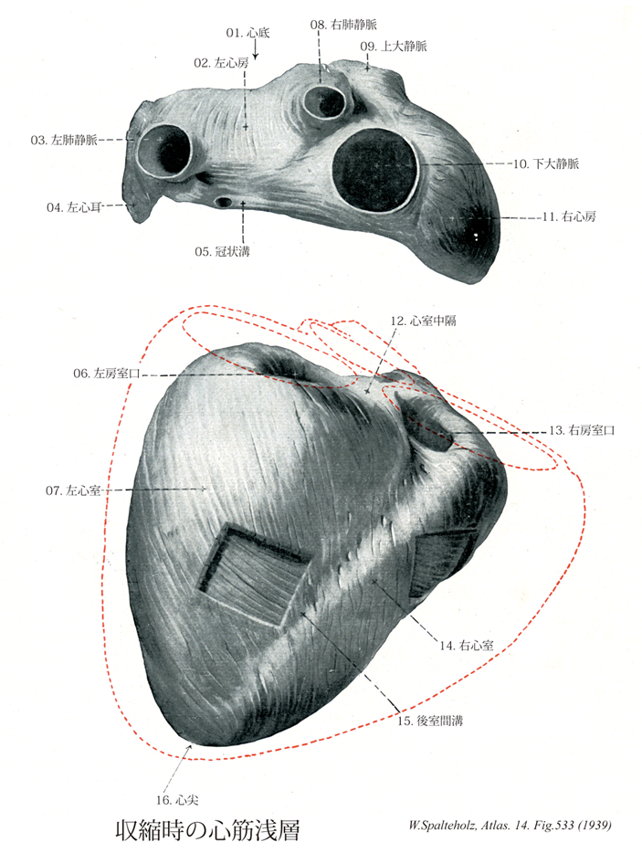
- 533_01【Base of heart心底 Basis cordis】 Broad aspect of the heart facing dorsally and to the right, located opposite to the apex of the nearly conical heart, it is mainly formed by the posterior wall of the left atrium. The pulmonary arteries and vasa privata arise and open here.
→(心底はほぼ円錐状の心臓の上側、心尖の反対側の広い面。主に左心房と右心房の一部とできる。横隔面からは冠状溝で境される。)
- 533_02【Left atrium左心房 Atrium cordis sinistrum; Atrium sinistrum】
→(左心房は心臓の後上部にあって、後面をつくっている。左心房は右心房よりもやや小さいが、壁はやや厚い。左心房の後壁の上部に、左右両肺からそれぞれ2本ずつ、前部で4本の肺静脈が開口している。左心房は前下方で房室口によって左心室に通じる。)
- 533_03【Left pulmonary veins左肺静脈 Venae pulmonales sinistrae】 The two left pulmonary veins which occasionally unite to form a single trunk.
→(2条。ときには合して1本の幹となる。 (Feneis))
- 533_04【Left auricle of atrium左心耳 Auricula atrii sinistra; Auricula sinistra cordis】 Outpouching of the atrium to the left of the pulmonary trunk.
→(左心耳は肺動脈幹の左方に中空指状に突出した左心房の一部。左心耳は右心耳に比べて格段に小さい。)
- 533_05【Coronary sulcus冠状溝 Sulcus coronarius】 Groove that runs around the heart, demarcating the borders between the atria and ventricles.
→(心房と心室の境には冠状溝とう溝があるが、冠状溝は冠状動脈と冠状静脈洞で埋められている。)
- 533_06【Left atrioventricular orifice; Left atrioventricular opening左房室口 Ostium atrioventricularis sinistrum】 Openings between the atria and ventricles.
→(左房室口は左心房と左心室の間で左心房弁がある。)
- 533_07【Left ventricle左心室 Ventriculus sinister】
→(左心室は心臓の左下部を占め、後上方にある左房室口で左心房と交通し、右上隅にある大動脈口によって大動脈につらなる。左心室の壁は右心室に比べ2~3倍厚い。心室中隔は、右心室に向かって膨隆しているので、心室を横断面でみると、左心室の内腔は円いのに対して、右心室の内腔は半月状である)
- 533_08【Right pulmonary veins右肺静脈 Venae pulmonales dextrae】 The two right pulmonary veins which occasionally unite to form a single trunk.
→(2本あるが、時に合流して1本の幹となる。 (Feneis))
- 533_09【Superior vena cava上大静脈 Vena cava superior; Vena cava cranialis】
→(上大静脈は上半身の血液を集める静脈で、上縦隔の中で左右の腕頭静脈が合してはじまり、途中で奇静脈を受け入れながら上行大動脈の右側を下行して右心房にそそぐ。)
- 533_10【Inferior vena cava下大静脈 Vena cava inferior; Vena cava caudalis】 It arises at the union of the right and left common iliac veins, lies on the right side of the aorta, and opens into the right atrium of the heart.
→(下大静脈は下肢および骨盤と腹部の器官の大部分から血液を受ける本幹で、第5腰椎体の右側で左右の総腸骨静脈の合流として始まり、このあと脊柱に沿って大動脈の右側を上行、肝臓の後面をこれに接して通過し、第八胸椎の高さで横隔膜の大静脈孔を貫いて胸腔に入り、ただちに右心房にそそぐ。下大静脈に流入する枝には総腸骨静脈、下横隔静脈、第3・第4腰静脈、肝静脈、腎静脈、右副腎静脈、右精巣静脈、右卵巣静脈、蔓状静脈叢などがある)
- 533_11【Right atrium右心房 Atrium cordis dextrum; Atrium dextrum】
→(右心房は心臓の右上部を占め、その後上部と後下部とに、それぞれ、上大静脈と下大静脈が注いでいる。)
- 533_12【Interventricular septum心室中隔 Septum interventriculare】 Partition dividing the right and left ventricles of the heart. It can be identified from externally by the anterior and posterior interventricular sulci.
→(心室中隔は左右の心室をわける壁で、外からは前後の室間溝でわかる。)
- 533_13【Right atrioventricular orifice; Right atrioventricular opening右房室口 Ostium atrioventriculare dextrum】 Openings between the atria and ventricles.
→(右房室口は右心房と右心室の間に開く。右房室口は輪状の線維性結合織(線維輪)で囲まれ、弁をもつ。)
- 533_14【Right ventricle右心室 Ventriculus dexter】
→(右心室は心臓の最下位部を占め、後上方にある右房室口で右心房と交通し、前上方にある肺動脈口で肺静脈に連なる。)
- 533_15【Posterior interventricular sulcus後室間溝 Sulcus interventricularis posterior; Sulcus interventricularis dorsalis】 Longitudinal groove on the diaphragmatic surface of the heart corresponding to the interventricular septum. It transmits the posterior branch of the interventricular branch of the right coronary artery.
→(後室間溝は横隔面で心室中隔に層とする縦溝。右冠状動脈の後室間枝が走る。)
- 533_16【Apex of heart心尖 Apex cordis】 Part of the heart directed downward and to the left. It is formed by the left ventricle.
→(心尖は心臓の下端部でやや尖っている。心尖は心臓の拍動とともに前胸壁にあたる。これを心尖拍動といい、体表で触れることができる。すなわち、一般に左側の第5肋間で、正中線から約4横指(約7cm)左方で触れる。この位置は弾性では左乳頭のすこし内下方である。小児ではやや高くかつ外方にある。)