Spalteholz HANDATLAS DER ANATOMIE DES MENSCHEN VON WERNER SPALTEHOLZ
メニューは解剖学(TA)にリンクしてあります。図の番号をクリックすると下記の説明へ、右側の用語をクリックすると解剖学(TA)にジャンプします。
771


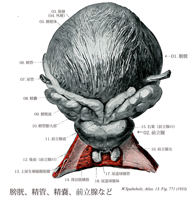
- 771_01【Urinary bladder; Bladder膀胱 Vesica urinaria】 Organ located beneath the peritoneum in the lesser pelvis posterior to the pubic symphysis. Its size varies depending on fullness, with the urge to evacuate the bladder occurring at about 350 ml. Even at maximum distension it remains below the level of the navel.
→(膀胱は腎臓で産生され尿管によって送られる尿を約350~500mlまたはそれ以上を一時的に貯える。平滑筋よりなり弾性に富む尿の貯留器官。膀胱は骨盤腔のもっとも前部にあり、恥骨の後ろに位置する。軽度に充満する時には、四面体を呈し、頂にあたるところを膀胱尖といい、錐体の底部にあたるところを膀胱底と呼ぶ。尖と底との間を膀胱体と呼ぶ。)
- 771_02【Prostate; Prostate gland; Prostatic glands前立腺 Prostata】 Chestnut-sized tubuloalveolar gland below the urinary bladder and having a smooth surface. It surrounds the urethra.
→(前立腺は膀胱底の下に密接し、骨盤底の上にのる30~50個の胞状管状腺である。男性の尿道起始部を取り囲んで栗の実に似た形をしている。強靱な結合組織の被膜に被われる。外周に近い部部分は色がやや暗く見え(いわゆる外腺exogland)、中心部は白っぽい(いわゆる内腺endogland)。Prostataはpro(前に)stata(立つもの)という意味である。古代ギリシャでは永来ヒトの前に立って護衛する人をprostatesといったそうである。前立腺を初めて記載し、prostatesと名付けたのは、アレキサンドリア学派のエラシストラトスErasistratus(BC310-250)である。前立腺が膀胱の前に立ってこれを守っていると考えたのだろう。日本では、以前には摂護腺と呼ばれていたが、ギリシャ語の直訳の前立腺が現在では正規の用語になっている。)
- 771_03【Muscular layer of urinary bladder; Muscular coat of urinary bladder筋層(膀胱の) Tunica muscularis vesicae】 The muscular coat of the urinary bladder mainly consists of interwoven bundles of smooth-muscle cells that conform to the degree of its distension. In the trigone region muscle fibers from the urinary bladder overlap with those of the ureter.
→(膀胱の筋層は膀胱壁の主部をつくる平滑筋層である。平滑筋は原則的に3層(内縦・中輪・外縦)に配列するが、全体的に網状を呈して3層の区別は必ずしも明瞭ではない。平滑筋は収縮によって膀胱内圧を圧し排尿に与えるので、全体として排尿筋とよばれる。膀胱頚では、筋層の中輪走筋が輪状にとり囲み、膀胱括約筋をつくるといわれたが、このような輪走筋は明らかでなく、膀胱三角の内縦層の平滑筋が膀胱頚から尿道に達している。このような縦走筋が収縮すると、尿道の始部は短くなり、かつその内腔は漏斗状にひろがって、内尿道口が開き尿の排出に与る。膀胱の平滑筋が弛緩すると、縦走筋も弛緩する。同時に膀胱頚にある輪状の弾性線維によって内尿道口は閉ざされるといわれる。)
- 771_04【External layer of muscular coat外層(膀胱筋層の) Stratum externum (Tunica muscularis vesicae)】
→()
- 771_05【Body of bladder膀胱体 Corpus vesicae】 Part of the bladder between the apex and fundus resting against the peritoneal cavity.
→()
- 771_06【Ductus deferens; Deferent duct精管 Ductus deferens; Vas deferens】 The course of the ca. 50 cm long ductus deferens is initially tortuous, then becomes straight. It is a continuation of the duct of epididymis, opening into the urethra.
→(精巣上体からはじまる精巣の分泌管で、精巣上体尾につづく精子を送る通路。精索中にある。全長約30cm(延ばせばその2倍)、膀胱底で紡錘状に膨れ、精管膨大部といい、内部に膨大部憩室を含む。膨大部の下端で、精嚢が精嚢排出管を経て合流し、これより遠位では精管は射精管と呼ばれ、尿道前立腺部後壁にある精丘の上で、尿道に開く。)
- 771_07【Ureter尿管 Ureter】 Urinary duct situated in the retroperitoneum. It connects the renal pelvis with the urinary bladder, measures 25-30 cm in length and is about 3 mm thick.
→(尿管は全長約25~27cmで、上半分は腹腔内を走り腹部といわれ、下半分は骨盤内にあり骨盤部といわれる。腎盂につづき、腎臓から膀胱に至る管。輪層と縦層の平滑筋に囲まれた移行上皮によって裏打ちされ、外部は外膜でおおわれている。腎門の内下側から出て、大腰筋の前面を斜めに内下方に向かい、精巣(卵巣)動脈の後ろで、これと交叉して下行する。第四腰椎の高さで、総腸骨動・静脈の前を横切って骨盤内に入る。ついで、骨盤の側壁に沿って走り、最後に前内方にまたがって骨盤邸の上面を走り膀胱に開く。尿管はつぎの3箇所にやや細い狭窄部をもつ。すなわち、1.腎盂から尿管への移行部(上端部)、2.腹部から骨盤部への移行部(この部は総腸骨動・静脈と交叉し、尿管は腹膜と癒着している、3.膀胱壁を貫く部(尿管は膀胱壁を斜めに貫き、長さは約2cm)の3箇所である。)
- 771_08【Seminal gland; Seminal vesicle精嚢;精嚢腺 Glandula vesiculosa; Glandula seminalis; Vesicula seminalis】 Thinwalled coiled tube about 5 cm in length.
→(精嚢は、精管膨大部のすぐ下方で、精管から外上方に膨出する嚢状性器官である。精嚢は膀胱底の後壁と直腸との間で、精管膨大部の外側にあり、長さ約3cm・重さ約2g、小指頭大である。精嚢の発達は男性ホルモン依存性で、思春期においてホルモン活性が高くなるとともに発達する。長い間精子を貯蔵すると考えられてきたが、正常には精子を貯えていない。老齢でホルモン産生が減退すると、精嚢は萎縮する。精嚢は腺で、導管は前立腺のすぐ上方で精管に合流する。)
- 771_09【Fundus of bladder; Fundus of urinary bladder膀胱底 Fundus vesicae】 Part of the bladder that rests against the pelvic floor and is attached to its subperitoneal connective tissue. It tapers off into the neck of bladder. The ureters open into its posterior wall.
→(尖と反対側に位置する後壁、とくに尿管の間にある下部。 (Feneis))
- 771_10【Ampulla of ductus deferens精管膨大部 Ampulla ductus deferentis】 Longitudinal expansion at the fundus of bladder.
→(膀胱の後側で、精管は紡錘状に膨らみ、精管膨大部といわれる。)
- 771_11【Base of prostate前立腺底;膀胱面(前立腺の) Basis prostatae】 Part of the gland that is fused with the fundus of bladder.
→()
- 771_12【Posterior surface of prostate後面;直腸面(前立腺の) Facies posterior; Facies rectalis】 Surface of the prostate facing the rectum.
→()
- 771_13【Superior fascia of urogenital diaphragm上尿生殖隔膜筋膜;内尿生殖隔膜筋膜 Fascia diaphragmatis urogenitalis superior; Fascia diaphragmatis urogenitalis interna】 Obsolete term. Current scientific opinion holds that there is no complete boundary to the deep perineal space.
→(深会陰横筋の坐骨直腸窩側にある筋膜。 (Feneis))
- 771_14Guthrie's muscle【Deep transverse perineal muscle♂深会陰横筋 Musculus transversus perinei profundus♂】 Trapezoidal sheet of muscle spread out in the pubic arch. I: Pudendal nerve.
→(深会陰横筋は男では尿道球の上に接し、坐骨枝と恥骨の下枝との合する所から起こって横走し、正中線で両側のものがたがいに結合する。起始は坐骨枝で停止は会陰部で対向する反対側の坐骨枝。作用として浅会陰横筋とともに会陰の横走筋を形成しており(縦走筋は球海綿体筋と外肛門括約筋)、会陰ならびに骨盤筋膜を支持して腹圧に対向する。男性では尿道球をも支持する。陰部神経(陰茎背神経、陰核背神経)から支配される。この筋は臨床上重要である。)
- 771_15【Right lobe of prostate右葉(前立腺の) Lobus dexter (Prostata)】
→()
- 771_16【Apex of prostate前立腺尖 Apex prostatae】 Anteroinferiorly directed tip of the prostate that surrounds the urethra, close to the superficial transverse perineal muscle.
→()
- 771_17【Duct of bulbo-urethral gland尿道球腺管;排出管(尿道球腺の) Ductus glandulae bulbourethralis; Ductus excretorius】 The 3-4 cm long excretory duct.
→(尿道球腺管は3~4cm長の腺排出管。)
- 771_18【body of bulbourethral gland尿道球腺体 Corpus glandulae bulbourethralis】
→(")
- 771_18aCowper's glands【Bulbourethral gland尿道球腺;カウパー腺 Glandula bulbourethralis】 Pea-sized mucous gland on the posterior end of the bulb of penis at the level of the deep transverse perineal muscle.
→(尿道球腺はカウパー腺ともよばれる。尿道海綿体球部の後上方、尿生殖隔膜内に位置する。女性の大前庭腺(バルトリン腺Bartholin's gland)に相当する。尿道球の後端両側に位置する径1cmほどのえんどう豆大の粘液腺で、導管は長く、3~4cm、尿道球腺管といい、前方へ走って尿道海綿体部のはじまりの部分で下面に開く。弾性の性的興奮時には、この線の分泌物部が尿道を潤す。1702年にイギリスの外科医・解剖学者William Cowper (1666-1709)が記載したことによるが、これに先だつ1684年にJ. Meryが報告している。)