Spalteholz HANDATLAS DER ANATOMIE DES MENSCHEN VON WERNER SPALTEHOLZ
メニューは解剖学(TA)にリンクしてあります。図の番号をクリックすると下記の説明へ、右側の用語をクリックすると解剖学(TA)にジャンプします。
778


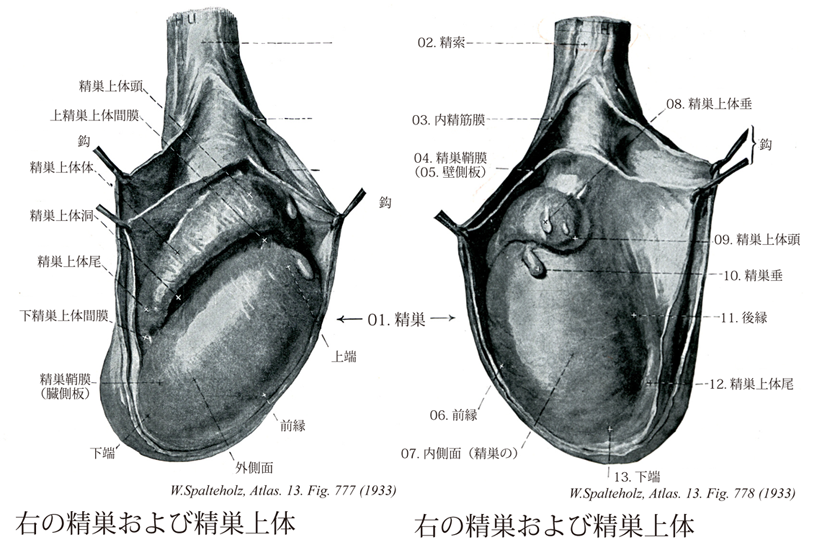
- 778_00【Epididymis精巣上体;副睾丸 Epididymis】 Lying on the posteromedial surface of the mediastinum of testis, it serves to store spermatozoa.
→(精巣上体は副睾丸ともいい、精子を精巣から精管へ送る通路である。精巣の上端から後縁にかけてこれに接し、共通の皮膜に囲まれる。重量的2g。精巣上端に近い方から、精巣上体頭、体、尾の3部を分け、精巣上体尾は精管に移行する。頭は迂曲する精巣輸出管を含み、各輸出管は結合組織で仕切られて、精巣上体小葉をつくる。最上位の精巣輸出管に他の管が合流し、1本になったものが精巣上体間で、これが頭からはじまってきわめて迂曲し、体尾を構成する。これがそのまま精管に移行する。)
- 778_01【Testis; Testicle精巣;睾丸 Testis; Orchis】 About 5 cm long.
→(精巣は睾丸ともいい、男性の性腺。精巣上体とともに陰嚢中にある。重量約8.4g。左の精巣は、通常は右の精巣よりやや大きい。上端、下端、外側面、内側面、前縁、後縁を区別する。表面は結合組織性の白膜におおわれ、白膜は実質内にやや膨隆して精巣縦隔をつくり、そこからさらに精巣中隔が伸び出して、精巣実質を約300の精巣小葉に分ける。小葉は迂曲する精細管(曲精細管)で占められる。精細管は精子を形成する部分で、精巣縦隔に近い部分では直精細管となり、これは縦隔内の網状の精巣網に合流、精巣網はさらに10~20本の精巣輸出管につながる。精巣付近には発生時の構造の遺残がいくつかみられる。精巣垂はミューラー管Muellerian ductの上端部の遺残である。また、精巣上体の頭部に同様の小体がある。これは精巣上体垂でウォルフ管Wolffian duct上端の中腎細管の遺残である。)
- 778_02【Spermatic cord精索 Funiculus spermaticus】 It consists of the ductus deferens, its accompanying vessels, nerves and connective tissues, as well as coverings.
→(精索は精管が血管、神経とともに皮膜に包まれ、精巣上体から深鼡径輪に達するまでの約11.5cm長の紐状の構造。蔓状静脈叢、精巣動脈、脂肪、平滑筋などを含む。精索と子宮円索とは共に鼡径管を通っているが、その由来は同じではない。精索(精管)に相当するものは女性ではほとんど退化して、わずかに卵巣状態(の縦管)として残り、子宮円索は男性の精巣導帯gubernaculum testis(精巣の下端と陰嚢の皮膚をつなぐ結合組織で、ハンター導帯Hunter's gubernaculumとも呼ぶ)に相当する。このように由来の異なるものが男女で同じ場所を通っている原因は、女性では卵巣下降descent of ovariesが子宮の高さで止まり、卵巣が腹腔外に出てこないからである。)
- 778_03【Internal spermatic fascia内精筋膜;精巣および精索鞘膜;総鞘膜 Fascia spermatica interna; Tunica vaginalis communis testis et funiculi spermatici】 Projection from the transversalis fascia through the inguinal canal that encloses the spermatic cord, epididymis, and testes.
→(腹横筋膜が手袋の指のようにのび、精巣拳筋の下で精巣、精巣上体、精管と脈管、神経をともに包んでいる。 (Feneis))
- 778_04【Tunica vaginalis testis; Tunica vaginalis of testis精巣鞘膜 Tunica vaginalis testis; Tunica vaginalis propria testis】 Serous covering of the testis. Vestige of the vaginal process of the peritoneum, it consists of the following layers.
→(陰嚢では内精筋膜の下層に白くて固い膜がある。これが精巣と精巣上体を包む精巣鞘膜の、壁側板である。この膜を縦に切開すると、精巣鞘膜の臓側板に被われた精巣精巣上体が現れる。)
- 778_05【Parietal layer of tunica vaginalis testis壁側板;睾丸膜;精巣周膜(精巣鞘膜の) Lamina parietalis (Tunicae vaginalis testis); Periorchium】 Layer that lines the inner surface of the tunica vaginalis and is reflected on the visceral layer at the posterior border of the epididymis and mediastinum of testis.
→(精巣鞘膜の外側葉。 (Feneis))
- 778_06【Anterior border of testis前縁;自由縁(精巣の) Margo anterior (Testis)】 Free anterior margin of the testis.
→(前方の自由縁。 (Feneis))
- 778_07【Medial surface of testis内側面(精巣の) Facies medialis testis】 Flattened, medialfacing surface of the testis.
→()
- 778_08【Appendix of epididymidis精巣上体垂 Appendix epididymidis】 Stalked vesicular appendix at the head of epididymis.
→(男性における中腎管の遺残物 中腎管の頭方端が精巣上体垂として存続することがあり、通常精巣上体の頭部に付着している(図13-32A)。精巣輸出管の尾方で、若干の中腎管が、中腎傍体paradidymisとよばれる小体として存続することがある。(ムーア人体発生学))
- 778_09【Head of epididymis精巣上体頭 Caput epididymidis】 Part of the epididymis formed by the efferent ductules.
→(主に精巣輸出管よりなる。 (Feneis))
- 778_10Morgagni, Hydatid of【Appendix of testis精巣垂 Appendix testis】 Vesicular appendage of the testis.
→(男性における中腎傍管の遺残物 中腎傍管の頭方端が、精巣の上端に付着する精巣垂として存続することがある(図13-32A)。尿道前立腺部に開口する小さな嚢胞上の構造物である前立腺小室prostatic utricleは、腟と相同である。前立腺の小室の上皮は、尿生殖洞の上皮から発生する。この上皮内には、ニューロン特異性エノラーゼおよびセロトニンを含む内分泌細胞が見つかっている(Wernert, 1990)。尿道前立腺部の後壁にある小さな隆起である精丘seminal colliculusは、成人における洞結節の派生物であり(Moore, 1992),女性の処女膜と相同物である(表13-1)。 女性における中腎傍管の遺残物 卵管の卵管采の形成に関与しない中腎傍管の頭方端部は、モルがニー嚢胞hydatid of Morganiとよばれる胞状構造物として存続することがある(図13-32C) (ムーア人体発生学))
- 778_11【Posterior border of testis後縁;間膜縁(精巣の) Margo posterior testis】 Posterior margin of the testis, attached to a fold of the serous membrane.
→(漿膜の折り返しヒダが付着している。 (FeneisD))
- 778_12【Tail of epididymis精巣上体尾 Cauda epididymidis】 Lower part of the duct of epididymis.
→(精巣上体管の弯曲からなる精巣上体の下部。 (Feneis))
- 778_13【Lower pole of testis; Inferior pole of testis; Testicular serosa下端;下極(精巣の) Extremitas inferior testis; Polus inferior testis】
→()