Spalteholz HANDATLAS DER ANATOMIE DES MENSCHEN VON WERNER SPALTEHOLZ
メニューは解剖学(TA)にリンクしてあります。図の番号をクリックすると下記の説明へ、右側の用語をクリックすると解剖学(TA)にジャンプします。
825


- 825_00【Spinal cord脊髄 Medulla spinalis】 It extends from the end of the medulla oblongata near the exit of the first spinal nerve to the beginning of the terminal filum at L1 or L2.
→(脊髄は頚部(頚髄)、胸部(胸髄)、腰部(腰髄)、仙骨部または脊髄円錐(仙髄と尾髄)とからなり、それぞれ髄節に分かれ、それに対応して31対の脊髄神経が出る。頚髄では8対の頚神経、胸髄では12対の胸神経、腰仙髄では各々5対の腰神経と仙骨神経とが出る。尾髄からは通常1対の尾骨神経が出る。上肢および下肢支配の神経の出る頚髄下部と腰髄下部は発達が著しく、太くなっており、それぞれ頸膨大、腰部大とよばれる。脊髄下端は細くなり脊髄円錐となっておわる。その高さは成人では第1ないし第2腰椎の高さに相当する。新生児、幼児では低く第3腰椎の高さでおわっている。脊髄円錐の先はさらに細く糸状の終糸となって尾骨の背面に付着している。終糸に沿って走る脊髄神経の束はその形状から馬尾とよばれている。脊髄外側面でその腹側と背側の正中には(前)正中裂および(後)正中溝とよばれる溝があり、脊髄を左右の半分に分けている。前者は後者より深く、そこには前脊髄動脈が走っている。左右の脊髄半の外側面には腹側の前外側溝と背側の後外側溝の二つの溝がある。頚髄の高さの背側面は中心部の灰白質とその周辺の白質から成る。灰白質はそれぞれ前角(柱)、中間質(帯)、後角(柱)がある。灰白質の中央を貫いて中心管が通る。上方は第四脳室に開き、下方は脊髄炎水の所では拡大して終室となる。白質は前外側溝と後外側行と②より腹側の前索と外側の側索および背側の後索の3部分に分けられる。頚髄の高さで後索は後中間溝により内・外の薄束と楔状束とに分けられる。)
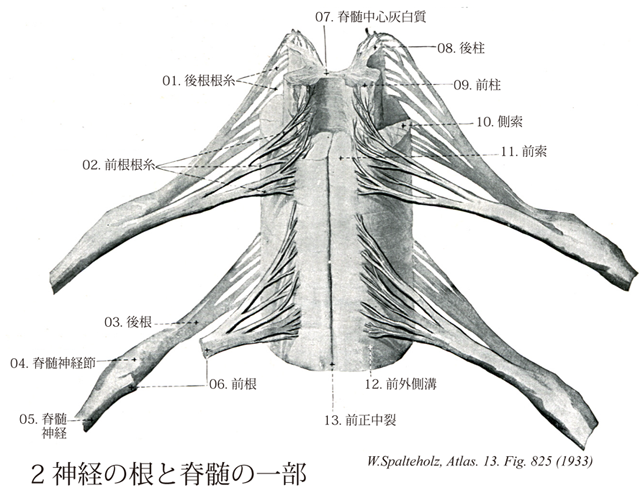
- 825_01【Posterior root fibers; Dorsal root fibers後根根糸;後根線維 Fila radicularia posterioris; Fila radicularia radicis posterioris】
→()
- 825_01a【Rootlets of spinal nerve根糸;根線維(脊髄神経の) Fila radicularia】 Fine root fibers that exit from the spinal cord and attach to the anterior and posterior roots of the individual spinal nerves.
→(脊髄神経の根糸は脊髄よりでる細い根線維。脊髄神経の前根および後根へと束になる。)
- 825_02【Anterior root fibers; Ventral root fibers前根根糸;前根線維(運動性の) Fila radicularia anterioris; Fila radicularia radicis anterioris】
→()
- 825_03【Posterior root of spinal nerve; Sensory root of spinal nerve; Dorsal root of spinal nerve後根;感覚根;背側根(脊髄神経の) Radix posterior; Radix dorsalis; Radices dorsalis; Radix sensoria (Nervus spinalis)】
→(脊髄神経の後根は脊髄神経節の神経芽細胞の中枢性の突起が集まってできる。後根の線維は脊髄にはいると長い上行枝と短い下行枝に分ける。上行枝と下行し共に灰白質の細胞とシナプス結合する。上行枝の一部は延髄の楔状束核(Burdach核)と薄束核(Goll核)に終止する。)
- 825_04【Spinal ganglion; Spinal sensory ganglion; Dorsal root ganglion脊髄神経節;感覚性脊髄神経節 Ganglion sensorium nervi spinalis; Ganglion spinale】 The ganglion belonging to the dorsal root.
→(脊髄神経の後根に存在する神経細胞体の集団を脊髄神経節(時に後根神経節と別称)という。これらの神経細胞体は知覚性ニューロンの物であり、その樹状突起は線維状を呈して長く、皮膚・筋・腱・関節包・内臓などに分布する。また細胞体から中枢側に向かう神経突起は後根内を走り脊髄に入る。)
- 825_05【Spinal nerves脊髄神経 Nervi spinales】 Nerves of the spinal cord. Portion of the nerve between the union of both roots and its bifurcation behind the intervertebral foramen: trunk of spinal nerve.
→(脊髄から出る神経。31対あり、頚神経8対、胸神経12対、腰神経5対、仙骨神経5対、尾骨神経1対に分けられる。それぞえ前根(運動根)、後根(知覚根)として脊髄から出る。後根には膨隆部すなわち脊髄神経節がある。2根は椎間孔で合流し混合脊髄神経となるが、すぐに前枝と後枝に別れる。前枝は体壁の前外側部と四肢に、後枝は前枝に比べると著しく細く、固有背筋と背部の皮膚に分布する。)
- 825_06【Anterior root of spinal nerve; Motor root; of spinal nerve; Ventral root of spinal nerve前根;運動根;腹側根(脊髄神経の) Radix anterior; Radices ventrales; Radix motoria (Nervus spinalis)】
→(脊髄神経の前根は脊髄の前角にある運動ニューロンの神経線維(運動線維)と側角にある自律神経系ニューロンの線維とからなる。いずれも遠心性線維である。)
- 825_07【Spinal area X; Spinal lamina X第X脊髄野;脊髄第X層;脊髄中心灰白質 Area spinalis X; Lamina spinalis X; Substantia grisea centralis】 Region around the central canal.
→(第Ⅹ層は脊髄中心管を囲む領域であり介在ニューロン、神経膠細胞、交叉性軸索がそこに含まれている。中心管を取り囲む灰白質は、Rexedによって第10領域10th areaまたはregionとして位置づけられた。これは後索と前白交連の間をなす部位で、中心管によって背側の後灰白交連と、腹側の前科白交連に分けられる。とくに中心管近傍は中心膠様質substantia gelatinosa centralisともよばれる。そこはこの部がグリアと無髄線維を主体に構成され、ゼラチン状に見えるからである(weigert1880)。Bok(1928)は、後柱と前柱の灰白質が中心灰白質に移行する部位に出現する細胞群を後および前角交連核nucleus cornucommissuralis posteriorとよんだ。)
- 825_08【Posterior column of spinal cord grey; Posterior grey column of spinal cord; Posterior column of spinal cord gray; Posterior gray column of spinal cord後柱;背側柱(脊髄の) Columna grisea posterior; Columna posterior (Medullae spinalis)】
→(脊髄の横断面で中心管を取り囲む灰白質は蝶の形を呈している。主に知覚性の細胞よりなる後角を立体的にみると、柱状となっているので後柱と呼ばれる。主に脊髄神経の後根線維の終止部となっている。後柱は脊髄の各々の外側半分にある灰白質の外側半分にある灰白質の外後方への著しい膨隆で、脊髄の横断面に現れる後角に相当する。主に知覚性の細胞よりなる。)
- 825_09【Anterior column; Ventral column; Ventral grey column of spinal cord前柱;腹側柱(脊髄の灰白柱の) Columna anterior; Columna grisea anterioir medullae spinalis】 Its motor neurons are mainly arranged in groups or nuclei.
→(脊髄の灰白柱の前柱はその基底部で外側に向かい突起を出す。主に運動性の神経細胞よりなる。)
- 825_10【Lateral funiculus of spinal cord側索(脊髄の) Funiculus lateralis (Medullae spinalis)】 White substance between the anterior and posterior horn, including their root fibers.
→(脊髄の側索は脊髄白質で前外側溝と後外側溝にはさまれた部分をいう。おおよそ歯状靱帯付着部と後根侵入部との間の部分に相当する。側索と前索の移行部は前側索と称される。側索には脊髄下行路(錐体側索路、赤核脊髄路、網様体脊髄路)、脊髄上行路(脊髄小脳路、脊髄視蓋路、脊髄視床路)および固有束が通る。下行路は灰白質近くの内側部を、上行路は外側表層近くを通る傾向にある。下行路のうち錐体側索路(外側皮質脊髄路)がもっとも背側を通り、その腹側を赤核脊髄路が下行する。さらに腹側でⅨ層の背外側近くを延髄網様体脊髄路が下行する。上行路では後脊髄小脳路が最も背外側の部分を通り、その腹側を前および吻側脊髄小脳路が上行する。脊髄網様体路、脊髄視蓋路を含む外側脊髄視床路は最も腹側の前側索を通る。これら以外に多数の上行性および下行性固有束の線維が混在している。また後角の後外側表層には後外側束がある。)
- 825_11【Anterior funiculus of spinal cord; Ventral funiculus of spinal cord前索;腹索(脊髄の) Funiculus anterior (Medullae spinalis)】 White substance between the anterior median fissure and anterior nerve root cells and fibers.
→(脊髄の白質で前外側溝から前正中裂までの部分をいう。脊髄下行路(錐体前索路、内側縦束、内側前庭脊髄路、橋網様体脊髄路、視蓋脊髄路)、上行路(前脊髄視床路)および固有束が通る。下行路の錐体前索路(前皮質脊髄路)は前正中裂に接して最内側部を通る。その外側には橋網様体脊髄路、内側前庭脊髄路、間質核脊髄路を含む内側縦束が位置し、さらにその外側を視蓋脊髄路が下行する。上行路の前脊髄視床路は前索の外側部を通る。その他、上行性あるいは下行性固有束が前索を通る。)
- 825_12【Anterolateral sulcus of spinal cord; Ventrolateral sulcus of spinal cord前外側溝;腹外側溝(脊髄の) Sulcus anterolateralis; Sulcus lateralis anterior (Medullae spinalis)】 Occasional indistinct groove from which the anterior nerve rootlets emerge.
→(脊髄の前外側溝は脊髄の腹側の脊髄神経前根の出口に見られることのある浅い溝。(Feneis))
- 825_13【Anterior median fissure of spinal cord; Ventral median fissure of spinal cord前正中裂;腹側正中裂(脊髄の) Fissura mediana anterior (Medullae spinalis)】 Deep, longitudinal, midline fissure on the anterior aspect of the spinal cord.
→(脊髄には、前面と後面とに正中を縦走する溝がみられる。前面の溝は深く前正中裂と呼ばれる。)