Spalteholz HANDATLAS DER ANATOMIE DES MENSCHEN VON WERNER SPALTEHOLZ
メニューは解剖学(TA)にリンクしてあります。図の番号をクリックすると下記の説明へ、右側の用語をクリックすると解剖学(TA)にジャンプします。
836


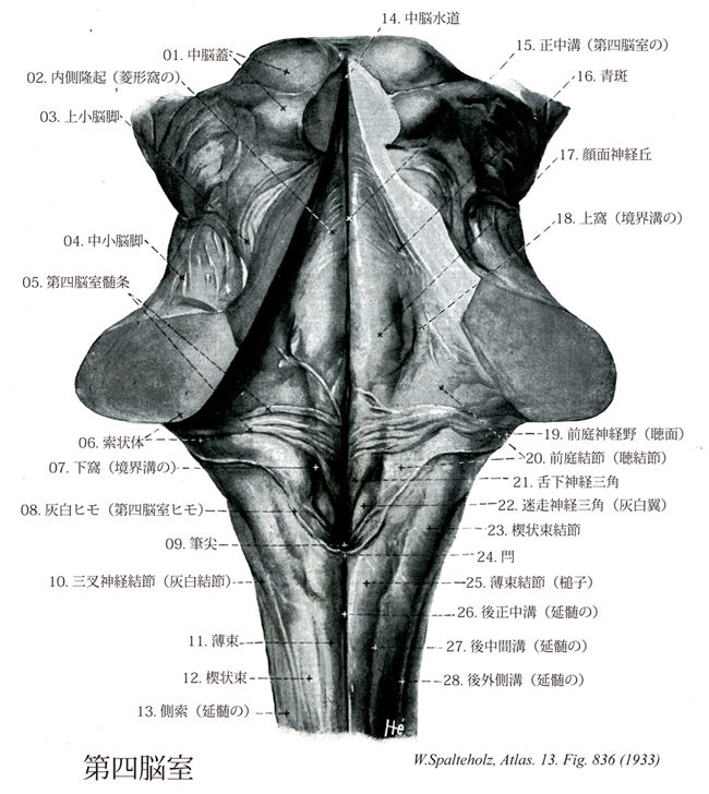
- 836_00【Fourth ventricle第四脳室 Ventriculus quartus】 Dilatation of the embryonic neural tube lumen in the rhombencephalon.
→(第四脳室は菱脳の中にできる脳室で、頭方は中脳水道に、尾方は中心管につづく。第四脳室はその上壁をなす第四脳室蓋と底部の菱形窩により囲まれる。第四脳室蓋の前方は左右の上小脳脚とその間にある薄い白質板の上髄帆とからなる。上髄帆は尾側に伸びて上髄帆小帯となる。第四脳室蓋の後方は下髄帆と第四脳室脈絡組織とからなる。前者は虫部小節と片葉との間にある薄い白質板で、その下面をおおう上衣細胞の尾方延長部は軟膜によっておおわれる。この軟膜が第四脳室脈絡組織(上衣細胞と粘膜とを脈絡組織と呼ぶ場合もある)で、そこに出入る血管とともに脈絡叢をつくる。第四脳室脈絡組織の延髄への付着部が第四脳室ヒモである。第四脳室は左右の第四脳室陥凹に開く第四脳室外側口(Lateral aperture)と尾方の第四脳正中口とによりクモ膜下腔と交通する。)
- 836_01【Tectum of midbrain中脳蓋;四丘体 Tectum mesencephali; Corpora quadrigemina】 Part of the mesencephaIon lying on the tegmentum of midbrain.
→(中脳蓋は中脳水道が通り、屋根状になった脳背側部。上丘と下丘が含まれる。)
- 836_02【Medial eminence of floor of fourth ventricle内側隆起(菱形窩の) Eminentia medialis fossae rhomboideae】 Elongated eminence between the median sulcus and sulcus limitans that is produced by the cranial nerve nuclei.
→(菱形窩の内側隆起は、以前は正中溝と境界溝の間にある細長い隆起(顔面神経小丘、舌下神経三角、迷走神経三角を含めて呼んでいた)。現在は、第4脳室底で顔面神経小丘より吻側の隆起だけをさす。)
- 836_03【Superior cerebellar peduncle上小脳脚;結合腕 Pedunculus cerebellaris superior; Brachium conjunctivum】 Part conveying fibers mostly from the dentate nucleus to the red nucleus and thalamus.
→(上小脳脚(結合腕Brachium conjunctivum)は主として小脳を出る線維からなる。その主体をなす線維は小脳視床路と小脳赤核路である。これらは主として歯状核から出て、腹内側方に進んで深部に入り、中脳下半で大部分交叉し、上小脳脚交叉(結合腕交叉)を作り、反対側の中脳被蓋を上行し、一部は赤核に終わるが(小脳赤核路)、一部はさらに視床の前外側腹側核に至る(小脳視床路)。なお上小脳脚の表面を前脊髄小脳路が逆行して小脳に入り、主としてその前葉に分布する。また鈎状束は室頂核から出て大部分交叉し、上小脳脚の背外側をへて鈎状に曲がり、下小脳脚内側部の上部に来て前庭神経各核にならびに橋、延髄の網様体内側部に分布する。)
- 836_04【Middle cerebellar peduncle中小脳脚;橋腕;橋小脳脚 Pedunculus cerebellaris medius; Brachium pontis】 Part conveying the transverse fibers of the pons, mainly neencephalic tracts, to the cerebellum.
→(中小脳脚(橋腕)は3対ある小脳脚のうち最大のもので、主として橋核から起始する線維からなり、橋底の正中線を越えて対側の背側に移り太い束となって橋被蓋の外側を乗り越えて小脳にはいる。少数の対側へ移らない線維もある。少数の側副線維が小脳核に達している以外ほとんどが橋小脳路線維からできている。)
- 836_05【Medullary striae of fourth ventricle第四脳室髄条;ピッコロミーニ氏髄条 Striae medullares ventriculi quarti】 Bundle of myelinated nerve fibers that passes from the arcuate nucleus to the cerebellum.
→(第四脳室髄条は後脳部と髄脳部の境界部の脳室底には数本の線維が正中溝を横切って横走する線維の束。下小脳脚まで追跡できる。髄条は、次の諸部分から起こる線維より成る複雑な線維束である。すなわち、①中隔核、②外側視索前野、③視床前核群である。海馬体および扁桃体複合核からの線維は中隔核に投射する。)
- 836_06【Restiform body索状体;髄小脳脚 Corpus restiforme; Corpus medullocerebellare】 Posterolateral group of afferent fibers passing to the cerebellum.
→(索状体は下小脳脚の大部分を占める外側部の純求心性線維束である。脊髄から小脳への脊髄小脳線維と延髄から小脳への楔状束小脳線維・網様体小脳線維を含む。)
- 836_07【Inferior fovea of sulcus limitans下窩(境界溝の) Fovea inferior sulci limitantis】 Pit at the tip of the trigone of vagus nerve.
→(第四脳室の境界溝の下窩は舌下神経三角や迷走神経三角(灰白翼)の尖端にある浅いくぼみ。菱形窩の左右を境する溝の中のわずかな陥凹)
- 836_08【Grey line; Taenia cinerea; Gray line灰白ヒモ;第四脳室ヒモ Taenia cinerea; Taenia ventriculi quarti; Tenia ventriculi quarti】 Line along the inferior part of the roof of the rhomboid fossa.
→()
- 836_09【Calamus scriptorius筆尖 Calamus scriptorius】
→(菱形窩の下端はややペン先の形をなしているために筆尖とよばれる。 菱形窩の下部 下方に向かって細くなり、とくに下端はペン先のように細く筆尖と呼ばれる。内側隆起の下部は頂点を下方に向けた三角形状を呈し、舌下神経三角trigonum nervi hypoglossiという(舌下神経核をいれるp.685)。そのすぐ外側には、やや灰白色を呈する三角形の部があり、迷走神経三角(灰白翼)trogonum nervi vagi (ala cinerea)と呼ぶ。迷走神経三角の下方で、薄束結節との間は最後野area postremaといわれ、神経膠と血管に富む特異な部位である。(解剖学講義))
- 836_10【Trigeminal tubercle三叉神経結節;灰白結節;三叉神経隆起 Tuberculum trigeminale; Tuberculum cinereum】 Elevation produced by the spinal nucleus of trigeminal nerve in the continuation of the spinal cord.
→(三叉神経隆起は延髄の背側面に見られる三叉神経脊髄路の表面の縦の小隆起である。楔状束結節の外側縁にある。)
- 836_11Goll's tract【Gracile fasciculus薄束;ゴル索;内側部(後索の) Fasciculus gracilis; Pars medialis funiculus posterior】 Medial portion of the posterior funiculus. It lies medial to the posterior intermediate sulcus and contains fibers of tactile sensation and deep sensibility from the lower half of the body (coccygeal vertebrae to T5).
→(薄束はゴル束ともよばれる。楔状束(ブルダッハ束)とともに脊髄後索をなす。両側の後索は胸髄上部と頚髄において後中間中隔によって2分される。この中隔はおよそ第六胸髄の高さで認められ、薄束(内側)と楔状束(外側)を分ける。薄束は脊髄全長にわたって存在し、仙髄部、腰髄部、下位6胸髄部の後根由来の長い上行枝を含む。薄束は後索の内側部にある。体の下半分から線維を含む。触覚と深部知覚を伝える。Goll, Frindrich (1829-1903)スイスの解剖学者、チューリヒ大学の教授。脊髄後索の内側部(薄束)について1860年に記述(「Beitrage zur feineren Anatomie・・・」;, Denk. Medchir. Ges. Kanton Zurich, 1860, 130-171).)
- 836_12Burdach's tract【Cuneate fasciculus楔状束;外側部;ブルダッハ索(後索の) Fasciculus cuneatus; Pars lateralis funiculus posterior】 Lateral portion of the posterior funiculus. It lies lateral to the posterior intermediate sulcus and contains fibers of tactile sensation and deep sensibility. It begins at the upper half of the body (T4-C1).
→(楔状束はブルダッハ束ともよばれる。楔状束は最初、およそ第六胸髄の高さで出現し、上位の6胸神経と前頚神経の後根の長い上行枝を含む。薄束と楔状束の神経線維は同側を上行し、後索の延髄中継核、すなわち薄束核と楔状束核に終わる。後索系には2部があり、薄束(Goll束)および楔状束(Burdach束)として脊髄の後索を上行する。これらの線維束は太い後根線維の直接の続きであって、延髄の後索核にまで達してシナプス結合する。後索系は主として四肢から起こる線維からなり、系統発生的に新しくて、ヒトでもっとも発達している。ヒトではこの線維の長さは長いもので約150cmである。楔状束は後索の外側部。身体の上半分から起こる線維を含む。ドイツの解剖・生理学者Karl Friedrich Burdach (1776-1847)の名を冠する。以前に同部についての報告はあったが、ブルダッハの正確な報告で知られるようになった。)
- 836_13【Lateral funiculus of medulla側索(延髄の) Funiculus lateralis (Medullae oblongatae)】 Continuation of the lateral funiculus of the spinal cord to the inferior olive.
→(脊髄の側索のつづき。オリーブまで達す。(Feneis))
- 836_14Sylvius, Aqueduct of【Aqueduct of midbrain; Cerebral aqueduct中脳水道;脳水道 Aqueductus mesencephali; Aqueductus cerebri】 Narrow canal in the mesencephalon between the third and fourth ventricles.
→(中脳水道はシルビウス水道ともよばれる。中脳では脳室系は細い管となり、間脳の第三脳室と菱脳の第四脳室とを結合する。これを中脳水道と称し、横断面は円形または底辺を背側に向けた角のとれた三角形をなし、中心灰白質によってかこまれる。その存在については古くから知られていたが、フランスの解剖学者Jacobus Sylvius (1478-1555)の著書(1555年)で初めて説明がなされた。)
- 836_15【Median sulcus of fourth ventricle正中溝(第四脳室の);菱形窩内溝 Sulcus medianus ventriculi quarti; Sulcus medianus fossae rhomboideae】 Midline groove running through the rhomboid fossa.
→(第四脳室の正中溝は菱形窩の正中を通る溝。)
- 836_16【Locus caeruleus; Locus coeruleus青斑 Locus caeruleus; Locus coeruleus】 Elongated collection of bluish-black cells lying below the lateral wall of the fourth ventricle.
→(青斑は中脳水道に近い菱形窩の最前部の外側にある細胞群で、三叉神経中脳路核の腹内側に位置する。肉眼的には第四脳室底において、上小脳脚の内側に吻尾側方向に伸びた青黒色の帯状のものとして認められる。これは細胞体に含まれるメラニン色素によるもので、そのため青斑には鉄色質の別名がある。青斑の細胞群は青斑核とよばれる。ノルアドレナリンを含む大型細胞の集団である。青斑核の腹側には同じような細胞が散在しており、青斑下核と呼ばれる。青斑核細胞の遠心性ノルアドレナリン線維は3群を形成する。①上行線維群:内側前脳束に入り扁桃核にいたるもの、帯状回線維となって帯状回、海馬台にいたるもの、その他梨状葉皮質、前頭葉新皮質に分布する線維からなる。②外側線維群:上小脳脚を通り小脳前葉の皮質の分子層、Purkinje細胞同区に分布する線維である。③下行線維群:これは広く脳幹に分布した後、脊髄前索、前側索を下行し、脊髄全長にわたって後角基部から前角にかけて分布する。求心性線維は次の領域からくる。視床下部(視索前野、後側、背側、外側視床下野)、中脳の中心灰白質、黒質、背側被蓋核、橋縫線核、その他脳幹に分布するカテコールアミンニューロンからの線維を受ける。機能は十分解明されていないが、REM睡眠と深い関係にある。)
- 836_17【Facial colliculus顔面神経丘 Colliculus facialis】 Rounded protuberance above the medullary stria of fourth ventricle. It is produced by the internal genu of facial nerve and the nucleus of abducens nerve.
→(顔面神経丘は第四脳室髄条の上方で、内側隆起は円みをおびた高まりをつくる。顔面神経丘は外転神経核と、これを取り囲むように走る顔面神経線維束とでできる。)
- 836_18【Superior fovea of sulcus limitans上窩(境界溝の) Fovea superior sulci limitantis】 Groove running lateral to the facial colliculus.
→(顔面神経丘の外側で顔面神経丘と前庭神経野との間に上窩というくぼみがある。)
- 836_19【Vestibular area; Acoustic tubercle; Acoustic area前庭神経野;聴面;前庭結節;聴結節 Area vestibularis; Area acustica; Tuberculum vestibularis; Tuberculum acusticum】 Area overlying the vestibular nuclei lateral to the sulcus limitans at the beginning of the lateral recess.
→(前庭神経野は境界溝の側方、外側陥凹のはじまるところの隆起で前庭神経核と蝸牛神経核の一部を容れる。)
- 836_20【Vestibular tubercle; Acoustic tubercle前庭結節;聴結節 Tuberculum vestibularis; Tuberculum acusticum】 Area overlying the vestibular nuclei lateral to the sulcus limitans at the beginning of the lateral recess.
→(前庭神経野は境界溝の側方、外側陥凹のはじまるところの隆起で前庭神経核と蝸牛神経核の一部を容れる。)
- 836_21【Hypoglossal trigone; Trigone of hypoglossal nerve舌下神経三角 Trigonum nervi hypoglossi】 Triangular elevation overlying the nucleus of the hypoglossal nerve. It is situated between the median sulcus and the sulcus limitans above the trigone of vagus nerve.
→(舌下神経三角は第四脳室底にある内側隆起の下部で先端を下方に向けた三角を呈する小隆起。その内部には舌下神経核を入れる。)
- 836_22【Vagal trigone; Trigone of vagus nerve; Ala cinerea迷走神経三角;灰白翼 Trigonum nervi vagi; Trigonum vagale; Ala cinerea】 Triangular elevation overlying the dorsal nucleus of the vagus nerve. It lies caudal to the trigone of hypoglossal nerve.
→(迷走神経三角(灰白翼)は舌下神経のすぐ外側にはほぼ三角形を呈する小隆起。これは表面からみると灰白色をしており、その深部には迷走神経背側核および孤束核がある。)
- 836_23【Cuneate tubercle楔状束結節;楔状束核結節 Tuberculum cuneatum; Tuberculum nuclei cuneati】 Enlargement at the end of the cuneate fasciculus produced by the cuneate nucleus.
→(楔状束結節は楔状束の延髄の上端にある細長い隆起、薄束結節の外側にある。また、後外側溝によって外側にある側索と隔てられている。内部に楔状束核をいれる。)
- 836_24【Obex閂;カンヌキ Obex】 Small bridge at the inferior end of the roof of rhomboid fossa.
→(閂は延髄の背側面の尾側レベルの正中線上の点で菱形窩または第四脳室の後角の境をなしている。すなわち横走する有髄神経線維を含む薄板によって閉じられている。閂には第四脳室脈絡組織が付着している。)
- 836_25【Gracile tubercle薄束結節;薄束核結節;槌子 Tuberculum gracile; Clava; Tuberculum nuclei gracilis】 Enlargement at the end of the gracile fasciculus that is produced by the gracile nucleus.
→(薄束結節は薄束のいくぶん肥厚した上端部で薄束核上にできる細長い隆起。)
- 836_26【Posterior median sulcus of medulla; Dorsal median sulcus of medulla後正中溝;背側正中溝(延髄の) Sulcus medianus posterior medullae oblongatae】 Continuation of the posterior median sulcus of the spinal cord.
→(延髄の後正中溝は脊髄より続く溝で、上方はカンヌキで終わる。)
- 836_27【Posterior intermediate sulcus of medulla; Dorsal intermediate sulcus of medulla後中間溝;背側中間溝(延髄の) Sulcus intermedius posterior (Medullae oblongatae)】
→(延髄の後中間溝は脊髄の正中溝の両側にある溝の延長部で薄束と楔状束との境界を外面から示す。)
- 836_28【Posterolateral sulcus of medulla; Dorsolateral sulcus of medulla後外側溝;背外側溝(延髄の) Sulcus posterolateralis; Sulcus lateralis posterior (Medullae oblongatae)】 Groove extending anterior to the cuneate fasciculus and ending anterior to the trigeminal tubercle.
→(延髄の後外側溝は第四脳室外側陥凹にいたる溝。舌咽神経(Ⅸ)、迷走神経(Ⅹ)および副神経(ⅩⅠ)の脳神経根がでる。)