Spalteholz HANDATLAS DER ANATOMIE DES MENSCHEN VON WERNER SPALTEHOLZ
メニューは解剖学(TA)にリンクしてあります。図の番号をクリックすると下記の説明へ、右側の用語をクリックすると解剖学(TA)にジャンプします。
030


- 030_00【Temporal bone側頭骨 Os temporale】 Bone located between the occipital, sphenoidal, and parietal bones. It consists of petrous, tympanic, and squamous parts.
→(側頭骨は頭蓋の底部および側面にある大きな不規則形の骨。頭蓋側壁の中央部と頭蓋底中央の両側部を作るばかりでなく、骨の中に平衡聴覚器(外耳道・中耳・内耳)を容れる大切な骨である。岩様部(乳突部と錐体)、鼓室部および鱗部の3部が癒合して単一の骨になるのは生後1年ほど経ってからである。3部が合するところの外面には大きい孔がある。これを外耳孔といい、その内方のつづきは外耳道によって鼓室に通ずる。また、外耳孔の上方で鱗部の外側前方に出る頬骨突起は頬骨に達して頬骨弓をつくる。下縁から咬筋が起こる。)
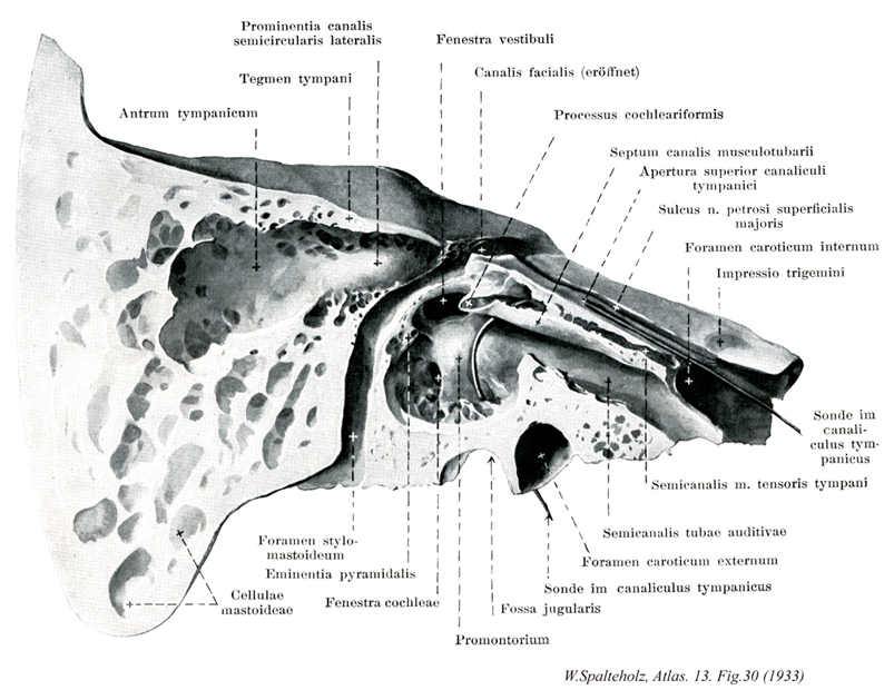
- 030_01【Prominence of lateral semicircular canal外側半規管隆起;外側半規管突隆 Prominentia canalis semicircularis lateralis】 Elevation above the prominence of the facial canal that is produced by the lateral semicircular canal.
→(顔面神経管隆起上方にある隆起。外側半規管によってできる。 (Feneis))
- 030_02【Tegmen tympani鼓室蓋 Tegmen tympani】 Roof of the tympanic cavity located lateral to the arcuate eminence.
→(弓状隆起の外前方、錐体鱗裂との間は、鼓室およびその前方につづく筋耳管管の上を被う部で骨質が薄く、これを鼓室蓋という。骨の発育藤生の場合や老人ではここに孔をみることがある。)
- 030_03【Mastoid antrum乳突洞 Antrum mastoideum; Antrum tympanicum】 Space continuous with the tympanic cavity to posterosuperior. The mastoid cells extend from here downward.
→(乳突洞は後方で乳突蜂巣とつながり、前方では洞孔を経て中耳の上鼓室陥凹とつながる側頭骨錐体部の空洞。)
- 030_04【Mastoid cells; *Mastoid air cells乳突蜂巣 Cellulae mastoideae】 Pneumatized cells that, like the tympanic cavity, are lined with squamous or cuboidal epithelium.
→(側頭骨乳様突起内にある多数の小さな相通じている腔。乳様突起洞あるいは鼓室洞に連なる。鼓室と同様、扁平または立方上皮で被われる。)
- 030_05【Stylomastoid foramen茎乳突孔 Foramen stylomastoideum】 External opening of the facial canal behind the styloid process and between the mastoid process and the jugular fossa.
→(錐体下面の後外側端は茎状突起の着く所で、これとそ後方の乳様突起との間にある茎乳突孔は顔面神経管の出口である。)
- 030_06【Pyramidal eminence錐体隆起 Eminentia pyramidalis】 Conical, bony prominence at the level of the oval window with a perforated summit. It contains the stapedius and gives its tendon exit through the opening at its tip.B
→(前庭窓の高さにある尖端に孔のあいた隆起。アブミ骨筋を有し、尖端の開口部より腱がでる。 (Feneis))
- 030_07【Round window; *Cochlear window蝸牛窓;正円窓 Fenestra cochleae】 Round opening at the end of the scala tympani that is closed off by the secondary tympanic membrane.
→(蝸牛窓は中耳の内壁にある孔で、蝸牛に開くが、生体ではあぶみ骨底にによって閉ざされている。結合組織性の第二鼓膜が張っている。第二鼓膜は臨床医に正円窓膜とよばれる薄い膜で、中耳側より内耳側に向かって鼓膜層、固有層、内層の3層から成っている。粘膜層は鼓室の粘膜層のつづきであり、固有層は結合組織からなり、内層は外リンパ隙に面する内皮から成るが、明瞭な細胞層を呈していない。また前庭の内側(鼓室と反体側)から側頭骨錐体の後面へ前庭膵管とよばれる細い管が通っている。前庭水管の中は膜迷路である内リンパ管が通っている。)
- 030_08【Oval window; Vestibular window前庭窓;卵円窓 Fenestra vestibuli】 Opening that is closed off by the base of the stapes.
→(前庭窓は鼓室の内側壁の卵形開口で、前庭に通じ生体ではこの窓にアブミ骨底がはまりこんでいる。前庭窓のすぐ下に岬角とよばれる隆起がある。)
- 030_09Fallopian canal【Facial canal; Facial nerve canal顔面神経管 Canalis nervi facialis】 Canal for transmission of the facial nerve. It begins at the internal acoustic opening and ends at the stylomastoid foramen.
→(顔面神経管は内耳孔に始まって茎乳突孔に終わる。顔面神経管は顔面神経の通路で、内耳道底にある横稜の顔面神経野にはじまり、まず蝸牛の外側に沿い殆ど水平位で前外方に進み、つぎにほぼ直角をなして後外方に曲がる。ここを顔面神経管膝という。ついで鼓室壁の前庭窓の上を通って少し外後方に進んだ後、下方へ向かって弓状に曲がり(この間、鼓室内側壁に顔面神経管隆起をつくる)、茎乳突孔に開く。この間は膝で1条の枝を出し、骨を貫いて前進し錐体前面の大錐体神経管裂孔を出て大錐体神経溝につづき、大錐体神経管がこれを通る。『フォロッピオ管』:側頭骨錐体にある顔面神経管。イタリアの解剖学者Gabriele Fallopio [Fallopius](1523-1563)によるもので、他にファロピウス管(卵管)にも名を残す。両者を混同しないため、顔面神経管はフォロッピオ管と呼ぶのがふつうである。)
- 030_10【Processus cochleariformisサジ状突起;匙状突起 Processus cochleariformis】 Spoon-shaped bony process above the promontory at the end of the canal for the tensor tympani. Together with a connective-tissue sling it serves as a pulley for the tensor tympani.
→(岬角の上方鼓膜張筋半規管中にある匙状の骨突起。結合組織性のワナとともに鼓膜張筋の支点の役目をする。 (Feneis))
- 030_11【Septum of musculotubal canal筋耳管管中隔;筋管総管中隔 Septum canalis musculotubarii】 Bony septum that divides the canals for the tensor tympani and the auditory tube.
→(筋耳管管中隔ははなはだ薄い骨が半載したチューブのような形となって鼓膜張筋半管を下から被っている。その後壁は鼓室の内側壁(迷路壁)の前上隅から後下に弓なりに隆起する顔面神経管隆起に沿って少し鼓室内に突出し、外方に向かって開口するサジ状突起に終わる。)
- 030_12【Internal opening of lesser petrosal nerve小錐体神経小管内口;鼓室上管 Apertura interna canaliculi nervi petrosi minoris; Apertura superior canaliculi tympanici】
→()
- 030_13【Groove for greater petrosal nerve大錐体神経溝;大浅錐体神経溝 Sulcus nervi petrosi majoris; Sulcus nervi petrosi superficialis majoris】 Groove for transmission of the greater petrosal nerve, extending anteromedially from the hiatus for the greater petrosal nerve to the foramen lacerum.
→(錐体前面の外方には錐体の長軸とほぼ平行に走る2小溝があって、その内上側が大錐体神経溝である。大錐体神経の通路である。)
- 030_14【Carotid canal頚動脈管 Canalis caroticus; Foramen caroticum internum】 Canal for the passage of the internal carotid artery.
→(錐体下面のほぼ中央に頚動脈管が開いている。頚動脈管は錐体内でほぼ直角に内側にまがったのち、前走して錐体先端で頭蓋腔内(中頭蓋窩)に開く。内頚動脈の通路。頚静脈孔と筋耳管管の間の下外方から始まる。 岩様部の管では最大で内頚動脈の通路である。)
- 030_15Meckel's groove【Trigeminal impression三叉神経圧痕 Impressio trigeminalis】 Shallow depression on the anterior surface of the apex of the petrous part of the temporal bone that lodges the trigeminal ganglion.
→(錐体前面の錐体尖の近くに指先で押したような浅い三叉神経圧痕がある。ここは三叉神経の根部と三叉神経節とがのるところである。)
- 030_16Jacobson's canaliculus【Tympanic canaliculus鼓室神経小管;ヤコブソン小管 Canaliculus tympanicus】 Tiny canal in the petrosal fossula for the passage of the tympanic nerve and the inferior tympanic artery.
→(鼓室神経小管は錐体小窩にある小管で鼓索神経の通路である。茎乳突孔の少し上で顔面神経管から分かれ、鼓室溝の後端の近くで鼓室中に出る。その後、鼓索神経は脂質の外側壁にある鼓膜の内側で粘膜に被われながら、ツチ骨柄とキヌタ骨長脚との間を前進し、鼓室の前上隅を貫いて錐体鼓室裂を通り、頭蓋底外面に出る。)
- 030_17【Canal for tensor tympani鼓膜張筋半管 Semicanalis musculi tensoris tympani】 Upper canal for the tensor tympani muscle.
→(筋耳管管は菲薄な筋耳管管中隔に上下の2部に分けられ、その上部の鼓膜張筋半管は鼓膜張筋を含む。)
- 030_18【Canal for auditory tube耳管半管 Semicanalis tubae auditivae; Semicanalis tubae auditoriae】 Lower canal for the auditory tube.
→(筋耳管管は菲薄な筋耳管管中隔に上下の2部に分けられ、その下部の耳管半管は耳管の骨部をつくる。)
- 030_19【External opening of carotid canal頚動脈管外口;外口(頚動脈管の) Apertura externa (Canalis caroticus)】 Opening in the external cranial base between the jugular foramen and the musculotubal canal.
→(頚静脈下の前内側には大きい頚静脈管外口がある。この頚静脈管外口の前から口蓋帆張筋の一部が起こる。)
- 030_20【Jugular fossa頚静脈窩;頚窩 Fossa jugularis】 Widening of the jugular foramen that contains the superior bulb of the jugular vein.
→(錐体下面の後縁に近い中部には弓状の大きく深い頚静脈窩がる。頚静脈上球を容れる。)
- 030_21【Promontory of tympanic cavity岬角(鼓室の) Promontorium (Tympani)】 Elevation produced by the basal turn of the cochlea.
→(蝸牛の基底回転によりできる隆起。 (Feneis))