Spalteholz HANDATLAS DER ANATOMIE DES MENSCHEN VON WERNER SPALTEHOLZ
メニューは解剖学(TA)にリンクしてあります。図の番号をクリックすると下記の説明へ、右側の用語をクリックすると解剖学(TA)にジャンプします。
405 


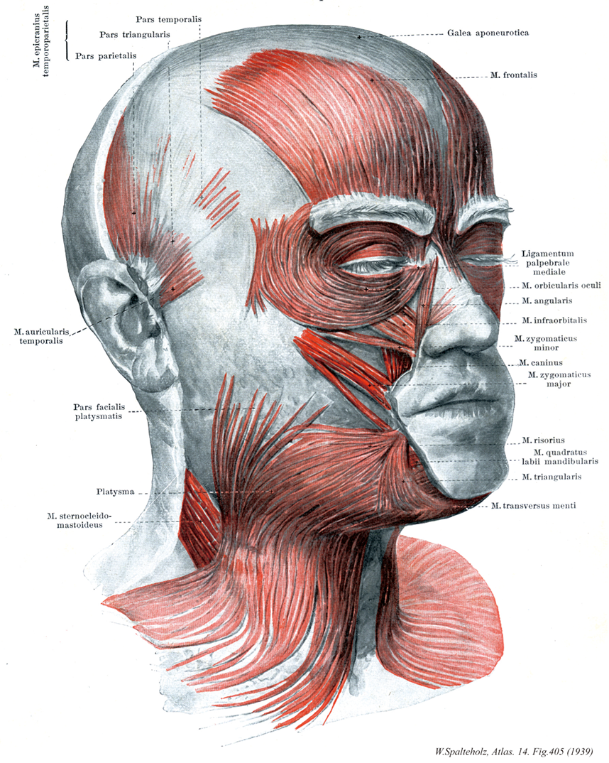
- 405_01【Temporoparietalis muscle側頭頭頂筋 Musculus temporoparietalis】 Muscle that extends from the auricular cartilage to the epicranial aponeurosis.
→(側頭頭長筋は側頭部から帽状腱膜まで広がる。本筋後部は特に上耳介筋と呼ばれ、耳介に付着している。参考:多くの哺乳類では発達し、前頭筋はここれからわかれたものという。日本人では56%に出現(Nishi)。)
- 405_02【Temporal part of temporoparietal muscle側頭部(側頭頭頂筋の) Pars temporalis(Musculus temporoparietalis)】
→(")
- 405_03【Triangular part of temporoparietal muscle三角部(側頭頭頂筋の) Pars triangularis(Musculus temporoparietalis)】
→(")
- 405_04【Parietal part of temporoparietal muscle頭頂部(側頭頭長筋) Pars parietalis(Musculus temporoparietalis)】
→()
- 405_05【Auricularis anterior muscle前耳介筋;側頭耳筋 Musculus auricularis anterior; Musculus auricularis temporalis】 Muscle lying in front of the auricle that passes from the temporal fascia to the spine of the helix. I: Facial nerve.
→(耳介の筋はヒトでは遺残であり、機能的意義を有しない。耳介根部の前方、上方および後方に付着する3筋が区別される。前耳介筋は側頭筋膜で起始する。参考:上、前、後耳介筋は退化的でで、ほとんど働きがない。しかし、練習によってかなり動かせることがある。なかでは、上耳介筋が最も幅が広い。前耳介筋は23%に欠如する(池田一二:福岡医大誌))
- 405_06【Facial part of platysma muscle顔面部(広頚筋) Pars facialis platysmatis】
→()
- 405_07【Platysma muscle広頚筋 Platysma】 Cutaneous muscle that extends (with anatomical variations) from above the mandible to the thorax. I: Facial nerve,
→(広頚筋は前頚部にある薄い膜状の皮筋で、第2咽頭弓(舌骨弓)に発生した筋原基に由来し、しかも頚部にとどまった浅顔面筋である。他の全ての浅顔面筋は頭部に完全に移り表情筋をつくる。広頚筋は極めて薄い筋性の板で、皮膚の直下にある。下顎骨縁から第2(3)肋骨の高さに広がり、さらに遠く肩峰に達する。広頚筋は頚筋膜浅葉の上に広がっていて、ここを走る外頚静脈の上を通る。上方で、筋束は下顎骨と顔面皮膚に付着する。無数の筋線維が表情筋の線維索と交錯している。下方で、広頚筋はさまざまな長さの線維束となって皮下組織に放散し、一部は真皮結合組織内に終わる。左右の筋の内側部のさまざまな長さの線維束となって皮下組織に放散し、一部は真皮結合組織内に終わる。左右の筋の内側部の線維は通常オトガイ下で互いに交錯するが、下方に向かうにつれて、互いに離れ、前頚部の三角形をした正中面は広頚筋に被われずに残る。参考:頚筋中唯一の皮筋で表情筋と同系である。皮膚とは固く、頚筋膜浅葉とはゆるくつく。顔面部は下唇下制筋とつづく。)
- 405_08【Sternocleidomastoid muscle胸鎖乳突筋 Musculus sternocleidomastoideus】 o: Two-headed muscle arising from the sternum and clavicle, i: Mastoid process; superior nuchal line. Rotates the face to the contralateral side and bends the head to the ipsilateral side. Bilateral contraction elevates the face. I: Accessory nerve, cervical plexus (C1-C2).
→(胸鎖乳突筋は側頚部にある強大な斜めに縦走する浅層の筋。胸骨柄前面と鎖骨の胸骨端から2頭をもっておこり、両頭は合して強い筋腹をつくって後上方に走り、乳様突起および後頭骨の上項線につく。作用は複雑で、両側が同時に働くとオトガイを上げて後頭部を片側が働けば頭を対側にまわすが、その浅オトガイが対側に向かって上り、頭は逆に同側に傾く。支配神経は副神経外枝と頚神経叢筋枝(C2, C3)であり、したがって僧帽筋と同系の筋である。また、第6咽頭弓に発生する鰓弓筋で、鎖骨上窩を囲む2頭(胸骨頭と鎖骨頭)をもって始まる。胸骨頭は胸骨柄の上縁から、鎖骨頭は鎖骨の胸骨端から起こる。筋膜は頚筋膜浅葉に鞘状に包まれており、斜め上方に向かって幾分螺旋状に回転しながら頚部外側面を横切り、よく発達した腱となって乳様突起と上項線に停止する。筋の表面は、起始部で腹側に、停止部で外側に向く。参考:副神経外枝の僧帽筋枝は、外枝がこの筋に入る前に分かれることと、筋内で分かれて再び外に現れることがある。胸鎖乳突筋はドイツ語ではKopfnicker(頭をこっくりとうなずかせる筋)と呼ばれるが、これは作用の点からは正しくない。この筋が片側だけ収縮すると、頭はその側へ傾き反対側を振り向いて、あたかも「首をかしげる」状態になる。また両側の物が同時に収縮すると、頭を胴体にめり込ませるように働くのえある。Musculus sternocleidomastoideusというラテン名はあまりにも長たらしいので、米英では多少簡略化してsternomastoid muscleともよぶ。片側の胸鎖乳突筋が先天的に短い場合、または出産時の外傷などによって瘢痕化して短縮すると、この筋の作用を考えればすぐわかるように、頭は病側へ傾くと共に健側にねじれたままの状態になるこれを斜径torticolis, wryneck(性格には筋性斜径)といい、かなり頻度の高いものである。略語(SCM))
- 405_09【Epicranial aponeurosis帽状腱膜;上頭蓋腱膜;頭外被腱膜 Galea aponeurotica; Aponeurosis epicranialis】 Hoodlike aponeurosis of both parts of the epicranius that can be moved against the periosteum. Its inferior surface extends from the highest nuchal line to the supraorbital margin and its lateral border nearly reaches the zygomatic arch.
→(学会では上頭蓋腱膜を採用する必要を認めていないが英語圏では一般にEpicranial aponeurosisを用いているのであえて併記した。帽状腱膜はヘルメット様の扁平腱で、頭蓋表筋の後頭前頭筋の後頭筋腹と前頭筋腹とを連結する腱膜で、側頭頭頂腱膜とともに頭骨外被を形成する。)
- 405_10【Frontal belly of occipitofrontalis muscle前腹(後頭前頭筋の);前頭筋 Venter frontalis (Musculus occipitofrontalis); Musculus frontalis】 Portion of the occipitofrontalis muscle that passes from the eyebrow region and glabella to the epicranial aponeurosis. It elevates the eyebrows and wrinkles the forehead.
→(参考:人類では帽状腱膜の移動性は少ないので前頭筋は主に眉を上げるためだけである。また後頭筋はその一般に退化的でその働きも弱い。)
- 405_11【Medial palpebral ligament内側眼瞼靱帯 Ligamentum palpebrale mediale】 Connective-tissue band connecting the medial palpebral commissure and medial wall of the orbit, lying immediately anterior to the fossa for the lacrimal sac.
→(内側眼瞼交連と内側眼窩壁との間の結合組織性結合。涙腺窩の直前にある。(Feneis))
- 405_12【Orbicularis oculi muscle; Orbicular muscle of eye眼輪筋 Musculus orbicularis oculi】 Ringlike sphincter muscle around the eye consisting of various parts. It acts to close the eyelids and support the flow of tears into the lacrimal sac and nose. I: Facial nerve.
→(眼輪筋の外側部にある(外側)眼瞼縫線は筋の後面と内側部のほかは不明瞭である。外眼角と頬骨を結ぶ外側眼瞼靱帯とは別物で、両者は内側部でゆるく結合するだけであるが、この縫線と靱帯はしばしば混同されている。外側部では眼輪筋と骨との結合がないので、眼を強く閉じると筋とともに皮膚が外眼角に引かれて目じりにヒダができるのである。なお、眼裂を開くのは主に上眼挙筋(眼筋)が行ない、大きく開くときは同時に前頭筋によって眉が上がることが多い。下眼瞼は弾力によって元に戻ると思われるが、その動きは小さい。まばたきのとき涙が吸い込まれることは、涙がこぼれそうになると無意識にまばたきを行うことからも判る。その機構として眼を閉じるとき眼輪筋の涙嚢部が涙嚢の壁を引いて広げるという節が有名である。しかし、一般に涙嚢部といわれる筋(Horner筋)が涙嚢と関係のないことは以前から記載があり(Eisler)涙の排出機構にも諸説があった。長嶋1954, 1955は涙道の圧をしらべ、瞼を開くときに涙小管腔が陰圧になって涙が吸い込まれ、瞼を閉じるとき涙小管は陽圧に涙嚢は陰圧になって涙は涙嚢に送られ、さらに瞼を開くとき涙嚢は陽圧となって鼻涙管に排出されるとした。その後の研究も合わせると、Horner筋は瞼閉じるとき涙小管を屈曲・圧迫するもので、これとは別に涙嚢を包む筋膜(涙嚢間膜)から起こるごく小さい筋束(Jones 1957)があって涙嚢を広げるという機構が有力である。このポンプ作用の主力はHoener筋と涙小管にあり、そのほか毛細管現象による吸引、重力による鼻腔への流下なども助けるという。なお、いわゆるHorner筋は涙小管を囲んで眼瞼縁に向かうが、Jonesの筋束は外輪筋眼瞼部の周辺部に合するもので、後者は従来見落とされていたが、または、涙嚢部と区別だれていなかったと思われる。)
- 405_13【Levator labii superioris alaeque nasi muscle上唇鼻翼挙筋;眼角筋 Musculus levator labii superioris alaeque nasi; Musculus angularis】 Muscle that originates from the frontal process of the maxilla and inserts into the upper lip and ala of the nose. I: Facial nerve.
→(上唇鼻翼挙筋は上顎骨前頭突起から愧死して、前述の筋と癒合して停止する。参考:"alaeque nasi"は"et alae nasi"と大体同じ意味。この2筋も、一般に単一筋板を作り、分離しがたい。さらに、小頬骨筋も合わせて3筋を上唇方形筋M. quadratus labii superiorisということがある。)
- 405_14【Levator labii superioris muscle上唇挙筋;眼窩下筋 Musculus levator labii superioris; Musculus infraorbitalis】 Muscle that arises above the infraorbital foramen and whose fibers insert into the orbicularis oris. I: Facial nerve.
→(上唇挙筋は眼窩下孔の上方で眼窩下縁から上唇に向かって内側下方へ走る。参考:“alaeque nasi”は“et alae nasi”と大体同じ意味。この2筋も、一般に単一筋板を作り、分離しがたい。さらに、小頬骨筋も合わせて3筋を上唇方形筋M. quadratus labii superiorisということがある。)
- 405_15【Zygomaticus minor muscle; Zygomatic minor muscle小頬骨筋 Musculus zygomaticus minor】 Muscle passing from the zygomatic bone to the upper lip. I: Facial nerve.
→(小頬骨筋は大胸骨筋の内側で起始して上唇に停止する。小頬骨筋は眼輪筋に密着する。小頬骨筋は、上唇挙筋、上唇鼻翼挙筋と協同して、鼻唇溝を深くする(“すすり泣き”)。)
- 405_16【Levator anguli oris muscle口角挙筋;犬歯筋 Musculus levator anguli oris; Musculus caninus】 Muscle passing from the canine fossa to the angle of the mouth. I: Facial nerve.
→(口角挙筋は犬歯上方および眼窩下孔で犬歯窩から口角まで走る。)
- 405_17【Zygomaticus major muscle; Greater zygomatic muscle大頬骨筋;頬骨筋 Musculus zygomaticus major】 Muscle extending from the zygomatic bone to the angle of the mouth and upper lip. I: Facial nerve.
→(大胸骨筋は頬骨弓から内側下方へ向かって上唇および口角まで走る。大頬骨筋は口角を引き挙げ、かつ口角を外方へ引く(狭義の“笑う筋”)。)
- 405_18Santorini's muscles【Risorius muscle笑筋 Musculus risorius】 Bundle of muscle fibers that are occasionally present, extending from the parotid fascia and skin of the cheek to the angle of the mouth. I: Facial nerve.
→(笑筋は、非常に繊細で変化に富む筋束であり、耳下腺筋膜および頬部皮膚から口角まで殆ど水平方向に走る。笑筋は頬の“笑窪”をつくり、しかも口裂を幅広くする。)
- 405_19【Depressor labii inferioris muscle下唇下制筋;下唇方形筋 Musculus depressor labii inferioris; Musculus quadratus labii mandibularis】 Muscle that lies deep to the depressor anguli oris and passes from the platysma superiorly and medially to the lower lip. I: Facial nerve.
→(下唇下制筋は広頸筋から斜めに下唇へ向かって、下外側から上内側へ放射する。下唇下制筋は口角下制筋によって被われ、下唇を下方および側方へ引く(不快の表情)。起始と走行:下顎骨の前面でオトガイ孔の下付近から起こり、その外側部は口角下制筋に被われる。斜めに内上方にはしる。参考:筋束の一部は広頸筋から移行し、また神経支配も共通である。そのため口角を外下方に引くとき、広頸筋も同時に収縮する。)
- 405_20【Depressor anguli oris muscle口角下制筋;オトガイ三角筋 Musculus depressor anguli oris; Musculus triangularis】 Muscle that passes from the anterior and lateral margins of the mandible to the angle of mouth. I: Facial nerve.
→(口角下制筋は下顎骨下縁から口角まで収斂しながら走る。表層に位置する口角下制筋は口角を下方へ引き、かつ鼻唇溝の上部を伸展させる。)
- 405_21【Transversus menti muscleオトガイ横筋 Musculus transversus menti】 Muscle fibers that cross between the right and left depressor anguli oris below the chin. I: Facial nerve.
→(オトガイ横筋は不規則筋で、左右の口角下制筋のオトガイ部の下を横に走る筋束。オトガイ部の皮膚にしわを寄せる。参考:欠けることは稀である。)