Spalteholz HANDATLAS DER ANATOMIE DES MENSCHEN VON WERNER SPALTEHOLZ
メニューは解剖学(TA)にリンクしてあります。図の番号をクリックすると下記の説明へ、右側の用語をクリックすると解剖学(TA)にジャンプします。
537 


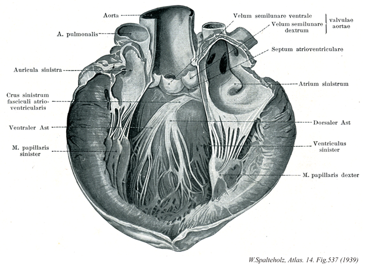
- 537_01【Aorta大動脈 Aorta】 Main artery supplying the body.
→(大動脈は体循環系の本幹をなす単一の太い動脈。上行大動脈・大動脈弓・胸大動脈および腹大動脈に区分される。後2者を総称して下行大動脈ともよぶ。上行大動脈は左心室の大動脈口よりはじまり心膜腔を出るまで約5~6cmの範囲をいう。肺動脈幹とともに心膜に包まれている。上行大動脈の起始部は3個の大動脈洞によるふくらみを呈し大動脈球とよばれ、左・右冠状動脈がここからでる。大動脈弓は上行大動脈につづく弯曲部であり(約5~6cm長)、肺動脈分岐部および左気管支をこえて左後方にまわり、第四胸椎体の左側で胸大動脈に移行する。大動脈弓の重要な枝として腕頭動脈・左総頚動脈・左鎖骨下動脈が出る。大動脈弓の凹弯する下面と肺動脈分岐部とのあいだを動脈間索が結ぶ。胸大動脈にまわり、第12胸椎の直前で横隔膜の大動脈裂肛の壁側枝として肋間動脈10対、また臓側枝として気管支動脈・食道動脈などを出す。腹大動脈は第12胸椎より第4腰椎までの前を下行したのち左右総頚動脈を出し、本幹のつづきは細い正中線骨動脈となって尾骨尖端に達している。壁側枝には各有対の下横隔動脈・腰動脈・総腸骨動脈があり、臓側枝には無対性の腹腔動脈・上腸間膜動脈・下腸間膜動脈また有対の副腎動脈・腎動脈・精巣(ないし卵巣)動脈がある。)
- 537_02【Pulmonary trunk; Pulmonary artery肺動脈幹;肺動脈 Truncus pulmonalis; Arteria pulmonarlis】 Arterial trunk that ascends in the pericardium. It divides into the right and left pulmonary arteries at the level of the reflection of the serous pericardium.
→(肺動脈幹は右心房と左右肺動脈の起始までの間で分岐前の肺動脈を明確にするために導入された。肺動脈幹は左の第3肋骨の胸骨付近の高さで右心部動脈円錐から起こる。長さは約5cmで心膜腔を通り、やや左側に向かい、頭背側で肺動脈幹のT字形の分岐となり、2本の肺動脈に分岐する。肺動脈幹の両側を2本の冠状動脈が走る。肺動脈幹の起始部は大動脈口の前左側に、中間部は上行大動脈の左側に、分岐部は大動脈弓の凹部に位置する。)
- 537_03【Left auricle of atrium左心耳 Auricula atrii sinistra; Auricula sinistra cordis】 Outpouching of the atrium to the left of the pulmonary trunk.
→(左心耳は肺動脈幹の左方に中空指状に突出した左心房の一部。左心耳は右心耳に比べて格段に小さい。)
- 537_04【Left bundle of atrioventricular bundle; Left branch of atrioventricular bundle左脚;左束枝(房室束の) Crus sinistrum fasciculi atrioventricularis】 Bundle that expands over the septum and extends to the base of the papillary muscles.
→(房室束の左脚は室中隔の左右を乳頭筋まではしる刺激伝導系の左脚で、そこからさらに分枝する。)
- 537_05【Anterior fascicle of atrioventricular bundle前束(房室束の) Fasciculus anterior (Fasciculus atrioventricularis)】
→()
- 537_06【Anterior papillary muscle of left ventricle前乳頭筋;左乳頭筋(左心室の) Musculus papillaris anterior; Musculus papillaris sinister (Ventriculus sinister)】 Larger, anterior papillary muscle arising from the lateral wall of the left ventricle.
→()
- 537_07【Aortic valve大動脈弁 Valva aortae】 Valve apparatus at the beginning of the aortic outflow tract.
→(大動脈弁は大動脈起始部にある3部分からなる弁。肺動脈口にある肺動脈弁と基本的に同様の形状をもち、3枚のポケットに似た半月状の弁、すなわち右半月弁、左半月弁、後半月弁からなり、冠状動脈の起始部と関連してそれぞれ右冠尖、左冠尖、無冠尖ともよばれる。)
- 537_08【Right semilunar cusp of aortic valeve; Right coronary cusp of aortic valve右半月弁;右冠尖(大動脈弁の) Valvula semilunaris dextra; Valvula coronaria dextra (Valva aortae)】
→()
- 537_09【Posterior semilunar cusp of aortic valve; Noncoronary cusp of aortic valve後半月弁;無冠尖(大動脈弁の) Valvula semilunaris posterior; Valvula non coronaria (Valva aortae)】
→()
- 537_10【Atrioventricular septum房室中隔 Septum atrioventriculare】 Portion of the membranous part of interventricular septum between the right atrium and left ventricle above the root of the septal cusp.
→(房室中隔は左心房と左心室の間の膜性部のなかで、房室弁起始より上方にある部分。)
- 537_11【Left atrium左心房 Atrium cordis sinistrum; Atrium sinistrum】
→(左心房は心臓の後上部にあって、後面をつくっている。左心房は右心房よりもやや小さいが、壁はやや厚い。左心房の後壁の上部に、左右両肺からそれぞれ2本ずつ、前部で4本の肺静脈が開口している。左心房は前下方で房室口によって左心室に通じる。)
- 537_12【Posterior fascicle of atrioventricular bundle後束(房室束の) Fasciculus posterior (Fasciculus atrioventricularis)】
→()
- 537_13【Left ventricle左心室 Ventriculus sinister】
→(左心室は心臓の左下部を占め、後上方にある左房室口で左心房と交通し、右上隅にある大動脈口によって大動脈につらなる。左心室の壁は右心室に比べ2~3倍厚い。心室中隔は、右心室に向かって膨隆しているので、心室を横断面でみると、左心室の内腔は円いのに対して、右心室の内腔は半月状である)
- 537_14【Posterior papillary muscle of left ventricle後乳頭筋;右乳頭筋(左心室の) Musculus papillaris posterior; Musculus papillaris dexter (Ventriculus sinister)】 It arises from between the interventricular septum and the lateral wall.
→()