Spalteholz HANDATLAS DER ANATOMIE DES MENSCHEN VON WERNER SPALTEHOLZ
メニューは解剖学(TA)にリンクしてあります。図の番号をクリックすると下記の説明へ、右側の用語をクリックすると解剖学(TA)にジャンプします。
555 


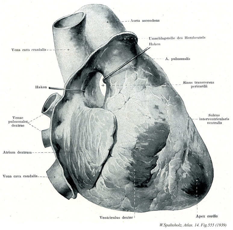
- 555_01【Superior vena cava上大静脈 Vena cava superior; Vena cava cranialis】
→(上大静脈は上半身の血液を集める静脈で、上縦隔の中で左右の腕頭静脈が合してはじまり、途中で奇静脈を受け入れながら上行大動脈の右側を下行して右心房にそそぐ。)
- 555_02【Right pulmonary veins右肺静脈 Venae pulmonales dextrae】 The two right pulmonary veins which occasionally unite to form a single trunk.
→(2本あるが、時に合流して1本の幹となる。 (Feneis))
- 555_02a【Pulmonary veins肺静脈 Venae pulmonales】 Blood vessels leading from the lungs to the heart.
→(肺静脈は正常では肺から上肺静脈と下肺静脈が、酸素に富む血管を肺胞壁の毛細血管網から左心房に運ぶ。また肺組織や細いまたは中等大の気管支の壁からも血液を受ける。両側の肺門では、肺静脈は肺門の前下縁近くに位置している。上・下肺静脈は左心房に開口する前に共通幹を形成することがある。)
- 555_03【Right atrium右心房 Atrium cordis dextrum; Atrium dextrum】
→(右心房は心臓の右上部を占め、その後上部と後下部とに、それぞれ、上大静脈と下大静脈が注いでいる。)
- 555_04【Inferior vena cava下大静脈 Vena cava inferior; Vena cava caudalis】 It arises at the union of the right and left common iliac veins, lies on the right side of the aorta, and opens into the right atrium of the heart.
→(下大静脈は下肢および骨盤と腹部の器官の大部分から血液を受ける本幹で、第5腰椎体の右側で左右の総腸骨静脈の合流として始まり、このあと脊柱に沿って大動脈の右側を上行、肝臓の後面をこれに接して通過し、第八胸椎の高さで横隔膜の大静脈孔を貫いて胸腔に入り、ただちに右心房にそそぐ。下大静脈に流入する枝には総腸骨静脈、下横隔静脈、第3・第4腰静脈、肝静脈、腎静脈、右副腎静脈、右精巣静脈、右卵巣静脈、蔓状静脈叢などがある)
- 555_05【Right ventricle右心室 Ventriculus dexter】
→(右心室は心臓の最下位部を占め、後上方にある右房室口で右心房と交通し、前上方にある肺動脈口で肺静脈に連なる。)
- 555_06【Ascending aorta上行大動脈;大動脈上行部 Pars ascendens aortae; Aorta ascendens】 Ascending part of the aorta up to its exit from the pericardium.
→(左心室からおこり、肺動脈幹の後ろを上行して大動脈弓にいたる5cmほどの部。基部の内腔は膨らんで大動脈洞(バルサルバ洞)をなし、ここから左右の冠状動脈が出る。(イラスト解剖学))
- 555_07【Pericardium心膜 Pericardium】 Lubricant-containing sheath enclosing the heart. It consists of a fibrous layer and a double-layered serous coat.
→(心膜は心臓と大血管起始部の被覆と活動のための膜。外層の線維性心膜fibrous pericardiumと内層の漿膜性心膜serous pericardiumの2層からなる閉鎖嚢。漿膜性心膜は心臓表面を直接おおう臓側板(心外膜)と線維性心膜の内面をおおう壁側板にわけられる。線維性心膜は強靱な膜で、大血管の壁につづき、心臓を固定・保持するとともに、その急激な過度の拡張を防ぐ。さらに心臓は心膜腔で囲まれ、潤滑な心膜性心膜で包まれるので、摩擦なく拍動することができる。)
- 555_08【Pulmonary trunk; Pulmonary artery肺動脈幹;肺動脈 Truncus pulmonalis; Arteria pulmonarlis】 Arterial trunk that ascends in the pericardium. It divides into the right and left pulmonary arteries at the level of the reflection of the serous pericardium.
→(肺動脈幹は右心房と左右肺動脈の起始までの間で分岐前の肺動脈を明確にするために導入された。肺動脈幹は左の第3肋骨の胸骨付近の高さで右心部動脈円錐から起こる。長さは約5cmで心膜腔を通り、やや左側に向かい、頭背側で肺動脈幹のT字形の分岐となり、2本の肺動脈に分岐する。肺動脈幹の両側を2本の冠状動脈が走る。肺動脈幹の起始部は大動脈口の前左側に、中間部は上行大動脈の左側に、分岐部は大動脈弓の凹部に位置する。)
- 555_09Thiele's canal【Transverse pericardial sinus心膜横洞 Sinus transversus pericardii】 Passageway in the pericardial cavity behind the ascending aorta and pulmonary trunk and in front of the veins.
→(心膜横洞は大動脈、肺動脈管の後方で静脈の前方を占める心膜腔の狭い部位。)
- 555_10【Anterior interventricular sulcus前室間溝 Sulcus interventricularis anterior】 Longitudinal groove on the anterior surface of the heart overlying the interventricular septum. It transmits the anterior branch of the interventricular branch of the left coronary artery.
→(前室間溝は心室中隔にあたる前方の縦溝で、前室間枝(冠状動脈の)が走る。)
- 555_11【Apex of heart心尖 Apex cordis】 Part of the heart directed downward and to the left. It is formed by the left ventricle.
→(心尖は心臓の下端部でやや尖っている。心尖は心臓の拍動とともに前胸壁にあたる。これを心尖拍動といい、体表で触れることができる。すなわち、一般に左側の第5肋間で、正中線から約4横指(約7cm)左方で触れる。この位置は弾性では左乳頭のすこし内下方である。小児ではやや高くかつ外方にある。)