Spalteholz HANDATLAS DER ANATOMIE DES MENSCHEN VON WERNER SPALTEHOLZ
メニューは解剖学(TA)にリンクしてあります。図の番号をクリックすると下記の説明へ、右側の用語をクリックすると解剖学(TA)にジャンプします。
1029


Rivinus' membrane
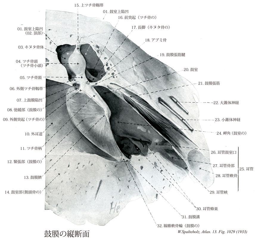
- 1029_00Rivinus' membrane【Tympanic membrane鼓膜 Membrana tympanica】 Membrane stretched diagonally at the end of the external acoustic meatus. It has a diameter of 9-11 mm.
→(鼓膜は外耳道と中耳すなわち鼓室との境にある直径約1cmのほぼ卵円形薄い膜。鼓膜は外耳道に対して傾斜し、外面を前下方に向けている。鼓膜の外面は平面でなく、内方に向かってやや陥凹している。鼓膜は生体で耳鏡を外耳道に挿入して観察すると、やや透明で、白色の線条がみられツチ骨柄によってできるツチ骨条といわれる。ツチ骨条の上端には外側突起の部分がツチ骨隆起である。その前後に前ツチ骨ヒダと後ツチ骨ヒダとがあり、緊張部と弛緩部との境界である。緊張部ではその前上方より鼓膜中央まで内面にツチ骨柄が付着するために鼓膜痔帯が内方に向かって漏斗状に陥入し、鼓膜臍を形成する。鼓膜は外側の皮膚面、固有層、内側の粘膜面の三者からなる。皮膚面は外耳道の皮膚のつづきで重層扁平上皮を有する。固有層は線維性結合組織からなり、その線維の走向により内外の2層を区別することができる。粘膜面は鼓室表面の粘膜のつづきであって、単層扁平上皮によりおおわれている。鼓膜の皮膚面には外耳道神経の枝が、また粘膜面には鼓室神経の枝が、それぞれ分布する。)
- 1029_01【Epitympanic recess鼓室上陥凹 Recessus epitympanicus】 Dome of the tympanic cavity above the upper border of the tympanic membrane that arches upward and laterally.
→(鼓膜上縁の上に、上外側へ向けてできた陥凹。 (Feneis))
- 1029_02【Cupular part of epitympanic recess頂部(鼓室上陥凹の) Pars cupularis recessi epitympanici】 Superior portion of the epitympanic recess.
→()
- 1029_03【Body of incusキヌタ骨体 Corpus incudis】 Portion that articulates via a saddle-shaped articular surface with the malleus.
→(鞍状の関節を介してツチ骨と接す。 (Feneis))
- 1029_04【Head of malleusツチ骨頭;ツチ骨小頭 Caput mallei; Capitulum mallei】 Portion bearing a convex facet for articulation with the body of incus.
→(ツチ骨頭は球状で、鼓室上陥凹にあり、後方でキヌタ骨と関節をつくる。)
- 1029_05【Neck of malleusツチ骨頚 Collum mallei】 Segment connecting the head and handle of malleus.
→(ツチ骨頚は頭の下の細い部で、鼓膜の弛緩部の高さにある。)
- 1029_06【Lateral ligament of malleus外側ツチ骨靱帯 Ligamentum mallei laterale】 Band connecting the neck of malleus with the upper margin of the tympanic notch.
→(ツチ骨頚を鼓膜切痕の上縁とむすぶ。 (Feneis))
- 1029_07【Superior recess of tympanic membrane上鼓膜陥凹;プルサク腔 Recessus superior membranae tympanicae】 Mucosal pouch bordered laterally by the pars flaccida of the tympanic membrane and medially by the head and neck of malleus as well as the body of incus.
→((Prussak腔):外側は鼓膜弛緩部、内側はツチ骨ヒダと鼓膜の間のヒダ。 (Feneis))
- 1029_08Shrapnell's membrane【Pars flaccida of tympanic membrane弛緩部;シュラプネル膜(鼓膜の) Pars flaccida membranae tympanicae】 Smaller, flaccid part of the tympanic membrane above the anterior and posterior malleolar folds.
→((Shrapnell膜):前および後ツチ骨ヒダの上方部。 (Feneis))
- 1029_09【Lateral process of malleus外側突起;短突起(ツチ骨の) Processus lateralis; Processus brevis (Malleus)】 Short, lateral process at the end of the handle of malleus. It produces the malleolar prominence in the tympanic membrane.
→(ツチ骨の外側突起はツチ骨頚から外側にでる突起である。)
- 1029_10【External acoustic meatus; External auditory meatus外耳道 Meatus acusticus externus】
→(外耳道は側頭骨の鼓室部を耳介から鼓膜へ至る通路で骨性部分。軟骨性外耳道からなる。)
- 1029_11【Handle of malleusツチ骨柄 Manubrium mallei】 Manubrium of malleus that is fused along its lateral surface with the tympanic membrane as far as the lateral process.
→(ツチ骨柄は細長く後下方にのびる部で、鼓膜の内面に付着する。)
- 1029_12【Pars tensa of tympanic membrane; Tens part of tympanic membrane緊張部(鼓膜の) Pars tensa membranae tympanicae】 Much larger portion of the tympanic membrane that is stretched within the tympanic ring.
→(鼓膜溝にある線維軟骨輪に付く鼓膜の大部分は、緊張しているので、緊張部といわれる。)
- 1029_13【Umbo of tympanic membrane鼓膜臍 Umbo membranae tympanicae】 Portion of the membrane lying at the tip of the handle of malleus that draws the tympanic membrane inward.
→(緊張部の中央は、内方に向かって最も深く凹み、鼓膜臍と呼ばれる。)
- 1029_14【Tympanic part of temporal bone鼓室部(側頭骨の) Pars tympanica (Os temporale)】 The part of the temporal bone that forms most of the wall of the bony acoustic meatus except for the posterior, superior portion.
→(鼓室部は外耳道の前下壁を作る半管状、不正四角形の薄い骨板で、初めは独立した結合組織(鼓室骨)として発達し、後に錐体の下面に癒着した小さい骨部である。鼓室部の前上縁は下顎窩(顎関節の関節窩)の後縁にある錐体鼓室裂であり、また鼓室部の下端は錐体の下面に接着して鋭い稜線を作り、その後外側方への延長は茎状突起の根元におおいかぶさっている。鼓室部という日本名は鼓室を包含するすべての骨部を指すかのような誤った印象をあたえるのでよくない。Tympanicaはギリシャ語のtympanon(ケトルドラムという楽器)に由来する形容詞で、語源からわかるように元来は「鼓膜に関係した」という意味である。)
- 1029_15【Superior ligament of malleus上ツチ骨靱帯;上ツチ骨小頭靱帯 Ligamentum mallei superius; ligamentum capituli mallei superius】 Band passing from the head of malleus to the roof of the epitympanic recess.
→(ツチ骨頭より鼓室上陥凹へいたる靱帯。 (Feneis))
- 1029_16Folian process; Rau's process【Anterior process of malleus前突起;長突起(ツチ骨の) Processus anterior; Processus longus (Malleus)】 Longer, very thin process. In the newborn it extends into the petrotympanic fissure. It regresses in the adult.
→(ツチ骨の前突起はツチ骨頚から前下方に出る小突起で、靱帯によって鼓室の前壁と結合する。)
- 1029_17【Long limb of incus長脚(キヌタ骨の) Crus longum incudis】 Limb descending nearly vertically behind the handle of malleus with the lenticular process at its end.
→(ツチ骨柄の後方でほぼ垂直下方へのび、尖端には豆状突起がある。 (Feneis))
- 1029_18【Stapesアブミ骨;鐙骨 Stapes】 Stirrup-shaped ossicle, the base of which is integrated into the oval window.
→(アブミ骨は3つの耳小骨のうち最小のもので、底部は前庭窓についている。アブミ骨頭、前脚、後脚、アブミ骨底を区別する。頭部はキヌタ骨の長肢の豆状突起と関節で結合する。)
- 1029_19【Tendon of tensor tympani muscle鼓膜張筋腱 Tendo musculus tensor tympani】
→()
- 1029_20【Tympanic cavity鼓室 Cavitas tympani; Tympanum】 Cleftlike pneumatized space between the bony labyrinth and the tympanic membrane.
→(側頭骨の錐体の中にあり、外耳道とは鼓膜によって境され、咽頭腔と耳管をもって交通する腔所である。鼓室の中には3個の耳小骨とその付属器があり、これらは鼓膜の振動を内耳に伝える役割を果たす。鼓室は臨床的に故障が起こりやすい場所で、中耳炎の炎症がひろくなると乳突洞を経て乳頭蜂巣へ波及し、または錐体尖の方にも及ぶ。鼓室の各壁(各面)が、どのような構造物に接しているかまとめると:上壁(骨壁を隔てて中頭蓋窩に接する)、下壁(骨壁を隔てて内頚静脈の頚静脈上丘に接する)、前壁(耳管の鼓室口がある)、内側壁(蝸牛の骨壁が岬角を作る)。)
- 1029_21Eustachian muscle【Tensor tympani muscle鼓膜張筋 Musculus tensor tympani】 Muscle lying in the canal for the tensor tympani above the auditory tube. Its tendon runs laterally at nearly a right angle around the processus cochleariformis and attaches to the base of the handle of malleus. I: Mandibular nerve.
→(耳管上方の鼓膜張筋半管中にある。腱はサジ状突起でほぼ直角に外側へまがり、ツチ骨柄の底部へつく。神:鼓膜張筋神経。 (Feneis))
- 1029_22【Greater petrosal nerve; Parasympathetic root of pterygopalatine ganglion大錐体神経;翼口蓋神経節の副交感神経根;大浅錐体神経 Nervus petrosus major; Radix parasympathica pterygopalatini; Nervus petrosus superficialis major】 Nerve leaving CN VII at the geniculate ganglion as a bundle of parasympathetic fibers. It reaches the anterior surface of the petrous pyramid, passes through the foramen lacerum, and travels with the deep petrosal nerve in the pterygoid canal to the pterygopalatine ganglion.
→(膝神経節から出て錐体の前上面を前にすすみ、破裂孔の軟骨を貫いて頭蓋底外面に出て、交感神経性の深錐体神経と合して翼突管神経をなし、翼口蓋神経節に入る。)
- 1029_23【Lesser petrosal nerve; Parasympathetic root of otic ganglion小錐体神経;耳神経節の副交感神経根;小浅錐体神経 Nervus petrosus minor; Radix parasympathica ganglii otici】 Nerve containing parasympathetic fibers from the glossopharyngeal nerve. It arises from the tympanic plexus, penetrates the anterior wall of the petrous part of temporal bone, and emerges from the middle cranial fossa through the sphenopetrosal fissure. Its fibers synapse in the otic ganglion.
→(鼓室神経叢よりでて耳神経節へいたる副交感神経神経。錐体前壁を貫き、蝶錐体裂を通り、卵円孔の下、下顎神経の内側で耳神経節へ入る。 (Feneis))
- 1029_24【Promontory of tympanic cavity岬角(鼓室の) Promontorium (Tympani)】 Elevation produced by the basal turn of the cochlea.
→(蝸牛の基底回転によりできる隆起。 (Feneis))
- 1029_25Eustachian tube【Pharyngotympanic tube; Auditory tube耳管;咽鼓管;咽頭鼓室管;オイスタヒイ管 Tuba auditiva; Tuba auditoria】 Nearly 4 cm long, partly cartilaginous, partly bony passageway connecting the middle ear and nasopharynx that serves to ventilate the tympanic cavity.
→(『ユースタキ管、欧氏管、オイスタヒ管』:日本ではオイスタキオ管とかユースタキ管ともいわれる。イタリアの解剖学者Bartolommeo Eustachio [Eustachius] (1524-1574)の名を冠するが、アリストテレスらも記載している。彼の名はオイスタヒ弁Eustachian valve(=下大静脈弁)にも残っている。耳管は鼓室と咽頭腔とを結ぶ長さ約33mmの管であって、前者へは耳管鼓室口をもって、後者へは耳管咽頭口をもって、それぞれ開く。耳管の内腔は平時は閉じているが、食物を飲み込むときには口蓋帆張筋などの働きにより開くようになっている。食物に限らず唾液をのみをごくりと飲み込む際にも同じこと起こる。耳管の粘膜は咽頭鼻部の粘膜のつづきとなっており、線毛上皮を有する。粘膜固有層内には耳管線(粘液を分泌する)の腺体やリンパ小節が含まれている。耳管粘膜に接する耳管壁部分は咽頭に近い部分では耳管軟骨により保護されている(耳管軟骨部)が、鼓室に近い部分(骨部)では骨膜となっている。)
- 1029_26【Tympanic opening of pharyngotympanic tube耳管鼓室口 Ostium tympanicum tubae auditivae; Ostium tympanicum tubae auditoriae】 Opening of the auditory tube in the anterior wall of the tympanic cavity. It usually lies slightly above the floor of the tympanic cavity.
→(耳管の鼓室前壁の開口部。鼓室底よりやや上方である。 (Feneis))
- 1029_27【Bony part of pharyngotympanic tube耳管骨部 Pars ossea tubae auditivae】 Lateral, posterior, superior bony part of auditory tube, comprising about one-third of its entire length. It lies beneath the canal for the tensor tympani and emerges between the carotid canal and foramen spinosum.
→(外側上方にある耳管の骨性部。耳管全長の約1/3にあたる。鼓膜張筋半管の下にあり、頚動脈管および棘孔の間に入口がある。 (Feneis))
- 1029_28【Cartilage of tube; Cartilage of pharyngotympanic tube耳管軟骨 Cartilago tubae auditivae; Cartilago tbae auditoriae】 Auditory tube cartilage that appears hook-shaped in cross-section. It decreases in height posterolaterally and is composed of elastic cartilage only in the angle between its two laminae.
→(横断面では鈎状の軟骨。外側後方へ低くなり、両軟骨板のなす角の部分のみ弾性軟骨でできている。 (Feneis))
- 1029_29【Isthmus of pharyngotympanic tube; Isthmus of auditory tube耳管峡 Isthmus tubae auditivae; Isthmus tubae auditoriae】 Narrow part of the auditory tube between the cartilaginous and bony parts.
→(骨部と軟骨部の間にある。 (Feneis))
- 1029_30【Tubal air cells; Cellulae pneumaticae tubae auditivae耳管蜂巣;耳管含気蜂巣 Cellulae pneumaticae】 Small depressions in the wall of the bony part of auditory tube.
→(耳管骨部の壁にある蜂巣。 (Feneis))
- 1029_31【Tympanic sulcus鼓膜溝;鼓室輪溝 Sulcus tympanicus; Sulcus anuli tympanici】 Groove that provides attachment to the tympanic membrane.
→(鼓膜が付着する溝。(Feneis))
- 1029_32【Fibrocartilaginous ring of tympanic membrane線維軟骨輪(鼓膜の) Anulus fibrocartilagineus membranae tympani】 Ring of tissue anchoring the tympanic membrane in the tympanic sulcus.
→(鼓膜溝中にある鼓膜の接着組織。(Feneis))