Spalteholz HANDATLAS DER ANATOMIE DES MENSCHEN VON WERNER SPALTEHOLZ
メニューは解剖学(TA)にリンクしてあります。図の番号をクリックすると下記の説明へ、右側の用語をクリックすると解剖学(TA)にジャンプします。
384


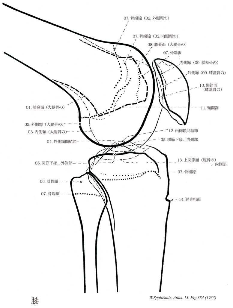
- 384_00【Knee膝;ヒザ Genu】
→(大腿と下腿の間の関節部。)
- 384_01【Popliteal surface of femur膝窩面;膝窩平面(大腿骨の) Facies poplitea femoris; Planum politeum】 Triangular area on the posterior aspect of the femur between the divergent lips of the linea aspera and the intercondylar line.
→(外側唇(外側顆上線)と内側唇(内側顆上線)の二またの間にはさまれた部分は長三角形の平坦な平面を示すので膝窩面と呼ばれる。)
- 384_02【Lateral condyle of femur外側顆;腓側顆(大腿骨の) Condylus lateralis; Condylus fibularis】 Rounded projection on the lateral aspect of the femur.
→(外側顆は幅が広くやや平坦で、その前端は少し前上方に突出する。)
- 384_03【Medial condyle of femur内側顆;脛側顆(大腿骨の) Condylus medialis; Condyus tibialis】 Rounded projection on the medial aspect of the femur forming part of the knee joint.
→(大腿骨体の下部(遠位部)はことに著しく厚く大きく、その下端は左右の肥厚した内側顆および外側顆となる。内側顆は狭く長く、凸面の張り出しが強い。)
- 384_04【Lateral intercondylar tubercle外側顆間結節;腓側顆間結節 Tuberculum intercondylare laterale; Tuberculum intercondylicum fibulare】 Protuberance from the lateral articular surface at the margin facing the intercondylar eminence.
→(窩間隆起はさらに内外の結節、すなわち内側顆間結節および外側顆間結節に分かれる。)
- 384_05【Infraglenoidal border関節下縁 Margo infraglenoidalis】
→()
- 384_06【Head of fibula腓骨頭;腓骨小頭 Caput fibulae; Capitulum fibulae】 Proximal end of the fibula.
→(腓骨の上端の膨らみは腓骨頭と呼ばる。腓骨頭の前面から長趾伸筋、長腓骨筋、後面からヒラメ筋の一部が起こり、外側面に大腿二頭筋が付着く。)
- 384_07【Epiphysial line; Epiphyseal line骨端線 Linea epiphysialis】 Line visible on radiographs and in sections of bone that marks the former site of the epiphysial plate.
→(X線像で骨端接合部が閉鎖した後に、1条の細い線が残って見えるが、これは骨端接合部瘢痕(骨端線)といわれる。)
- 384_08【Patellar surface of femur膝蓋面(大腿骨の) Facies patellaris femoris】 Surface that articulates with the patella.
→(内側顆と外側顆の下面を被う関節面の前端は互いにつながって膝蓋面をつくる。膝蓋面は中央に縦溝のあるややくぼんだ面で、膝蓋骨の後面に対向する。)
- 384_09【Patella膝蓋骨 Patella】 The kneecap, which is embedded in the tendon of the quadriceps femoris muscle.
→(膝関節の前面にあり、尖端を下方に向けた扁平な栗の実によく似た骨で、幅広い上端部が膝蓋骨底で、尖った下端部が膝蓋骨尖である。大腿四頭筋腱中に発生した種子骨とみなされ、上縁には大腿直筋と中間広筋の内側縁には内側広筋の、外側縁には外側広筋のそれぞれの腱が付着する。前面は凸面状で、大腿四頭筋腱による縦に走る小隆起を伴う粗面をなし、小血管孔がある。後面には、上方の広い卵形の平滑な面と、下方の小さい逆三角形の粗な面がある。平滑な面は大腿骨の膝蓋面に対する関節面をなし、中央部にある縦方向の隆起によって小さい内側部と大きい外側部に分けられる。下方の粗面の下端には膝蓋靱帯が付着するが、粗面の上方部には脂肪組織が入り、脛骨と膝蓋骨とを隔てる。ラテン語のPatera(皿・円板状の)の縮小形。)
- 384_10【Articular surface of patella関節面(膝蓋骨の) Facies articularis patellae】 Cartilage-covered articular surface facing the femur.
→(膝蓋骨の後面は大腿骨の膝蓋面に対向する滑らかな関節面である。)
- 384_11【Intercondylar fossa顆間窩 Fossa intercondylaris】 Notch on the posterior aspect of the femur between the medial and lateral condyles.
→(後方に向かって突き出た内側顆と外側顆の間に深い顆間窩がある(両顆の後方に突出した部の上方から腓腹筋の内・外側頭の内側に接して足底筋がおこる。)
- 384_12【Medial intercondylar tubercle内側顆間結節;脛側顆間結節 Tuberculum intercondylare mediale; Tuberculum intercondylicum tibiale】 Protuberance from the medial articular surface at the margin facing the intercondylar eminence.
→(窩間隆起はさらに内外の結節、すなわち内側顆間結節および外側顆間結節に分かれる。)
- 384_13【Superior articular surface of tibia上関節面;近位関節面(脛骨の) Facies articularis superior tibiae; Facies articulares proximales】 Articular surfaces of the tibia that form part of the knee joint.
→(内側顆および外側顆のいずれの上面にも卵円形でわずかにくぼんでいる滑らかな上関節面があり、大腿骨の内側顆および外側顆に対向している。)
- 384_14【Tibial tuberosity脛骨粗面 Tuberositas tibiae】 Roughened area near the upper part of the anterior border of the tibia giving attachment to the patellar ligament.
→(脛骨体の前縁の上端はS状に曲がる縁で脛骨粗面(上半部に膝蓋靱帯がつく)となって結節状に隆起している。脛骨粗面の上半のやや平滑なところは膝蓋靱帯の着く所である。)
