Spalteholz HANDATLAS DER ANATOMIE DES MENSCHEN VON WERNER SPALTEHOLZ
メニューは解剖学(TA)にリンクしてあります。図の番号をクリックすると下記の説明へ、右側の用語をクリックすると解剖学(TA)にジャンプします。
413 


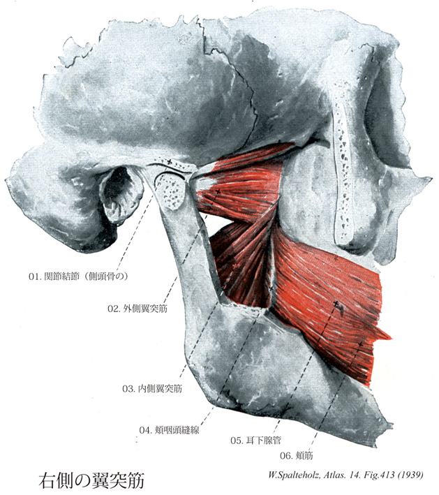
- 413_01【Articular tubercle of temporal bone関節結節(側頭骨の) Tuberculum articulare ossis temporalis】 Rounded projection anterior to the mandibular fossa.
→(顎関節の直前で頬骨突起基部の下面に高まる関節結節は下顎窩の前方の境となるが、生体では関節結節と関節面が一続きの軟骨で被われて関節包の内にあり、顎関節の関節窩となる。)
- 413_02【Lateral pterygoid muscle外側翼突筋 Musculus pterygoideus lateralis】 o: Lateral surface of lateral plate of pterygoid process and inferior surface of greater wing of sphenoid, i: Two-headed (variant: three-headed) at disco-capsular system of temporomandibular joint and pterygoid fovea. I: Mandibular nerve.
→(外側翼突筋は2頭からなる。上頭は蝶形骨大翼の下面から起こる。下頭は蝶形骨翼状突起外側板に起始する。下頭は側頭下窩を通過して、下顎骨関節突起(翼突筋窩に)停止し、上頭もまた関節円板および関節包に付着する。三叉神経の下顎神経の外側翼突筋神経より支配を受ける。作用として下顎骨を引く。片側が働けば下顎骨前部は対側に働く。)
- 413_03【Medial pterygoid muscle内側翼突筋 Musculus pterygoideus medialis】 o: Pterygoid fossa and the maxillary tuberosity. i: Pterygoid tuberosity on inner side of the angle of the mandible, passing obliquely downward and backward. Synergist of the temporal and masseter muscles. I: Mandibular nerve.
→(内側翼突筋は蝶形骨の翼突窩で起始して、下顎角内面に停止する。したがって、この筋は、下顎骨の外面側を走る咬筋浅部と同様な走行方向で下顎骨の内側面を走る。両筋は作用方向は同一であり、したがって協力筋である。)
- 413_04【Pterygomandibular raphe翼突下顎縫線;頬咽頭縫線 Raphe pterygomandibularis; Raphe buccipharyngica】 Tendinous line between the pterygoid hamulus and the retromolar fossa of the mandible. It divides the buccinator from the constrictor muscles of pharynx.
→(翼突下顎縫線は翼突鈎と下顎の間の腱。頬筋と咽頭収縮筋とを分けている。)
- 413_05Stensen's (Stenon) duct【Parotid duct耳下腺管 Ductus parotideus】 Excretory duct that extends around the anterior border of the masseter, usually over the buccal fat pad, and opens opposite to the upper second molar tooth.
→(耳下腺管はステンセン管ともよばれる。または、ステノン管ともよばれ、日本ではステノ氏孔などともいう。耳下腺管は頬骨弓の下方約2cmの部を水平に走り、頬筋を貫いて上顎第2大臼歯対側の口腔粘膜に開口する。デンマークの解剖学者Niels Steno [Nicholas Stensen] (1638-1686)によって、1661年頃に発見された。後年、ステンセンはローマカトリックの司教となっている。)
- 413_06【Buccinator muscle頬筋 Musculus buccinator】 Muscle arising from the pterygomandibular raphe and adjacent areas of the maxilla and mandible to the height of the first molar teeth, and inserting into the orbicularis oris at the angle of the mouth. It forms the cheek, moves food from the oral vestibule between the dental arcades during mastication, prevents entrapment of the mucous membrane of the mouth, and is active during laughing and crying. I: Facial nerve.
→(頬筋は頬の筋性土台に該当し、口角部で口輪筋に付着する。頬筋は弓状に上顎骨歯槽突起の臼歯部、かつ下顎骨歯槽突起から起こる。上および下顎間は腱性の翼突下顎放線によって橋渡しされ、この放線もまた頬筋の起始である。上咽頭収縮筋の一部がこの放線の後部で起始する。口角付近で、線維索が交叉するので、頬の上方に位置する部分は下唇に広範囲わたって達することもあるし、達しないこともある頬筋は上顎の第2大臼歯のレベルで耳下腺管によって貫通され、しかも本筋は脂肪体からこれを隔てる浅筋膜(頬咽頭筋膜)を有する唯一の顔面筋である。頬筋は上・下歯列弓および頬粘膜間に入り込んだ植物片を再度歯列弓間に押し戻し、咀嚼および植物片のかたちづくりに重要な役割を果たしている。本筋は口腔前庭を圧縮して、空気あるいは液体を口裂を通してふき出す(泡をふき出す、口笛をふく、吐き出す:“トランペット吹きの筋”)。両側の頬筋の収縮はは口角の外側部をくぼませる。参考:この筋は頬粘膜に密に結合しているが、皮膚との間は脂肪組織で隔てられている。上顎第2大臼歯の高さで耳下腺管に貫かれる。)