Spalteholz HANDATLAS DER ANATOMIE DES MENSCHEN VON WERNER SPALTEHOLZ
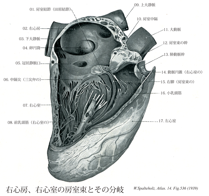
メニューは解剖学(TA)にリンクしてあります。図の番号をクリックすると下記の説明へ、右側の用語をクリックすると解剖学(TA)にジャンプします。
536 


Aschoff-Tawara node; Tawara, Node of
- 536_01Aschoff-Tawara node; Tawara, Node of【Atrioventricular node; Tahara node房室結節(田原結節) Nodus atrioventricularis】 Specialized muscle tissue in the interatrial septum below the fossa ovalis and in front of the opening of coronary sinus. It transmits the myogenically conducted impulse received from the sinu-atrial node after a period of latency to the ventricles via the bundle of His (and right and left bundles of His). If sinu-atrial node function is disrupted, it ca. act as a secondary center for generation of cardiac impulses.
→(田原の結節ともよばれてる。房室結節は心房中隔で、卵円窩の下方、冠状静脈洞口の前方にある特殊心筋組織の小塊。洞房結節から筋性に伝搬された刺激を、一定の潜伏時間をおいてHis束およびその脚を通して心室に伝える。洞房結節が機能的に脱落した際には、房室結節は二次的な刺激形成中枢として心拍動の維持にあたることができる。九州大学の田原順(1873-1938)が1901年、東京帝国大学を卒業、1903年ドイツに留学。アールブルグ大学でアショッフの元で研究中、心筋の房室間の刺激伝導路系を発見。その起始部の小筋塊を田原結節と命名("Das Reizleitungssystem des Saugetierherzens", 1906)。帰国後、九州帝国大学教授となる。世界に通じる数少ない日本人の名前の一つである。)
- 536_02【Right atrium右心房 Atrium cordis dextrum; Atrium dextrum】
→(右心房は心臓の右上部を占め、その後上部と後下部とに、それぞれ、上大静脈と下大静脈が注いでいる。)
- 536_03【Inferior vena cava下大静脈 Vena cava inferior; Vena cava caudalis】 It arises at the union of the right and left common iliac veins, lies on the right side of the aorta, and opens into the right atrium of the heart.
→(下大静脈は下肢および骨盤と腹部の器官の大部分から血液を受ける本幹で、第5腰椎体の右側で左右の総腸骨静脈の合流として始まり、このあと脊柱に沿って大動脈の右側を上行、肝臓の後面をこれに接して通過し、第八胸椎の高さで横隔膜の大静脈孔を貫いて胸腔に入り、ただちに右心房にそそぐ。下大静脈に流入する枝には総腸骨静脈、下横隔静脈、第3・第4腰静脈、肝静脈、腎静脈、右副腎静脈、右精巣静脈、右卵巣静脈、蔓状静脈叢などがある)
- 536_04【Fossa ovalis; Oval fossa卵円窩 Fossa ovalis】 Depression in the interatrial septum, which represents the closed foramen ovale (open during fetal development).
→(卵円窩は心房中隔で、胎生時の卵円孔の跡の凹み。)
- 536_05【Opening of coronary sinus冠状静脈口;冠状静脈洞口 Ostium sinus coronarii】
→(心臓を栄養した後の血液をもどす冠状静脈洞の開口。下大静脈口の内側に位置する。(イラスト解剖学))
- 536_06【Septal cusp of tricuspid valve中隔尖(三尖弁の) Cuspis septalis (Valva tricuspidalis)】 Cusp or leaflet arising from the interventricular septum.
→(三尖弁の中隔尖は三尖弁の内部にあり、中隔からおこる弁尖端。)
- 536_07【Right ventricle右心室 Ventriculus dexter】
→(右心室は心臓の最下位部を占め、後上方にある右房室口で右心房と交通し、前上方にある肺動脈口で肺静脈に連なる。)
- 536_08【Anterior papillary muscle of right ventricle前乳頭筋(右心室の) Musculus papillaris anterior (Ventriculus dexter)】 Largest papillary muscle, located anteriorly and often overlying the septomarginal trabecula. It is connected with the anterior and posterior cusps.
→(右心室の前乳頭筋は前方にあり、大きい。)
- 536_09【Superior vena cava上大静脈 Vena cava superior; Vena cava cranialis】
→(上大静脈は上半身の血液を集める静脈で、上縦隔の中で左右の腕頭静脈が合してはじまり、途中で奇静脈を受け入れながら上行大動脈の右側を下行して右心房にそそぐ。)
- 536_10【Atrioventricular septum房室中隔 Septum atrioventriculare】 Portion of the membranous part of interventricular septum between the right atrium and left ventricle above the root of the septal cusp.
→(房室中隔は左心房と左心室の間の膜性部のなかで、房室弁起始より上方にある部分。)
- 536_11【Aorta大動脈 Aorta】 Main artery supplying the body.
→(大動脈は体循環系の本幹をなす単一の太い動脈。上行大動脈・大動脈弓・胸大動脈および腹大動脈に区分される。後2者を総称して下行大動脈ともよぶ。上行大動脈は左心室の大動脈口よりはじまり心膜腔を出るまで約5~6cmの範囲をいう。肺動脈幹とともに心膜に包まれている。上行大動脈の起始部は3個の大動脈洞によるふくらみを呈し大動脈球とよばれ、左・右冠状動脈がここからでる。大動脈弓は上行大動脈につづく弯曲部であり(約5~6cm長)、肺動脈分岐部および左気管支をこえて左後方にまわり、第四胸椎体の左側で胸大動脈に移行する。大動脈弓の重要な枝として腕頭動脈・左総頚動脈・左鎖骨下動脈が出る。大動脈弓の凹弯する下面と肺動脈分岐部とのあいだを動脈間索が結ぶ。胸大動脈にまわり、第12胸椎の直前で横隔膜の大動脈裂肛の壁側枝として肋間動脈10対、また臓側枝として気管支動脈・食道動脈などを出す。腹大動脈は第12胸椎より第4腰椎までの前を下行したのち左右総頚動脈を出し、本幹のつづきは細い正中線骨動脈となって尾骨尖端に達している。壁側枝には各有対の下横隔動脈・腰動脈・総腸骨動脈があり、臓側枝には無対性の腹腔動脈・上腸間膜動脈・下腸間膜動脈また有対の副腎動脈・腎動脈・精巣(ないし卵巣)動脈がある。)
- 536_12【Trunk (Bundle of His)房室束の幹;His束の幹 Truncus (Fasciculus atrioventricularis)】
→()
- 536_13【Pulmonary trunk; Pulmonary artery肺動脈幹;肺動脈 Truncus pulmonalis; Arteria pulmonarlis】 Arterial trunk that ascends in the pericardium. It divides into the right and left pulmonary arteries at the level of the reflection of the serous pericardium.
→(肺動脈幹は右心房と左右肺動脈の起始までの間で分岐前の肺動脈を明確にするために導入された。肺動脈幹は左の第3肋骨の胸骨付近の高さで右心部動脈円錐から起こる。長さは約5cmで心膜腔を通り、やや左側に向かい、頭背側で肺動脈幹のT字形の分岐となり、2本の肺動脈に分岐する。肺動脈幹の両側を2本の冠状動脈が走る。肺動脈幹の起始部は大動脈口の前左側に、中間部は上行大動脈の左側に、分岐部は大動脈弓の凹部に位置する。)
- 536_14【Conus arteriosus; Infundibulum of right ventricle動脈円錐;漏斗(右心室の) Conus arteriosus; Infundibulum】 Funnelshaped smooth-walled outflow tract leading to the pulmonary trunk.
→(右心室の動脈円錐は肺動脈管へ開く前の、漏斗状で平滑な壁からなる流出路。発生学的には心球にあたる。)
- 536_15【Right bundle of atrioventricular bundle; Right branch of atrioventricular bundle右脚;右束枝(房室束の) Crus dextrum fasciculi atrioventricularis】 Bundle that runs in an arch into the septomarginal trabecula and continues to the anterior papillary muscle.
→(房室束の右脚は室中隔の左右を乳頭筋まではしる刺激伝導系の右脚で、そこからさらに分枝する。)
- 536_16【Little papillary muscles小乳頭筋 Musculi papillares parvi】
→(")
- 536_17【Left ventricle左心室 Ventriculus sinister】
→(左心室は心臓の左下部を占め、後上方にある左房室口で左心房と交通し、右上隅にある大動脈口によって大動脈につらなる。左心室の壁は右心室に比べ2~3倍厚い。心室中隔は、右心室に向かって膨隆しているので、心室を横断面でみると、左心室の内腔は円いのに対して、右心室の内腔は半月状である)
