Spalteholz HANDATLAS DER ANATOMIE DES MENSCHEN VON WERNER SPALTEHOLZ
メニューは解剖学(TA)にリンクしてあります。図の番号をクリックすると下記の説明へ、右側の用語をクリックすると解剖学(TA)にジャンプします。
556 


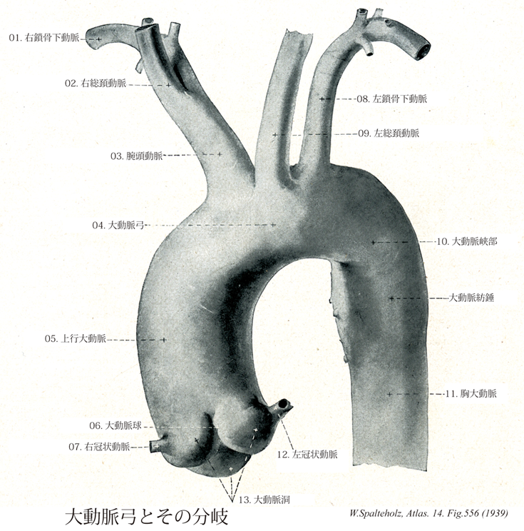
- 556_01【Right subclavian artery右鎖骨下動脈 Arteria subclavia dextra】
→()
- 556_01a【Subclavian artery鎖骨下動脈 Arteria subclavia】 Artery that passes with the roots of brachial plexus between the anterior and middle scalene muscles through the scalene space, over the first rib in the groove for the subclavian artery. From the lateral border of the first rib, it continues as the axillary artery.
→(鎖骨下動脈は上肢の主幹動脈の根部をなし、右側は腕頭動脈から、左側は大動脈弓からそれぞれ分かれてはじまり、前斜角筋の後方を通って第1肋骨外側縁で腋窩動脈につづく。胸・頚・上肢移行部の動脈として、多彩な分枝と変異に富むことを特徴とする。分枝はつぎの通りである。椎骨動脈、内胸動脈、甲状頚動脈、肋頚動脈、下行肩甲動脈に分枝し、第一肋骨を越えたところで腋窩動脈となる。)
- 556_02【Right common carotid artery右総頚動脈 Arteria carotis communis dextra】
→(総頚動脈は頭部に血液を送る血管の主幹。右は腕頭動脈の枝、左は大動脈弓の上行部より出る。そのため左総頚動脈は右のものよりも4~5cm長い。総頚動脈は枝を出さず、気管・喉頭の両側を上行し、甲状軟骨上縁の高さで音叉のような形をなし内・外頚動脈に分かれる。分岐部の後側には頚動脈小体が存在する。また分岐部のないし内頚動脈始部の壁はやや薄く膨隆しており(頚動脈洞)、舌咽神経の枝を介し血圧を感受するという。)
- 556_02a【Common carotid artery総頚動脈 Arteria carotis communis】 Artery of the neck without any branches. It runs on both sides of the trachea and larynx and passes deep to the sternocleidomastoid. It arises on the right from the brachiocephalic trunk and on the left from the aortic arch.
→(総頚動脈は頭部に血液を送る血管の主幹。右は腕頭動脈の枝、左は大動脈弓の上行部より出る。そのため左総頚動脈は右のものよりも4~5cm長い。総頚動脈は枝を出さず、気管・喉頭の両側を上行し、甲状軟骨上縁の高さで音叉のような形をなし内・外頚動脈に分かれる。分岐部の後側には頚動脈小体が存在する。また分岐部のないし内頚動脈始部の壁はやや薄く膨隆しており(頚動脈洞)、舌咽神経の枝を介し血圧を感受するという。)
- 556_03【Brachiocephalic trunk腕頭動脈 Truncus brachiocephalicus】 It arises at the beginning of the aortic arch and divides behind the right sternoclavicular joint into the right subclavian artery and right common carotid artery.
→(大動脈弓から最初にでる動脈で、右胸鎖関節の後ろで鎖骨下動脈と右総頚動脈に分れる。しばしば最下甲状腺動脈を出す。)
- 556_04【Arch of aorta; Aortic arch大動脈弓 Arcus aortae】 It is located between the ascending and descending aorta. Its roof extends to the first rib at the left border of the sternum.
→(大動脈弓は上行大動脈につづく弯曲部であり(約5~6cm長)、肺動脈分岐部および左気管支をこえて左後方にまわり、第四胸椎体の左側で胸大動脈に移行する。)
- 556_05【Ascending aorta上行大動脈;大動脈上行部 Pars ascendens aortae; Aorta ascendens】 Ascending part of the aorta up to its exit from the pericardium.
→(左心室からおこり、肺動脈幹の後ろを上行して大動脈弓にいたる5cmほどの部。基部の内腔は膨らんで大動脈洞(バルサルバ洞)をなし、ここから左右の冠状動脈が出る。(イラスト解剖学))
- 556_06【Aortic bulb大動脈球 Bulbus aortae】 Onion-shaped dilatation visible on the outer surface of the aorta. It is produced by the aortic sinus.
→(球根様に大動脈基部が膨大した部分で、大動脈洞にる。 (Feneis))
- 556_07【Right coronary artery右冠状動脈 Arteria coronaria dextra; Arteria coronaria cordis dextra】 It travels in the right coronary sulcus and arises near the right aortic sinus.
→(右冠状動脈は大動脈の起始部(大動脈洞)の前面から起こり、肺動脈と右心耳との間を走って冠状溝に達し、この溝を右まわりに走って心臓の後面に進む。冠状溝を走る間に右辺縁枝を下方に出すが、その主脈は後室間溝を後面で下行する太い動脈(後室間枝)となって心尖に向かって下降する。)
- 556_08【Left subclavian artery左鎖骨下動脈 Arteria subclavia sinistra】
→(鎖骨下動脈は上肢の主幹動脈の根部をなし、右側は腕頭動脈から、左側は大動脈弓からそれぞれ分かれてはじまり、前斜角筋の後方を通って第1肋骨外側縁で腋窩動脈につづく。胸・頚・上肢移行部の動脈として、多彩な分枝と変異に富むことを特徴とする。分枝はつぎの通りである。椎骨動脈、内胸動脈、甲状頚動脈、肋頚動脈、下行肩甲動脈に分枝し、第一肋骨を越えたところで腋窩動脈となる。)
- 556_09【Left common carotid artery左総頚動脈 Arteria carotis communis sinistra】
→(総頚動脈は頭部に血液を送る血管の主幹。右は腕頭動脈の枝、左は大動脈弓の上行部より出る。そのため左総頚動脈は右のものよりも4~5cm長い。総頚動脈は枝を出さず、気管・喉頭の両側を上行し、甲状軟骨上縁の高さで音叉のような形をなし内・外頚動脈に分かれる。分岐部の後側には頚動脈小体が存在する。また分岐部のないし内頚動脈始部の壁はやや薄く膨隆しており(頚動脈洞)、舌咽神経の枝を介し血圧を感受するという。)
- 556_10【Aortic isthmus大動脈峡部 Isthmus aortae】 Constriction of variable size behind the ligamentum arteriosum. In the fetus it is a narrowed portion of the aortic arch between the exit of the left subclavian artery and the ductus arteriosus. It can remain as an aortic isthmus stenosis.
→(大動脈峡部は左鎖骨下動脈と動脈管索の間で大動脈が狭くなった部。胎生期の大動脈弓は、左鎖骨下動脈起始の遠位で動脈間合流の近位にあたる部分が若干くびれている。)
- 556_11【Thoracic aorta胸大動脈;大動脈胸部 Pars thoracica aortae; Aorta thoracica】 Part of the aorta descending to the aortic hiatus of the diaphragm at the level of the twelfth thoracic vertebra.
→(胸大動脈は、大動脈弓の延長である。第4胸椎体の下縁の左側で始まり、第5から第12胸椎の左側で後縦隔を下行する。下行しながら、正中面に近付き、食道と脊柱の左側に沿って走るが、食道の後方、脊柱の前を走るようになる。胸大動脈は横隔膜を貫いた直後に腹大動脈という名前に変わる。胸大動脈と腹大動脈とを総称して下行大動脈という。)
- 556_12【Left coronary artery左冠状動脈 Arteria coronaria sinistra; Arteria coronaria cordis sinistra】 It arises near the left aortic sinus.
→(左冠状動脈は右心耳と肺動脈との間から前面に現れて、前室間溝を大心臓静脈の主脈と共に下降する(前室間枝)。これから分かれるかなり大きい枝が冠状溝に沿って後面へ回り(回旋枝)、右冠状動脈の枝と吻合する。この回旋枝からは、左辺縁枝が下方に出る。)
- 556_13Valsalva, Sinus of【Aortic sinus; Aortic lumen大動脈洞 Sinus aortae】 Calotte-like dilatations of the aortic lumen at the level of the three aortic valves.
→(バルサルバ洞とも呼ばれる。上行大動脈は基部に左・右・後3つの大動脈弓をもち、この内腔を大動脈洞という。左右の大動脈洞から冠状動脈がでる。イタリアの解剖学者Antonio Maria Valsalva (1666-1723)の名を冠する。)