Spalteholz HANDATLAS DER ANATOMIE DES MENSCHEN VON WERNER SPALTEHOLZ
メニューは解剖学(TA)にリンクしてあります。図の番号をクリックすると下記の説明へ、右側の用語をクリックすると解剖学(TA)にジャンプします。
578 


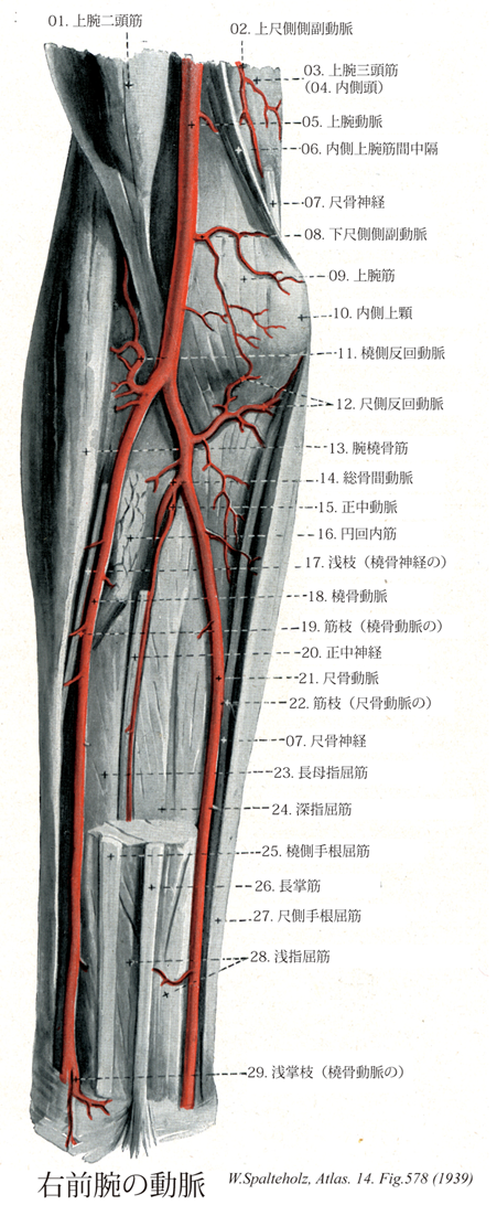
- 578_01【Biceps brachii muscle上腕二頭筋 Musculus biceps brachii】 Two-headed muscle that attaches on the radial tuberosity and extends with the aponeurosis brachii toward the ulna to blend into the antebrachial fascia. It acts in elbow joint flexion and forearm supination. I: Musculocutaneous nerve.
→(上腕二頭筋は、長頭が関節上結節に起始し、短頭は烏口突起に起始する。二頭筋の長頭(長いのは腱の部分のみ)は上腕骨頭を越え、結節間滑膜鞘に包まれて、結節間溝へ入る。共通の筋腹の終止腱は、肘窩の奥で、橈側粗面に停止する。腱性の帯である上腕二頭筋腱膜は終止腱から分かれ、前腕筋膜に放散している。肘関節を屈曲すると、上腕二頭筋は特に突出する。なぜならば、この筋は関節から離れて、上腕筋によって前に押し出されるからである。機能として肘関節に作用して前腕をまげる。上腕前面に力こぶをつくる。筋腹の内外両側の溝をそれぞれ内側二頭筋溝および外側二頭筋溝という。前者を尺側皮静脈、後者を橈側皮静脈が走る。長頭の件は滑膜に包まれながら肩関節腔を貫く。また上腕骨の結節間溝を通るところでは、結節間滑液鞘に包まれる。)
- 578_02【Superior ulnar collateral artery上尺側側副動脈;近位尺側側副動脈 Arteria collateralis ulnaris superior; Arteria collateralis ulnaris proximalis】 Artery that often arises near the deep artery of arm. It passes with the ulnar nerve to the cubital anastomosis.
→(上尺側側副動脈は上腕のほぼ中央の高さで本幹より分岐する。尺骨神経に伴行して、内側上腕筋間中隔の後面に沿って、上腕三角筋内側頭の表層を下行する細長い動脈で、上腕骨内側上顆と肘頭の間に達し、ここから肘関節動脈網へ入る。)
- 578_03【Triceps brachii muscle上腕三頭筋 Musculus triceps brachii】 Three-headed arm muscle with a common attachment on the olecranon and the posterior wall of the joint capsule. Extends the elbow. I: Radial nerve.
→(肘を伸ばす筋。3つの起始のうち、軽く伸展する時は内側頭が働き、強く伸展する時には長頭や外側頭も協同する。長頭は肩甲骨の関節下結節、外側頭は上腕骨上部の後面、内側頭は上腕骨体の後面からおこり、合したのち尺骨の肘頭につく。なお、この筋は肩関節の内転にも働く。神経支配:橈骨神経(C5,C7,C8).動脈:上腕深動脈。(イラスト解剖学))
- 578_04【Medial head of triceps brachii muscle; Deep head of triceps brachii muscle内側頭;深頭;尺側頭;上腕骨粗線(上腕三頭筋の) Caput mediale; Caput profundum; Caput ulnare; Linea aspera humeri (Musculus triceps brachii)】 o: Posterior surface of humerus, medial and distal to the groove for the radial nerve.
→(内側頭は上腕骨の後面、橈骨神経溝の内側遠位および両側間中隔(とりわけ、内側)から起こる。)
- 578_05【Brachial artery上腕動脈 Arteria brachialis】 Continuation of the axillary artery that passes from the inferior border of the pectoralis major in the medial bicipital groove to its division into the radial and ulnar arteries.
→(上腕動脈は大円筋の停止腱の下縁の高さで腋窩動脈よりつづいてはじまり、上腕前面の内側部で上腕二頭筋の内側(内側二頭筋溝)に沿って、正中神経および上腕静脈とともに下行し、肘関節の前面のやや遠位で橈骨動脈と尺骨動脈に分かれる。)
- 578_06【Medial intermuscular septum of arm; Medial brachial intermuscular septum内側上腕筋間中隔;尺側上腕筋間中隔 Septum intermusculare brachii mediale; Septum intermusculare brachii ulnaris】 Tendinous sheet that attaches the brachial fascia to the medial border of the humerus and gives origin to muscle fibers.
→(内側上腕筋間中隔は上腕骨内側縁と上腕筋膜の間にある腱性の筋起始板。)
- 578_07【Ulnar nerve尺骨神経 Nervus ulnaris】 Nerve arising from the medial cord that initially travels in the medial bicipital groove, pierces the medial intermuscular septum of the arm, and, after traversing the groove for the ulnar nerve, penetrates the flexor carpi ulnaris.
→(腕神経叢の枝であり、上腕の内側後部を下り肘頭の内(尺)側に達してから前面に近づき、尺側手根筋と深指屈筋(尺骨半)への筋枝を出したのち前腕を下りながら途中で手背尺側半の皮膚に分布する背側指神経および手掌尺側半の皮膚に分布する一つの皮枝を出す。手掌部に達した尺骨神経の本幹は短掌筋、小指外転筋、短小指屈筋、小指対立筋、尺側の虫様筋、短母指屈筋の深頭、母指内転筋、および骨間筋への筋枝を出すほか、総掌側指神経とその末梢側のつづきである固有掌側指神経になり小指および薬指の表面をおおう皮膚に分布する。)
- 578_08【Inferior ulnar collateral artery下尺側側副動脈;遠位尺側側副動脈 Arteria collateralis ulnaris inferior; Arteria collateralis ulnaris distalis】 Artery that often arises above over the medial epicondyle of humerus. It passes on the brachialis through the medial intermuscular septum and to the cubital anastomosis.
→(下尺側側副動脈は肘関節の少し上方で本幹より分岐し、上腕筋の表層を内側を走り、一部の枝は上腕骨内側上顆の前面で尺側反回動脈の前枝と吻合し、他は内側上腕筋間中隔を貫いて内側上顆の後面に出て肘関節動脈へ入る。)
- 578_09Casserio's muscle【Brachialis muscle上腕筋 Musculus brachialis】 o: Anterior surface of the humerus below the deltoid tuberosity. i: Tuberosity of ulna. Flexes the elbow joint. I: Musculocutaneous nerve.
→(上腕筋は上腕骨前面の三角筋粗面(均一な三角筋-上腕筋系の骨付着部と考えらえている)より遠位部で起こり、尺骨粗面に停止する。[臨床]上腕筋は、上腕骨上に直に接しているため、筋を上腕骨に圧迫するような外力が加わるとか、上腕骨の(顆上)過伸展骨折(伸展骨折)の際に、骨折端によって穿通され、容易に損傷される。損傷した筋組織の部位に生じる結合組織の瘢痕は、収縮し、上腕筋の短縮が起こることがある。このような場合、腕は肘関節を伸展することが不可能になる。)
- 578_10【Medial epicondyle of humerus; Medial humeral epicondyle内側上顆;尺側上顆(上腕骨の) Epicondylus medialis humeri; Epicondylus ulnaris humeri】 Protuberance that gives origin to the flexor muscles of the forearm.
→(上腕骨の下端部では、まず内側方に内側上顆が、外側方に外側上顆が突出している。内側上顆は指先でつまめるほどのおおきな突出であるが、外側上顆の突出は軽度である。)
- 578_11【Radial recurrent artery橈側反回動脈 Arteria recurrens radialis】 Recurrent artery that extends from the cubital fossa, passing medial to the radial nerve, to the radial collateral artery and cubital anastomosis.
→(橈側反回動脈は肘関節のすぐ遠位で分岐し、橈骨神経に沿って腕橈骨筋と上腕筋の間を上行枝、これらに筋枝を与えたのち、上腕骨外側上顆の前面で橈側側副動脈と吻合する。)
- 578_12【Ulnar recurrent artery尺側反回動脈 Arteria recurrens ulnaris】 Branch of the ulnar artery from the cubital fossa where it supplies the muscle, bone, and joint capsule.
→(尺側反回動脈は肘関節のすぐ遠位で本幹より分岐し、直ちに前枝・後枝に二分するが、これらの枝は別々に本幹より分岐することも多い。)
- 578_13【Brachioradialis muscle腕橈骨筋 Musculus brachioradialis】 o: Lateral supracondylar ridge of humerus, lateral intermuscular septum, i: Radial styloid process. Flexes the forearm from the intermediate position between pronation and supination. I: Radial nerve.
→(腕橈骨筋は橈骨の外側縁に位置し、上腕骨の外側縁、外側上顆の上と外側上腕筋間中隔から起始する。腕橈骨筋は橈骨の茎状突起基底部に停止する。)
- 578_14【Common interosseous artery総骨間動脈 Arteria interossea communis】 Trunk formed by the following vessels given off by the ulnar artery or branch arising from the division of the brachial artery (embryological) at the superior border of the pronator teres.
→(総骨間動脈は尺骨動脈の起始近く、橈骨結節の遠位で本幹より分岐し、背側に向かい約1cm走ったのち、前腕骨間膜の付近で2枝に分かれる。)
- 578_15【Median artery; Artery comitans of median nerve正中動脈;正中神経伴行動脈 Arteria comitans nervi mediani; Arteria mediana】 It accompanies the median nerve, which supplies the forearm muscles.
→(正中神経伴行動脈(正中動脈)は前骨間動脈の起始近くで分岐する枝で細いが、発生学的には前骨間動脈からつづく前腕の動脈の本幹であるという。比較解剖学的には食肉類や有蹄類では、正中動脈が前腕の動脈の主体をなし、尺骨動脈と橈骨動脈はたんなる側枝にすぎないが、霊長類ではじめて尺骨動脈と橈骨動脈が主幹となる。正中神経に伴行して浅指屈筋と深指屈筋の間を下行枝、この神経と周囲の筋へ分岐して前腕の領域でおわるが、ときに強大化して手掌の浅掌動脈弓の形成に加わることがある(約8%)。)
- 578_16【Pronator teres muscle円回内筋 Musculus pronator teres】 Muscle that attaches on the pronator tuberosity of the radius. It flexes the elbow joint, acting as a pronator. I: Median nerve.
→(円回内筋の上腕頭は、前腕の浅層の屈筋群とともに上腕骨の内側上顆と内側上腕筋間中隔から起こる。発達の弱い尺骨頭は鈎状突起と尺骨粗面の間の尺骨内側面から起こる。円回内筋は尺骨と橈骨の上を斜走し、橈骨の前縁を回って、回外筋の停止部の下方の橈骨の前縁を回って、回外筋の停止部の下方の橈骨外側面につく。円回内筋は腕橈骨筋とともに肘窩の遠位側の境界となる。正中神経は円回内筋の上腕頭と尺骨頭の間を通る。)
- 578_17【Superficial branch of radial nerve浅枝(橈骨神経の) Ramus superficialis nervus radialis】 Superficial cutaneous branch that runs along the brachioradialis with the radial artery, and then crosses under its accompanying muscle to reach the dorsum of hand and the fingers.
→(橈骨神経の浅枝は深枝とともに橈骨神経の最終枝で、腕橈骨筋に沿って橈骨動脈とともに走り、母指・示指・中指・第四指外側半の背面近位部の皮膚、および手背近位部の皮膚に分布する。)
- 578_18【Radial artery橈骨動脈 Arteria radialis】 Continuation of the brachial artery from its bifurcation, or first branch (embryological). It runs on the radial side between the brachioradialis and flexor carpi radialis over the pronator teres to the wrist (pulse palpation site). From there it ascends behind the trapezoid to the lateral aspect of the dorsum of hand where it courses to palmar. It extends between the two heads of the first dorsal interossei to reach to the deep palmar arch.
→(橈骨頚の高さで上腕動脈より分岐する動脈。前腕の橈側(外側)に沿って手根部まで下行し、次いで手根の外側(母指側)をまわって背側に達し、母指と第2指の中手骨間隙にある第1背側骨間筋の両頭の間を通って手掌に出る。前腕近位部では腕橈骨筋におおわれて走るが、遠位部では表在性になり、皮膚と前腕筋膜のおおわれるのみとなる。ことに手根部の近くではきわめて浅く、またその深層は橈骨下端に接するため、この部で脈拍を触れるのが容易である。この部は橈側手根筋のすぐ外側にあたる。手根から手背にいたる経路は、長母指外転筋、短母指伸筋、そして長母指伸筋のしたで、いわゆるtabatiereを通る。)
- 578_19【Muscular branch of radial artery筋枝(橈骨動脈の) Ramus muscularis (Arteria radialis)】
→()
- 578_20【Median nerve正中神経 Nervus medianus】 Nerve formed by the medial and lateral cords.
→(内側および外側神経束よりなる(C6-T1)。(Feneis))
- 578_21【Ulnar artery尺骨動脈 Arteria ulnaris】 Branch arising from the bifurcation of the brachial artery. It runs beneath the pronator teres toward the ulna, then accompanies the flexor carpi ulnaris on the flexor digitorum profundus to the wrist; from there it runs radially from the pisiform with the ulnar nerve to the palm, where it forms the superficial palmar arch.
→(尺骨動脈は上腕動脈の2終枝の一つ。肘関節の少し遠位で分岐し、このあたりでは橈骨動脈より太い。前腕の近位1/2における走行は深在性で、円回内筋・橈側手根屈筋・長掌筋・浅指屈筋の深層を斜め尺側に向かって下行し、前腕のほぼ中央でその尺側縁の浅層に出る。これからあと、尺骨神経と尺骨静脈(2条)に伴行して深指屈筋の表層で、これと尺側手根屈筋および浅指屈筋との間を手根にむけて下行する。次いで豆状骨の橈側を通って手掌に入り、屈筋支帯の表層で浅掌動脈弓をつくっておわる。)
- 578_22【Muscular branches of ulnar artery筋枝(尺骨動脈の) Ramus muscularis (Arteria ulnaris)】
→()
- 578_23【Flexor pollicis longus muscle長母指屈筋(手の) Musculus flexor pollicis longus】 o: Anterior surface of the radius, distal to the radial tuberosity. i: Base of distal phalanx of thumb. Flexes hand and phalanges of thumb. Radial abduction. I: Median nerve.
→(長母指屈筋は系統発生学的には独立した深指屈筋の一部である。その起始部は橈骨粗面から方形回内筋の上縁までの橈骨の前面、および骨間膜にまで広がっている。その腱は手根間を通り、短母指屈筋の2頭の間に入り込み、母指の末節骨底に付く。)
- 578_24【Flexor digitorum profundus muscle深指屈筋 Musculus flexor digitorum profundus】 o: Upper twothirds of anterior surface of ulna, i: Bases of the distal phalanges of the second through fifth fingers. Flexes wrist and interphalangeal joints. I: Median and ulnar nerves.
→(深指屈筋は尺骨(近位2/3)の前面と内側面、骨間膜、および前腕筋膜の広い範囲から起きる。その大きな筋腹は尺骨の前内側面を包み、浅指屈筋の滑走面となっている。4つの腱は手根管で互いに並び、浅指屈筋の腱を貫通し、第2~第5指の末節骨底に達する。)
- 578_25【Flexor carpi radialis muscle橈側手根屈筋 Musculus flexor carpi radialis】 o: Medial epicondyle of humerus, antebrachial fascia, i: Base of second metacarpal. Flexes and pronates elbow joint. Flexion and radial abduction of wrist joint. I: Median nerve.
→(橈側手根屈筋は上腕骨の内側上顆で浅層の屈筋群の共通起始部と、また同様に前腕屈筋および隣接する筋とを分けている結合組織中隔から起こる。筋は第2(しばしば第3)中手骨底に付く。羽状筋である橈側手根屈筋の長い腱は斜め下方に走り、橈骨の遠位1/3で橈骨動静脈の屈側に進み、屈筋支帯のしたのそれぞれ独自の管を通る。)
- 578_26【Palmaris longus muscle長掌筋 Musculus palmaris longus】 o: Medial epicondyle of humerus. i: Palmar aponeurosis. It tenses the aponeurosis and flexes the hand. I: Median nerve.
→(長掌筋は橈側手根屈筋と尺側手根屈筋の間に位置する。短い筋腹は長い腱に移行し、腱は橈側手根屈筋の腱の内側を手掌に下り、浅枝屈筋の浅層に進む。手を曲げると、屈筋支帯の上を通る腱が皮膚を通して観察できる。腱は手掌腱膜に移行し、広がる。手掌筋は(約20%で)欠如することも、2頭または2筋の発達もありうる。)
- 578_27【Flexor carpi ulnaris muscle尺側手根屈筋 Musculus flexor carpi ulnaris】 Muscle that attaches on the pisiform, via the pisohamate ligament on the hamate, and via the pisometacarpal ligament on the fifth metacarpal. Flexes and abducts the hand toward the ulna.
→(尺側手根屈筋は屈筋群の尺側外縁を形成している。上腕頭は上腕骨の内側上顆と内側上腕筋間中隔から起こる。尺骨頭は肘頭、尺骨後縁の近位2/3と前腕筋膜から起こる。2頭は腱性の帯で結合しており、この下を尺骨神経が前腕の屈側に向かう。筋の腱は(尺骨神経、尺骨動・静脈の)尺側前腕路の内側の境をなす。この腱は手根管を通らず、豆状骨に停止した後、豆鈎靱帯と豆中手靱帯で有鈎骨と第5中手骨に至る。筋の腱中に種子骨として豆状骨の停止部があるため、回転軸からの距離が増し、尺側手根屈筋は手掌を曲げるのに有利なトルク(回転力)を得られる。)
- 578_28【Flexor digitorum superficialis muscle浅指屈筋 Musculus flexor digitorum superficialis】 Muscle that attaches on the middle phalanx of the second through fifth fingers. Flexes the wrist and proximal interphalangeal joints. 1: Median nerve.
→(浅指屈筋は2頭筋で、前述した屈筋よりやや深部に位置するので、これらの筋は浅指屈筋を部分的におおっている。上腕尺骨頭は上腕骨の内側上顆の浅屈筋群の起始部と鈎状突起から起きる。橈骨頭は円回内筋の停止部下方の橈骨の前面の細長い部分から起こる。その終末腱は第2~第4中節骨に付く。浅指屈筋は屈筋支帯近くで広がり、2個の浅筋膜(中指と薬指へ)と2個の伸筋腹(示指と小指へ)に不完全に分かれる。それらの腱は手根管を通り、基節骨の上で2分し、その間を深指屈筋の腱が末節腱に通り抜ける(そこで、浅指屈筋を“被貫通屈筋”、深指屈筋を“貫通屈筋”と呼ぶ。腱は第2~第5中節骨の両側縁に付く。浅指屈筋の深層腱線維は最初に(近位部で)分かれる。骨に直背側部分を形成する。この管(鞘)の壁側は長軸方向に走る浅指屈筋の分岐した腱で作られている。不完全で短い手掌部分は腱の近位浅層の不分岐部でつくられる。)
- 578_29【Superficial palmar branch of radial artery浅掌枝(橈骨動脈の) Ramus palmaris superficialis (Arteria radialis)】 It supplies the muscles of the thenar eminence. It is often connected with the superficial palmar arch.
→(橈骨動脈の浅掌枝は本幹が手根の外側をまわって手背に向かう地点で分岐する浅枝で、母指球筋の表層、またはこれを貫いて手掌に出て、尺骨動脈枝と友に浅掌動脈弓をつくる。)