Spalteholz HANDATLAS DER ANATOMIE DES MENSCHEN VON WERNER SPALTEHOLZ
メニューは解剖学(TA)にリンクしてあります。図の番号をクリックすると下記の説明へ、右側の用語をクリックすると解剖学(TA)にジャンプします。
606 


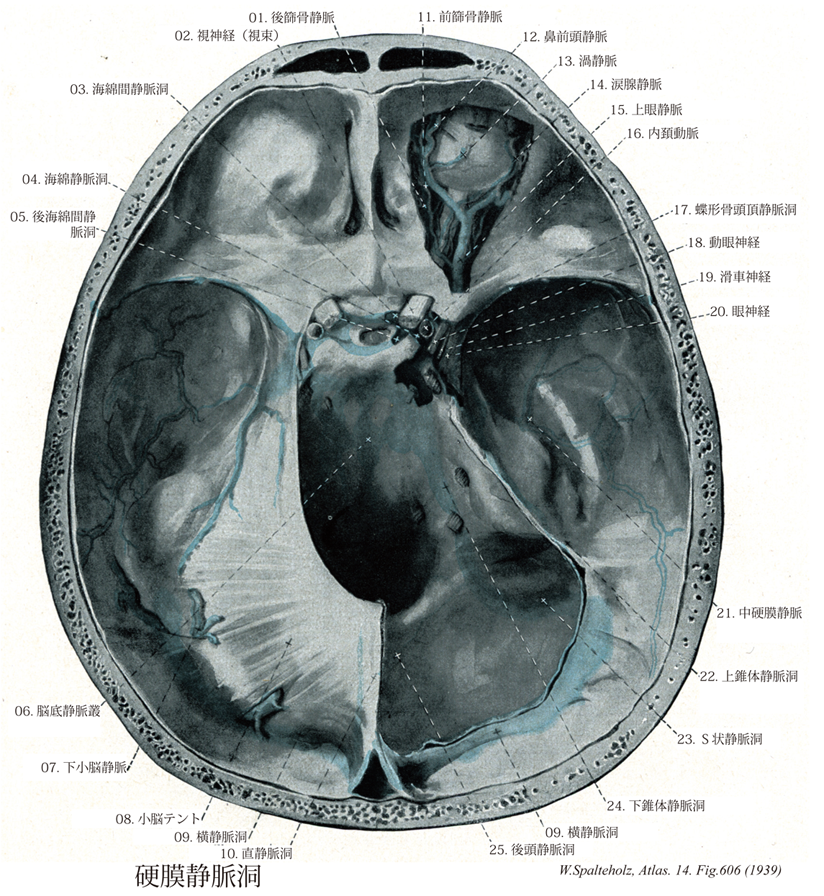
- 606_01【Posterior ethmoidal vein後篩骨静脈 Vena ethmoidalis posterior】
→()
- 606_01a【Ethmoidal veins篩骨静脈 Venae ethmoidales】 Branches draining the ethmoidal cells.
→(篩骨静脈は前・後篩骨動脈に伴行し上眼静脈に流入する静脈。篩骨洞からの血液を運ぶ。)
- 606_02【Optic nerve [II]視神経;視束[脳神経II] Nervus opticus; Fasciculus opicus [II]】 Nerve emerging medial to the posterior pole of the eyeball and extending to the optic chiasma.
→(視神経は脳神経の1つとして扱われてはいるが、実は前脳胞の延長部である。眼球網膜の第8層である神経細胞層中にある多極神経細胞から出る神経線維が集まって出来る神経である。すなわち杆状体細胞および錐体状細胞よりの興奮は網膜の内顆粒層の双極細胞に伝わり、それがさらに神経細胞層の細胞に連絡し、この神経細胞の出す神経突起である線維はまず眼球の後極よりやや内下方の一ヶ所に集まって、視神経円板を作り、強大な神経幹となり、網膜の続きである視神経鞘に囲まれて後内側に向かう。眼球から約15~20mm隔ったたところで、眼動脈の枝である網膜中心動脈およびこれに伴う静脈が外側から入り込み、その中軸を通って網膜に分布する。左右両側の視神経は眼窩後端の視神経管を通って頭蓋腔に入り、次第に相近づいて蝶形骨体上の視神経溝でほぼ半交叉をして視交叉を作り、そのつづきは視索と名が変わって間脳の外側膝状体および中脳の上丘などの第一次視覚中枢に達して、ここで終わる。網膜が眼胚から発達するので経路に相応する。ヒトの視神経は眼球網膜の神経細胞層中にある多極神経細胞から出る100万本以上の神経線維からなる。すなわち、杆状体細胞および錐体状細胞よりの興奮は網膜の内顆粒層の双極細胞に伝わり、それがさらに神経細胞層の細胞に連絡し、この神経細胞の出す神経突起である線維はまず眼球の後極よりやや内下方の一ヶ所に集まって、視神経円板を作り、強大な神経幹となり、網膜の続きである視神経鞘に囲まれて後内側に向かう。眼球から約15~20mm隔ったたところで、眼動脈の枝である網膜中心動脈およびこれに伴う静脈が外側から入り込み、その中軸を通って網膜に分布する。左右両側の視神経は眼窩後端の視神経管を通って頭蓋腔に入り、次第に相近づいて蝶形骨体上の視神経溝でほぼ半交叉をして視交叉を作り、そのつづきは視索と名が変わって間脳の外側膝状体および中脳の上丘などの第一次視覚中枢に達して、ここで終わる。)
- 606_03【Intercavernous sinus海綿間静脈洞 Sinus intercavernosus】
→(左右の海綿静脈洞を前方および後方で吻合させる静脈洞で、下垂体の後ろを前方に進み海綿静脈洞とともに輪状静脈洞を形成する)
- 606_03a【Anterior intercavernous sinus前海綿間静脈洞 Sinus intercavernosus anterior】 Connection between the right and left cavernous sinuses anterior to the pituitary gland.
→(前海綿間静脈洞は下垂体の前で左右の海綿静脈洞が結合するもの。)
- 606_04【Cavernous sinus海綿静脈洞 Sinus cavernosus】 Spongy structure of expanded veins on both sides of the sella turcica into which the ophthalmic veins and other veins empty. The carotid artery and abducent nerve lie within it and cranial nerves III, IV, VI, and V2 travel in its lateral side wall.
→(海綿静脈洞は静脈間が網目に吻合して大きい不規則な網状構造をしている。この海綿静脈洞は蝶形骨洞、トルコ鞍、下垂体などの両側にある静脈洞、上眼窩裂から錐体乳突部の岩様部まで広がっている。海綿静脈洞は、内頚動脈と外転神経をとり囲む。静脈洞の外側壁には動眼神経、滑車神経、三叉神経の枝である眼神経と上顎神経が存在する。左右の海綿静脈洞は脳底静脈叢および下垂体前面にある前海綿間静脈叢と後面にある後海綿間静脈叢により対側の静脈洞と連絡する。眼静脈と蝶形骨頭頂静脈洞は、海綿静脈洞に注ぎ込む。海綿静脈洞は、後方に向かい上錐体静脈洞と下錐体静脈洞に入り、上錐体動脈洞は横静脈洞に、下垂体静脈洞は、短い静脈網によって翼突筋静脈叢や喉頭静脈叢とも連絡する。)
- 606_05【Posterior intercavernous sinus後海綿間静脈洞 Sinus intercavernosus posterior】 Connection between the right and left cavernous sinuses posterior to the pituitary gland.
→(後海綿間静脈洞は下垂体の後で左右の海綿静脈洞が結合するもの。)
- 606_06【Basilar plexus; Basilar venous plexus脳底静脈叢 Plexus venosus basilaris; Plexus basilaris】 Venous plexus on the clivus that is connected with the cavernous and petrosal sinuses, as well as venous plexuses of the vertebral canal.
→(脳底静脈叢は海綿静脈洞、錐体静脈洞、および脊柱管静脈叢に引き続いて蝶形骨斜台の上に位置する静脈叢。)
- 606_07【Inferior cerebellar veisns下小脳静脈 Venae cerebelli inferiores; Venae cerebellares inferiores】
→()
- 606_07a【Cerebellar veins小脳静脈 Venae cerebelli】
→(小脳静脈には、主に小脳の上面に分布している上小脳静脈と下面を通る下小脳静脈がある。)
- 606_08【Tentorium cerebelli; Cerebellar tentorium小脳テント;小脳天幕 Tentorium cerebelli】 Dural septum stretched over the cerebellum between the superior border of petrous part of temporal bone and the transverse sinus. It supports the occipital lobe.
→(小脳テントは後頭骨の内後頭隆起とその左右にのびる横洞溝からおこる脳硬膜は小脳上面に広がって中脳を扼し、前方は蝶形骨の前床突起に付着し、側方には側頭骨の錐体上縁に付着する。小脳の上面をおおうところから小脳テントの名がある。つまり、頭蓋腔は小脳テントにより大脳を入れる上ならびに中頭蓋窩と、小脳、橋、延髄を入れる下頭蓋窩に二分される。そして同時に小脳テントは大脳と橋、延髄を結合する中脳が通れるだけの間隙をつくっているわけで、中脳を扼している小脳テントの部分をテント切痕とよぶ。)
- 606_09【Transverse sinus横静脈洞;横洞 Sinus transversus】 It commences at the confluence of sinuses and passes laterally to the sigmoid sinus.
→(二つの横静脈洞は静脈洞交会から起こり後頭骨の横洞溝の中を、外側に向かってから前方に走る。そして左右のそれぞれの横静脈洞は、後頭骨と側頭骨の岩様部との縫合部でS状静脈洞となって下方に曲がり後方に向かう。横静脈洞には上錐体静脈洞、下大脳静脈、下小脳静脈、板間静脈などが注ぐ。)
- 606_10【Straight sinus直静脈洞 Sinus rectus】 It commences at the union of the great cerebral vein and inferior sagittal sinus and runs within the root of the falx cerebri at its junction with the tentorium cerebelli to the confluence of sinuses.
→(直静脈洞は、小脳テントに付着する大脳鎌のところ後走する静脈洞。下矢状静脈洞と大大脳静脈が合流してでき、一般にやや左側に偏して走り、横静脈洞に合し、静脈洞交会に注ぎ込む。)
- 606_11【Anterior ethmoidal vein前篩骨静脈 Vena ethmoidalis anterior】
→()
- 606_12【Nasofrontal vein鼻前頭静脈 Vena nasofrontalis】 Connection between the ophthalmic vein and the union of the supratrochlear vein with the angular vein.
→(鼻前頭静脈は滑車上静脈と眼角静脈の合一部を上眼静脈とむすぶ。)
- 606_13Ruysch' veins【Vorticose veins; Choroid veins渦静脈;眼球脈絡膜静脈;大脈絡膜静脈 Venae vorticosae; Venae chorioideae majores】 Four or five branches from the choroid of the eyeball that penetrate the sdera laterally.
→(渦静脈は毛様体血管系の静脈は眼球の赤道部で集まり、4本の渦静脈(眼球脈絡膜動脈)となって眼球をさる。)
- 606_14【Lacrimal vein涙腺静脈 Vena lacrimalis】 Branch draining the lacrimal gland.
→(涙腺静脈は涙腺からの血液を集め、涙腺動脈と友に眼窩を通り、後方へ走り上眼静脈へそそぐ小静脈。)
- 606_15【Superior ophthalmic vein上眼静脈 Vena ophthalmica superior】 Vein arising medially above the eyeball with the nasofrontal vein and passing through the superior orbital fissure to the cavernous sinus.
→(上眼静脈は眼動脈に沿って走る。すなわち内眼角でおこり、この部で顔面静脈や眼角静脈と吻合し、眼窩上壁を内側に沿って走行する。おおよそ眼動脈の分布域からの静脈を集める。上眼窩裂を通って頭蓋腔に入り海綿静脈洞に注ぐ。)
- 606_16【Internal carotid artery内頚動脈 Arteria carotis interna】 It passes from the carotid bifurcation, without any branches, to the cranial base, continuing in the carotid canal to its terminal division into the middle and anterior cerebral arteries.
→(内頚動脈は、総頚動脈から起こり、頚部では頭蓋底にいたるまでは枝を出さない。ついで頚動脈管をへて中大脳動脈と前大脳動脈に分枝するまでをいう。内頚動脈は頚部、側頭骨錐体部(岩様部)、海綿静脈洞部、大脳部の4つの部分に分けられる。この内頚動脈の海綿静脈洞部と大脳部とは、特別な形態を呈するので、「頚動脈サイフォン」とよばれている。内頚動脈の主な枝として、眼動脈、後交通動脈、前脈絡叢動脈がでる。内頚動脈は、視交叉の外側で小さな前大脳動脈と大きな中大脳動脈とに分岐する。中大脳動脈は内頚動脈の直接の続きで終枝と考えられる。)
- 606_17【Sphenoparietal sinus蝶形骨頭頂静脈洞;蝶形頭頂静脈洞 Sinus sphenoparietalis】 Sinus that runs beneath the lesser wing of sphenoid to the cavernous sinus.
→(蝶形骨頭頂静脈洞は浅中大脳葉脈の続きで、蝶形骨の小翼に沿って内側下方へ走り内頚静脈へ流入する。小脳下面、延髄、内耳からの血流を受けている。)
- 606_18【Oculomotor nerve [III]動眼神経[脳神経III] Nervus oculomotorius [III]】 Nerve containing motor and parasympathetic fibers that exits the oculomotor sulcus and passes through the superior orbital fissure into the orbit.
→(動眼神経の主成分は動眼神経主核から出る体性運動性のもので外側直筋および上斜筋以外の眼筋を支配する。このほかに副交感性の動眼神経副核[Edinger-Westphal核]から出る線維が加わる。以上の2核から出る線維は多数の根をつくって大脳脚内側溝から出て1神経幹となり、滑車神経、外転神経および眼神経とともに、蝶形骨体の両側にある海綿静脈洞の上壁に沿ってすすみ、上眼窩裂を通って眼窩内に入り、上下の2枝に分かれる。上枝は上瞼挙筋および上直筋に、下枝は内側直筋、下直筋および下斜筋に分布する。また下枝からはきわめて短い動眼神経からの根が出て、毛様体神経節に入るが、これは動眼神経副核から出て、下枝を通って毛様体神経節に入る副交感線維にほかならない。)
- 606_19【Trochlear nerve [IV]滑車神経[脳神経IV] Nervus trochlearis [IV]】 Nerve exiting on the dorsal side, caudal to the tectal plate. It supplies the superior oblique muscle.
→(滑車神経は脳神経中最少のもので、滑車神経核からでて上斜筋を支配する鈍体性運動性神経である。この神経は脳の背側から脳をでる唯一の脳神経で、下丘のすぐ後方で、上小脳脚と上髄帆小帯との間から出て、大脳脚をめぐり、(側頭骨)錐体尖の近くで硬膜を貫いて海綿静脈洞の上壁に達し、動眼神経の外側から上側に向かって前進し、上眼窩裂を通って眼窩内に入り、上直筋、上眼瞼挙筋起始部の上を前内側にすすんで、上斜筋に分布する。)
- 606_20【Ophthalmic nerve; Ophthalmic division [Va; V1]眼神経 [三叉神経第1枝] Nervus ophthalmicus [Va; V1]】 First division of the trigeminal nerve, which passes through the superior orbital fissure.
→(眼神経は第五脳神経の第一枝(CN V1)。蝶形骨体上の海綿静脈洞の外側に沿って前方にすすみ、上眼窩裂を通って眼窩に入る。つぎの枝(①涙腺神経、②前頭神経、③鼻毛様体神経)に分かれる。また眼筋にいたる動眼、滑車、外転の3神経および交感神経との間に交通がある。)
- 606_21【Middle meningeal veins中硬膜静脈 Venae meningeae mediae】 Veins accompanying the middle meningeal artery.
→(中硬膜静脈は中硬膜動脈の伴行静脈。)
- 606_22【Superior petrosal sinus上錐体静脈洞 Sinus petrosus superior】 It passes from the cavernous sinus along the superior border of the petrous part of temporal bone to the sigmoid sinus.
→(上錐体静脈洞は側頭骨錐体部の上縁で天幕付着部を走る。海綿静脈洞の後部と横静脈洞とをつないでいる。錐体静脈、下大脳静脈などからの血流を受けている。)
- 606_23【Sigmoid sinusS状静脈洞 Sinus sigmoideus】 It exits the lateral wall of the cranium as a continuation of the transverse sinus and courses in an S-shape to the jugular foramen.
→(S状静脈洞は横静脈洞に引き続いて側頭骨乳突部内面を下内側に屈曲して走り、頚静脈孔で内頚静脈につづく。)
- 606_24【Inferior petrosal sinus下錐体静脈洞 Sinus petrosus inferior】 It runs from the cavernous sinus along the posterior inferior border of the petrous part of temporal bone to the jugular foramen.
→(下錐体静脈洞は海綿静脈洞の後下部に始まり、斜台と側頭骨錐体部との間の溝を後下方へ走り内頚静脈へ流入する。小脳下面、延髄、内耳からの血流を受けている。)
- 606_25【Occipital sinus後頭静脈洞;後頭洞 Sinus occipitalis】 It commences with a venous plexus at the foramen magnum and passes within the root of the falx cerebelli to the confluence of sinuses.
→(小脳鎌付着部に沿って正中線上を走る静脈洞で、上方は静脈洞交会または横静脈洞の左右どちらか(主に右側)に流入する。下方は内椎骨静脈叢、辺縁静脈洞と連続している。)