Spalteholz HANDATLAS DER ANATOMIE DES MENSCHEN VON WERNER SPALTEHOLZ
メニューは解剖学(TA)にリンクしてあります。図の番号をクリックすると下記の説明へ、右側の用語をクリックすると解剖学(TA)にジャンプします。
724


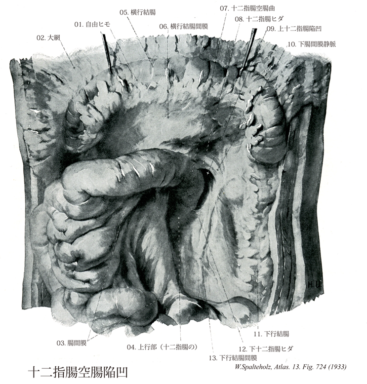
- 724_01【Free taenia; Free tenia自由ヒモ Taenia libera】 Free tenia between the mesocolic and omental teniae.
→(自由ヒモは間膜ヒモと大網ヒモの間にヒモ。)
- 724_02【Greater omentum大網 Omentum majus】 It extends from the greater curvature of stomach, draping over the intestinal loops like an apron. It is fused with the transverse colon and mesocolon.
→(大網は胃の大弯から広がっている腸管の表面にエプロンのように下垂している二重の腹膜葉。横行結腸および結腸間膜に癒合しているが自由に移動できる。横行結腸を越え、下腹部の小腸の迂曲部の前まで広がることがよくある。他の例では腹膜腔の陥凹部に強く引き込まれていることがある。大網のうち胃の大弯と横行結腸との間に広がる部分は胃結腸間膜と呼ばれる。胃に分布する大弯の血管弓が大網の付着部に存在する。大網は左方で脾臓の脾門に続き、この部分を胃脾間膜と呼ぶ。大網には脂肪と免疫系の細胞が多く、乳斑を形成している。大網は腹膜腔内の炎症による被包に巻き込まれることがよくあるが、その場合癒着したり、腹部臓器とともに大きくなったりする。)
- 724_03【Mesentery腸間膜;小腸間膜 Mesenterium】 Dorsal peritoneal fold enclosing vessels and nerves and protecting the supply of the small intestine against vascular torsion.
→(腸間膜は腸管が腹壁から遊離して存在する場合に、腹膜はその部の腹壁を離れて腸管の表面に達し、これを包んだのち再び腹壁にもどる。このため後腹壁と腸管の間に、往復2葉の腹膜が合した膜が生じ、大動脈と腸管の連絡路を提供する。この2葉の膜を腸間膜(広義)または総背側腸間膜と称し、部位により胃間膜、腸間膜(狭義)、結腸間膜などに区分する。①胃間膜mesogastriumは背側のみならず腹側にも間膜がある。②腸間膜は小腸間膜ともいい、空腸と回腸に付属し、その基部すなわち腸間膜根は第2腰椎の左側から右腸骨窩に斜走し、わずかに約15cmの長さをもつにすぎない。ここからおこった間膜はしだいに複雑なヒダを形成し、小腸への付着縁では数mの長さをもつに至る。③結腸間膜:発声の始め結腸前部にわたって存在するが、上行結腸間膜と下行結腸間膜は後壁腹側膜と癒着してしまうので、横行結腸とS状結腸だけに間膜が遺残する。横行結腸間膜はその基部が第2腰椎高さで膵下縁を横走し、大網後葉と付着して網嚢の底部を形成する。S状結腸間膜は腹腔の左下部にあり、その逆波逆V字形をなす。なお、盲腸は間膜を欠くが、虫垂は回腸終末部と連絡するヒダを有し、それを虫垂間膜と称する。)
- 724_04【Ascending part of duodenum上行部(十二指腸の) Pars ascendens (Duodenum)】 Segment to the left of the head of pancreas ascending to the duodenojejunal flexure.
→(十二指腸の上行部は水平につづき、左上方に向かって斜めに上行する部である。長さ約5cm。第2腰椎の左側で、急に前方に屈曲して(十二指腸空腸曲)、空腸に移行する。)
- 724_05【Transverse colon横行結腸 Colon transversum】 Intraperitoneal part of the colon between the hepatic and splenic flexures.
→(横行結腸は右結腸曲から左方に走り、やや上行して脾臓の下端で左結腸曲に達するまでの約30~50cmの結腸。その外表面は腹膜によって完全におおわれ、長い横行結腸間膜によって後腹壁に付着している。前腹壁との間には大網がある。横行結腸は広い横行結腸間膜をもつので、大きな可動性をもつ。横行結腸は前下方に球状を呈して横走し、その中央部は下垂する。その最下位は背臥位でほぼ臍の高さにあるが、直立位でとくに充満する時には下腹部さらに骨盤にまで下垂する。広い横行結腸間膜によって、腹膜腔は上・下2部に分けられる。)
- 724_06【Transverse mesocolon横行結腸間膜 Mesocolon transversum】 Peritoneal fold attached to the transverse colon. It arises anterior to the head of pancreas and along the inferior border of the body of pancreas. It is fused with the posterior layer of the greater omentum.
→(横行結腸間膜は膵臓の前面から起こる広い腹膜ヒダで、横行結腸の後壁に達し、これを包む。また、左結腸曲は横隔膜と腹膜ヒダで結ばれる。)
- 724_07【Duodenojejunal flexure十二指腸空腸曲 Flexura duodenojejunalis】 Flexure between the duodenum and jejunum.
→(十二指腸と空腸の間の弯曲。(Feneis))
- 724_08【Superior duodenal fold; Duodenojejunal fold上十二指腸ヒダ;十二指腸空腸ヒダ;上十二指腸結腸間膜ヒダ Plica duodenalis superior; Plica duodenojejunalis; Plica duodenomesocolica cranialis】 Peritoneal fold on the left side of the duodenojejunal flexure in front of the superior duodenal fossa. It contains the inferior mesenteric vein.
→(十二指腸空腸曲の左方で上十二指腸陥凹の前方にある腹膜ヒダ。下腸間膜静脈を包む。 (Feneis))
- 724_09【Superior duodenal fossa; Superior duodenal recess上十二指腸陥凹;上十二指腸結腸間膜陥凹;十二指腸空腸陥凹 Recessus duodenalis superior; Recessus duodenomesocolicus cranialis; Reccessus duodenojejunalis】 Peritoneal recess behind the superior duodenal fold.
→()
- 724_10【Inferior mesenteric vein下腸間膜静脈 Vena mesenterica inferior】 Branch extending from the left one-third of the colon to the superior part of the rectum and emptying into the splenic vein.
→(左結腸1/3から直腸上部まで達する脾静脈の枝。 (Feneis))
- 724_11【Descending colon下行結腸 Colon descendens】 Retroperitoneal segment of the colon extending along the left side of the body between the splenic flexure and sigmoid colon.
→(下行結腸は左結腸曲から下行し、左腸骨窩においてS状結腸へ移行する。長さ25~30cmで、左結腸曲からほぼ垂直に下行し、左結腸窩でS状結腸に移行する。下行結腸は、上行結腸に比べて、細く、前方には大網・小腸があり、後方には左腎臓の外側縁・腰方形筋・腸骨筋・大腰筋が接する。上行結腸と同様腸間膜を欠き後腹壁に固定されている。下行結腸に沿って結腸傍溝が走る。とくに外側の傍溝は下方で骨盤腔に連なり、上方では横隔結腸ヒダで境される。)
- 724_12【Inferior duodenal fold; Duodenomesocolic fold下十二指腸ヒダ;十二指腸結腸間膜ヒダ;下十二指腸結腸間膜ヒダ Plica duodenalis inferior; Plica duodenomesocolica; Plica duodenomesocolica caudalis】 Peritoneal fold below the duodenojejunal flexure.
→()
- 724_13【Descending mesocolon下行結腸間膜 Mesocolon descendens】 Peritoneal fold attached to the descending colon. It usually fuses with the posterior wall of the abdomen in the fourth month of embryonic development.
→(胎生4ヶ月に後腹壁と癒着する。(Feneis))