Spalteholz HANDATLAS DER ANATOMIE DES MENSCHEN VON WERNER SPALTEHOLZ
メニューは解剖学(TA)にリンクしてあります。図の番号をクリックすると下記の説明へ、右側の用語をクリックすると解剖学(TA)にジャンプします。
726


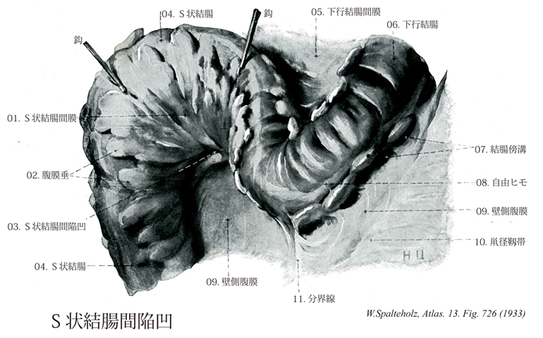
- 726_01【Sigmoid mesocolon; MesosigmoidS状結腸間膜 Mesocolon sigmoideum; Mesosigmoideum】 Peritoneal fold attached to the sigmoid colon.
→(S状結腸は腹膜で包まれる。腹膜はS状結腸間膜となる。間膜はS状結腸を後腹壁につり下げている。)
- 726_02【Omental appendices; Epiploic appendices; Fatty appendices of colon腹膜垂;大網垂;結腸脂肪垂 Appendices omentales; Appendices adiposae coli; Appendices epiploicae】 Appendages consisting of adipose tissue in the subserous layer along the free and omental tenia.
→(腹膜垂[epiploic appendages、羅appendices epiploicae]大網ヒモと自由ヒモのところで結腸結腸(腹膜)のところどころにみられる黄色、葉状の下垂物。漿膜中皮の下層の脂肪細胞が局所的に集積することによる。結腸の形態学的な特徴の1つ。[医学書院医学大辞典:相磯貞和])
- 726_03【Intersigmoid recessS状結腸間陥凹 Recessus intersigmoideus】 Peritoneal recess on the left side of the body in the angle of the root of the sigmoid mesocolon. The ureter can be palpated here.
→(S状結腸間膜が後腹壁に付着するところは逆V状を呈し、左下方に凹む、この凹みはS状結腸間陥凹といわれる。)
- 726_04【Sigmoid colonS状結腸 Colon sigmoideum】 Intraperitoneal segment of the colon between the descending colon and rectum.
→(S状結腸は骨盤上口と第3仙椎との間で、S状の曲線を描いて下行する部分。下行結腸下端からS字状に屈曲下行して内下方へ向かい、第3仙椎の前方で直腸へ移行する。長さ30~45cmである。ほぼ左側の腸骨稜の高さに始まり、左外腸骨動脈の前を不規則なS状をえがいて下行し、直腸に移行する。S状結腸の走行は一般に不整なS状で、2箇所で弯曲する。すなわち、まず骨盤の左側壁に接して下行し、ついで弯曲し小骨盤内を内上方に走り、再び弯曲して下走し、仙骨前面で第3仙椎の高さにおいて直腸に移行する。S状結腸は、成人では骨盤内にあるが、小児では骨盤がなお小さいので腹部にある。)
- 726_05【Descending mesocolon下行結腸間膜 Mesocolon descendens】 Peritoneal fold attached to the descending colon. It usually fuses with the posterior wall of the abdomen in the fourth month of embryonic development.
→(胎生4ヶ月に後腹壁と癒着する。(Feneis))
- 726_06【Descending colon下行結腸 Colon descendens】 Retroperitoneal segment of the colon extending along the left side of the body between the splenic flexure and sigmoid colon.
→(下行結腸は左結腸曲から下行し、左腸骨窩においてS状結腸へ移行する。長さ25~30cmで、左結腸曲からほぼ垂直に下行し、左結腸窩でS状結腸に移行する。下行結腸は、上行結腸に比べて、細く、前方には大網・小腸があり、後方には左腎臓の外側縁・腰方形筋・腸骨筋・大腰筋が接する。上行結腸と同様腸間膜を欠き後腹壁に固定されている。下行結腸に沿って結腸傍溝が走る。とくに外側の傍溝は下方で骨盤腔に連なり、上方では横隔結腸ヒダで境される。)
- 726_07【Paracolic gutters結腸傍溝;結腸傍陥凹 Sulci paracolici; Recessus paracoliei】 Occasional grooves on the left side of the descending colon.
→(下行結腸の左方にときにみられる溝。 (Feneis))
- 726_08【Free taenia; Free tenia自由ヒモ Taenia libera】 Free tenia between the mesocolic and omental teniae.
→(自由ヒモは間膜ヒモと大網ヒモの間にヒモ。)
- 726_09【Parietal peritoneum壁側腹膜 Peritoneum parietale】 Peritoneum lining the abdominal wall.
→(壁側腹膜は腹壁の腹膜。)
- 726_10Fallopian ligament; Poupart's ligament; Vesalius' ligament【Inguinal ligament鼡径靱帯;鼡径弓 Ligamentum inguinale; Arcus inguinalis】 Inferior end of the aponeurosis of the external oblique. It passes from the anterior superior iliac spine to the pubic tubercle.
→(鼡径靱帯は上前腸骨棘と恥骨結節との間に張る靱帯で、前面における体幹と下肢の境界である。外腹斜筋の停止腱膜のつくる腱弓の発達したものである。恥骨櫛は恥骨結節のやや後部から後外側にのびているので、鼡径靱帯は恥骨櫛よりもやや前方にある。鼡径靱帯の内側端の一部は分かれて後走し、恥骨櫛は恥骨結節のやや後部から後外側にのびているので、鼡径靱帯は恥骨櫛よりもやや前方にある。鼡径靱帯の内側端の一部は分かれて後走し、恥骨櫛内側部に達する。これを裂孔靱帯といい、鼡径管下壁の形成に関与する。裂孔靱帯外側縁が恥骨櫛に沿ってのびているものを恥骨櫛靱帯という。また、浅鼡径輪の外側脚をを作る外腹斜筋腱膜線維が鼡径靱帯内側端に到達した後、上内側に方向をかえて反転し、腹直筋鞘前葉をつくる内腹斜筋の前面に向かって線維を送る。これを反転靱帯といい、鼡径管内側端に到達した後、上内側に方向をかえて反転し、腹直筋鞘前葉をつくる内腹斜筋の前面に向かって線維を送る。これを反転靱帯といい、鼡径管内側端で、その後壁の形成に関与する。プーパルの靱帯とも呼ばれる。Poupart, Francois (1616-1708)フランスの外科医、ルイ14世の侍医。プーパルの靱帯(鼡径靱帯)を既述(""Suspenseurs del'-abdomen"", Hist. Acad. Roy. Sci., Paris, 1730, 51)、プーパル線は鼡径靱帯の中心と鎖骨とを結ぶ線。)
- 726_11【Linea terminalis of pelvis分界線;骨盤縁;腸骨恥骨線;終端線(骨盤の) Linea terminalis (Pelvis)】 Line extending from the promontory of the sacrum along the arcuate line, the pecten pubis, to the superior border of the pubic symphysis. It marks the boundary between the greater and lesser pelvis as well as the plane of the pelvic inlet.
→(腸骨恥骨線iliopectineal lineともよばれる。仙骨上縁の前端にある岬角から仙骨外側部を経て寛骨の弓状線に至り、更に恥骨櫛を経て恥骨結合上縁に達する稜線をたどることができる。これが分界線と呼ばれる。腸骨窩の下方の境界をなす。大骨盤から小骨盤を分ける。)
