Spalteholz HANDATLAS DER ANATOMIE DES MENSCHEN VON WERNER SPALTEHOLZ
メニューは解剖学(TA)にリンクしてあります。図の番号をクリックすると下記の説明へ、右側の用語をクリックすると解剖学(TA)にジャンプします。
750


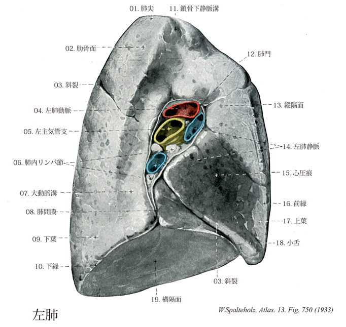
- 750_00【Right lung左肺 Pulmo sinister】 Larger of the two lungs.
→(左肺は上・下の2葉から成っている。左肺の上葉の前内側縁には心切痕とう切れ込みがあり、その下の部分は下のような形をしているので小舌と呼ばれる。)
- 750_00a【Lungs肺 Pulmones】 The lungs occupy the greater part of the thoracic cavity.
→(肺は胸腔をみたす1対の半円錐形の実質臓器で、呼吸器系の主部をなす。肺においてガス交換が呼吸気と血液の間で行われる。右肺(1200cc, 600g)は左肺(1100cc, 500g)よりやや大きい。肺尖・肺底・肋骨面を区別する。肺尖は鎖骨の2~3cm上方に達する。肺底は横隔面に相当し、横隔膜の円蓋にしたがって陥凹する。肋骨面は胸郭の形にしたがって膨隆する。内側面は左右の胸膜腔を隔てる縦隔に向かう面であって、全体としてややくぼむが心臓に接する部分は深いくぼみをなす。このくぼみを心圧痕といい、とくに左肺に著しい。内側面のうち後方の胸椎に接する部分を椎骨部といい、椎骨部と前述の心圧痕以外の内側面の部分を狭い意味で縦隔部という。縦隔部のうち、ほぼ中央部の肺胸膜におおわれない部分を肺門といい、肺門に出入りする気管支、肺動静脈などは結合織により束ねられて肺根をなし、肺胸膜から縦隔胸膜へ移行する胸膜におおわれるため滑沢であるが、後上方から前下方に走る深い切れ込み(斜裂)がある。右肺ではそのほかに、肋骨面の腋窩腺で斜裂から分かれ、第4肋骨に沿ってほとんど水平に走る切れ込み(水平裂)があり、上葉と中葉が分けられる。各葉の相接する面を葉間面という。左肺を前から見ると上葉に心臓の存在による切れ込みをみる。これを左肺心切痕といい、その下方の上葉前下端の小さい突出部を左肺小舌という。幼児の肺は淡紅色を呈するが、年とともに吸入された塵埃、煙の炭疽粒子などにより、暗赤色に変わる。肺は複合胞状腺の形態を示し、喉頭・気管・気管支およびその枝が導管、肺胞が腺胞に相当する。気管支は葉気管支、区気管支、区気管支枝、細気管支と何回も分支する。細気管支の直径は1mm以下になり、この部にいたると粘膜上皮は多列繊毛円柱上皮から単層の円柱上皮となり、軟骨輪輪は不規則な軟骨小片となる。細気管支はさらに枝分かれして呼吸細気管支になると、気管軟骨はなくなり、上皮は単層立方上皮となる。壁のところどころから肺胞もふくらみ出ている。気道の末端は肺胞管で、多数の肺胞がこの管からふくらみ出ている。その行きどまりを肺胞嚢とよぶ。肺の栄養血管は気管支動静脈で、気管分岐部付近で胸大動脈から直接デル。栄養血管は細葉を最小単位として取り囲む。機能血管である肺動脈は右三室から出て気管支系とともに肺実質内に分布する。胎生期には肺動脈と大動脈弓との間に連絡(動脈管)があるが、出生後閉塞して動脈幹索となる。ガス交換を行った後の血液は肺静脈に集められ左心房に還る。)
- 750_01【Apex of lung肺尖 Apex pulmonis】 Tip of the lung which extends into the superior thoracic aperture.
→(一部胸郭上口に位置している。 (Feneis))
- 750_02【Costal surface of lung肋骨面(肺の) Facies costalis pulmonis】 Surface of the lung adjacent to the ribs.
→()
- 750_03【Oblique fissure of lung斜裂;葉間裂;葉間切痕(肺の) Fissura obliqua; Fissura interlobaris; Incisura interlobaris】 Oblique cleft between the inferior and superior lobes of left lung, and between the lower, middle, and superior lobes of right lung. It runs paravertebrally from the fourth rib to the sixth rib in the middavicular line.
→(肺の斜裂は左右両肺にみられ、後上方から前下方に向かって斜めに走る。斜裂は肺後面において、肺尖の約6cm下方(第3胸椎の棘突起の高さ)で始まり、前下方に斜走し、下縁の内側部に達する。肺の内側面でみると、斜裂は後上方から前下方に肺門を斜めに横切る。左肺では下葉と上葉の間、右肺では下葉と中葉の間にある斜めの裂隙。したがって、斜裂は、脊柱側方で第四肋骨から前下方へ下り、鎖骨中間線で第6肋骨までいたる。)
- 750_04【Left pulmonary artery左肺動脈;左枝(肺動脈の) Arteria pulmonalis sinistra; Ramus sinister】 Artery lying in front of the descending aorta. On radiographs it appears as a 「pulmonary arch」 below the 「aortic arch.」
→(左肺動脈は肺動脈幹の2分枝のうち短い方の枝で、心外膜を貫いて左肺門にはいる。多数の枝を出して気管支区や気管支下区に分布するが個人差は大である。典型的には上葉動脈の枝として肺尖動脈(A1)、前区動脈、後区動脈(後2者は上行枝・下行枝を出す)、肺舌動脈の枝として上舌動脈・下舌動脈、下葉動脈の枝として下葉上動脈(A6)と肺底区の枝(前肺底動脈、後肺底動脈、外側肺底動脈、内側肺底動脈)を出す。)
- 750_05【Left main bronchus; Left bronchus左主気管支;左気管支 Bronchi principales sinister; Bronchus principalis sinister】
→(左主気管支は直接気管からでる左の気管支幹部。)
- 750_06【Intrapulmonary nodes; Intrapulmonary lymph nodes肺内リンパ節;肺リンパ節 Nodi lymphoidei intrapulmonales】 Lymph nodes located at the exit sites of the segmental bronchi anc in the lung tissue.
→(背内のリンパ管は気管支の分枝に沿って末梢から次第に合流しつつ、肺門に向かって走る。肺内で、リンパ管は小さいリンパ節を経過する。このようなリンパ節は一般に気管支の分枝によてできるぶ分岐角内に存在し肺リンパ節といわれる。)
- 750_07【Aortic groove; Groove for descending aorta大動脈溝;下行大動脈溝;肺の下行大動脈に対する溝 Sulcus aorticus】
→()
- 750_08【Pulmonary ligament肺間膜;縦隔肺ヒダ Ligamentum pulmonale; Plica mediastinopulmonalis】 Double fold of pulmonary pleura that passes inferiorly from the right and left sides of the hilum to the mediastinal pleura. Between the two folds, the lung is in contact with the mediastinal connective tissue without a pleural covering.
→(肺門から下方へ張る縦隔胸膜のヒダで、肺胸膜上につく。 (Feneis))
- 750_09【Inferior lobe; Lower lobe of lung下葉(肺の) Lobus inferior pulmonis】 It mainly extends dorsally. Its superior border runs obliquely from posterosuperior to anteroinferior. It begins paravertebrally at the fourth rib and ends at the intersection of the middavicular line and the sixth rib.
→(後側に主な拡がりをもつ。その上限界上後方から下前方へ斜めに走り、第四肋骨から脊柱側方で鎖骨中間線を第六肋骨が切る線までいたる。 (Feneis))
- 750_10【Inferior border of lung; Inferior margin of lung下縁(肺の) Margo inferior pulmonis】 Sharp margin at the junction of the costal and diaphragmatic surfaces. It is less sharp at the transition of the diaphragmatic surface into the mediastinal surface.
→(肋骨面と横隔面が合うところにある鋭い縁。横隔面が内側面へ移行する縁はそれほど鋭くない。 (Feneis))
- 750_11【Groove for subclavian vein鎖骨下静脈溝 Sulcus venae subclaviae】 Groove on the first rib anterior to the scalene tubercle for the passage of the subclavian vein.
→(鎖骨下静脈口は前斜角筋結節を挟んでその後に鎖骨下動脈溝(鎖骨下動脈が乗る)があり、その前には鎖骨下静脈溝がある。)
- 750_12【Hilum of lung; Hilus of lung肺門 Hilum pulmonis】 Site of entry and exit of bronchi, vessels, and nerves on the mediastinal surface of the lung. In general, the bronchi are located dorsally, the pulmonary arteries ventral and cranial to the bronchi, and the pulmonary veins ventral and caudal to the bronchi. In the right hilum the superior lobar bronchus is above the pulmonary artery, hence the term 「eparterial」 bronchus.
→(気管支、肺動・静脈、リンパ管などが肺に出入りする部分。内側面の中央で、およそ第6胸椎の高さにある。なお、出入りする血管などをまとめて肺根root of the lungという。また、肺門周辺にはリンパ節(気管支肺リンパ節)が発達し、肺内からのリンパを集める。(イラスト解剖学))
- 750_13【Mediastinal surface of lung; Mediastinal part of inferior lobe縦隔面;内側面(肺の) Facies mediastinalis; Facies medialis】 Surface of the lung in contact with the mediastinum, located anterior to the vertebral part of the costal surface.
→(椎骨部の前方にあり、縦隔に接する面。 (Feneis))
- 750_14【Left pulmonary veins左肺静脈 Venae pulmonales sinistrae】 The two left pulmonary veins which occasionally unite to form a single trunk.
→(2条。ときには合して1本の幹となる。 (Feneis))
- 750_15【Cardiac impression on lung心圧痕(肺の) Impressio cardiaca pulmonis】 Depression on the medial surface of both lungs produced by the heart.
→(両肺の内側面にある心臓による凹み。 (Feneis))
- 750_16【Anterior border of lung; Anterior margin of lung前縁(肺の) Margo anterior pulmonis】 Sharp, anterior margin at the junction of the mediastinal and costal surfaces.
→(縦隔面と肋骨面が前方で合うところにある鋭い縁。 (Feneis))
- 750_17【Superior lobe of lung; Upper lobe of lung上葉(肺の) Lobus superior pulmonis】 Superior lobe that extends posteriorly to the fourth rib. In the right lung, its inferior border runs anteriorly at about the level of the fourth rib. In the left, its inferior border extends to the osseocartilaginous border of the sixth rib.
→(後方では第四肋骨まで達する。右側では、その下端は第四肋骨にほぼ沿って、前方へいたる。左側では、第六肋骨の骨軟骨境界まで達する。 (Feneis))
- 750_18【Lingula of left lung; Lingula of superior lobe of left lung小舌;左肺の小舌 Lingula pulmonis sinistri】 Projection between the cardiac notch of the left lung and the inferior border of the left superior lobe.
→(左上葉の心切痕と下縁との間にある角。 (Feneis))
- 750_19【Diaphragmatic surface of lung; Diaphragmatic surface of inferior lobe横隔面(肺の) Facies diaphragmatica (pulmonis)】 Concave inferior surface of the lung that lies on the diaphragm.
→()