Spalteholz HANDATLAS DER ANATOMIE DES MENSCHEN VON WERNER SPALTEHOLZ
メニューは解剖学(TA)にリンクしてあります。図の番号をクリックすると下記の説明へ、右側の用語をクリックすると解剖学(TA)にジャンプします。
587 


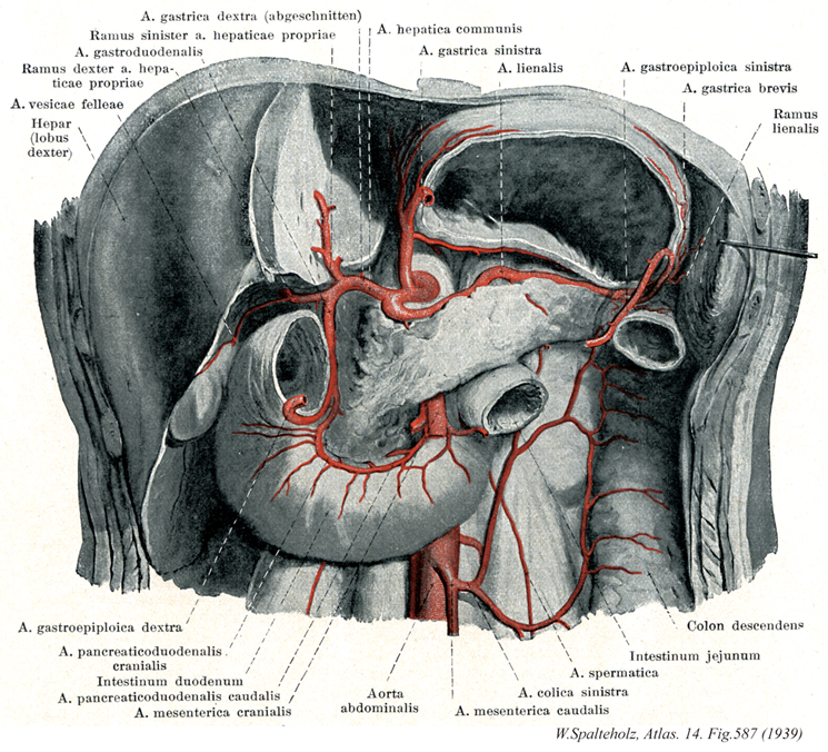
- 587_01【Right gastric artery右胃動脈 Arteria gastrica dextra】 It passes along the lesser curvature of stomach to the left gastric artery.
→(右胃動脈は幽門の上縁で総肝動脈から起こり、小弯に沿って左方に走って、胃の右下部に分布する。ほぼ角切痕の高さで左胃動脈と交通する。)
- 587_02【Left branch of hepatic artery; Left hepatic artery左枝;左肝動脈(肝動脈の) Ramus sinister arteria hepaticae】 Left branch of the hepatic artery proper supplying the left lobe of liver.
→(肝動脈の左枝は固有肝動脈の最終左枝で肝臓の左葉に分布する。)
- 587_03【Gastroduodenal artery胃十二指腸動脈 Arteria gastroduodenalis】 Branch of the common hepatic artery. It usually lies behind the pylorus and divides at its inferior border.
→(胃十二指腸動脈は幽門の後でその下縁において前方の上膵十二指腸動脈と、右胃大網動脈に分かれる。)
- 587_04【Right branch of proper hepatic artery右枝(固有肝動脈の) Ramus dexter (Arteria hepatica propria)】
→()
- 587_05【Cystic artery胆嚢動脈 Arteria cystica; Arteria vesicae felleae】 It divides and passes to the anterior and posterior surfaces of the gallbladder.
→(胆嚢動脈は肝動脈の右枝より起こり、胆嚢、肝の臓側面に分布する。)
- 587_06【Liver肝臓 Hepar】 Organ located in the upper right side of the abdomen in the hypochondrium. Its inferior border runs from the upper left to the lower right through the epigastric region. In healthy subjects its border does not reach below the costal margin. It moves with respiration and is thus palpable.
→(肝臓は身体内の最大の腺であり多様な機能を営むが、それを①胆汁の生産と分泌(腸管内へ)を行う、②炭水化物、脂肪、蛋白の代謝活動、③胃腸管から血液中に進入した最近や異物を細くする、とう3点に要約することができる。(1)位置と形状:肝臓は右上腹部ある巨大な消化腺で、重さは男で1,400g、女で1,200gほどある。色は暗赤褐色で、これは充満する血液によるものである。肝臓の表面が平滑で光沢に富むのは腹膜(の臓側葉)におおわれているからである。肝臓の上面は横隔膜の下面に接して丸く膨らみ、横隔面と呼ばれる。横隔膜上の心臓に対応して、浅い心圧痕をみる。からだの正中にほぼ相当して、横隔面を大きい右半と小さい左半に二分する肝鎌状間膜が走る。これは肝臓の表面を被う腹膜が左右から翻転しながら寄り合い、その間に線維性の結合組織をいれるもので、肝臓を横隔膜から吊り下げる役をしている。このようにして横隔膜と肝臓は平滑な腹膜で自由に滑り動くようになっているが、後部のせまい領域では、両者が線維性結合組織によって密着して活動性に欠ける。肝臓表面のこの領域を無漿膜野(裸の領域Area nuda--腹膜に包まれていない--の意)という。無漿膜野は前方へ細く張り出して肝鎌状間膜につづき、左右へ細く伸びて左三角間膜と右三角間膜になる。左三角間膜の端は、肝臓の左上端を横隔膜につなぐ索をなして線維付属(Appendix fibrosa hepatis)とよばれる。肝臓の上面と下面の境界は前方でうすくするどい縁をなし、下縁(または前縁)とよばれる。上腹部を斜め右下方へ走る一線をなし、触診することができる。これと右肋骨弓の交点に胆嚢の底が腹壁直下に頭を出している。下縁の正中部には肝円索切痕とよぶ切れこみがあって、肝鎌状間膜をはさんでいる。肝臓の下面は上腹部の内臓に面するので、臓側面とよばれる。ここには矢状方向に走る2条のくぼみと、それを横に結ぶくぼみがHの字をなしている。Hの左縦線は前方の半分が肝円索をいえる肝円索裂、後方の半分が静脈管索をいれる静脈管索裂である。Hの右の縦線には前方に、胆嚢の上面をおさめる胆嚢窩があり、後方に大静脈をおさめる大静脈溝がある。H字の横線に当たる溝は肝門で、門脈、固有肝動脈、肝管のほか多数のリンパ管と若干の神経が通っている。肝鎌状間膜、肝円索裂、静脈管索裂によって、肝臓は大きい右葉と小さい左葉に分けられる。肝臓の臓側面では、右葉(広義)が胆嚢窩、大静脈溝、肝門によって狭義の右葉、中央前方の方形葉、中央後方の尾状葉に分けられる。尾状葉は全科法へ乳頭突起を出し、前右方へ、肝門の後縁に沿って尾状突起を出す。乳頭突起に対峙して左葉から小綱隆起が張り出し、両者の間に小綱をはさむ。(2)肝臓の構築:肝臓の表面は大部分腹膜をかぶり、その下に線維性の結合組織がある。この結合組織は大血管とともに肝臓内に侵入し、血管周囲線維鞘をつくる。ギリソン鞘(Glisson's sheath)ともよばれる。肝臓の実質は径1mm前後の短六(ないし五)角柱の肝小葉を構造単位として成り立っているが、肝門からはいる肝固有動脈と門脈の枝はグリソン鞘を伴って、この肝小葉の稜線(三つの肝小葉の合するところ)に沿って走るこの動静脈を小葉間動・静脈とよぶ。肝小葉の角柱の中心を貫いて中心静脈という太い毛細血管が走り、その周囲に肝細胞の板が放射状に配列する。肝細胞板(hepatic cell plates)は分岐し、吻合し、あなをもち、すきまに洞様毛細血管(sinusoidal capillaries)をいれている。小葉間動静脈の枝は小葉の洞様毛細血管に注ぎ、中心静脈から、小葉下静脈(Vena sublobularis)とよばれる小静脈を経て下大静脈へと流れていく。肝細胞板の中に、肝細胞のあいだを縫って走る細管系が毛細胆管(bile capillary)であって、肝細胞の産生する胆汁を運ぶものである。毛細胆管は肝小葉のへりで小葉間胆管とよばれる小導管に注ぎ、グリソン鞘の中を合流しつつ肝門へ向かう。(3)肝臓と血管:肝臓は門脈の番人というべき器官である。すなわち消化管から送られてくる血液中に余分の糖分があればグリコゲンとして貯え、有害物質があれば分解、解毒する。脾臓から送られる破壊血液のヘモグロビンをビリルビンに変えて胆汁中に排泄する。門間区によって運ばれてくる膵臓のホルモンは、肝細胞でのグリコゲンの産生とブドウ糖への分解を調節する。しかし、門脈血は酸素に乏しい静脈血であるから、肝臓は動脈血を固有動脈にあおがねばならない。胎生期においては、臍から前腹壁を上行して肝臓の下面に達する臍静脈(Vena umbilicalis)が、肝門で門脈と合して、そのまま肝臓の下面を後方へ走り、下大静脈に注ぐ。細静脈と下大静脈のこの短絡路を静脈管またはアランチウス(Arantius)の管と称する。生後、胎生期の循環路は閉鎖し、結合組織索として残る。臍静脈の遺残が肝円索、静脈管の遺残が静脈管索である。 (解剖学事典 朝倉書店より引用) 肝臓の生理 肝臓は重要な機能を営む器官であり、肝臓を楔状すると12時間前後で低血糖で死亡するといわれている(動物実験では70%の肝切除でも数週で機能が正常になるといわれている)。)
- 587_07【Right lobe of liver右葉(肝臓の) Lobus hepatis dexter; Lobus dexter (Hepar)】 Traditionally the part of the liver to the right of the attachment of the falciform ligament on the diaphragm.
→(肝臓の右葉は厚く大きく肝臓の約4/5を占める。左葉との境は下大静脈と胆嚢底をむすぶ線に一致する。)
- 587_08【Right gastro-omental artery; Right gastro-epiploic artery右胃大網動脈 Arteria gastroomentalis dextra; Arteria gastroepiploica dextra】 It arises at the level of the inferior border of the pylorus and passes to the left as the continuation of the gastroduodenal artery in the greater omentum at a variable distance to the greater curvature of stomach. It extends to the left gastro-omental artery, with which it anastomoses.
→(右胃大網動脈は脾動脈から起こり、胃脾間膜内を前走して、大弯に沿った部に分布する。)
- 587_09【Superior pancreaticoduodenal artery上膵十二指腸動脈 Arteria pancreaticoduodenalis superior】
→()
- 587_10【Duodenum十二指腸 Duodenum; Intestinum duodenum】 The ca. 25-30 cm long segment of the small intestine between the pylorus and duodenojejunal flexure.
→(十二指腸は胃の幽門から十二指腸空腸曲まで約25cmの腸管。十二指腸Duodenumは12で、intestinum duodenum digitorumの意味。長さが指を12本横にならべた幅に等しいことによる。第1腰椎の椎体右縁の前方ではじまり、C字状に屈曲して膵臓の頭を取り囲む。腸間膜を欠き、後腹膜臓器の一つであり、胆管、膵管が開口するなど他の小腸とは異なる。十二指腸には4部が区別される。上部は幽門につづく5cmの長さの部で、上背外側へはしる。最初の2.5cmは可動性。上縁には小綱が付着する。上十二指腸曲において、ほぼ下方へ屈曲し、下行部(約8cm)へ移行する。その半ばで後内側壁に一条の十二指腸ヒダがり、その下端に大十二指腸乳頭が隆起し、ここに総胆管と膵管が共通に開口する。その上方2~3cmの部に小十二指腸乳頭があることが多く、副膵管の開口をみる。下行部は下十二指腸曲で左方へ屈曲し、水平部(下部、約8cm)へ移行し、第3腰椎体左縁に達し、左上方へ屈曲し、上行部へつづく。この部は約5cm走行したのち、第2腰椎の左方で急に前方に曲がり空腸へ移行する。この部を十二指腸空腸曲という。この曲がりは、横隔膜直下の後大動脈壁から下降する十二指腸提筋で固定されている。十二指腸の前半、ほぼ大小十二指腸乳頭までには、よく発達した十二指腸腺がある。複合管状胞状腺で、分泌物は粘液性でアルカリ性を示すことから胃酸から粘膜を保護するのではないかといわれる。)
- 587_11【Inferior pancreaticoduodenal artery下膵十二指腸動脈 Arteria pancreaticoduodenalis inferior】 Artery arising behind the pancreas and running between the duodenum and pancreas to the superior pancreaticoduodenal arteries. It supplies the head of pancreas and duodenum.
→(下膵十二指腸動脈は上腸間膜動脈よりおこり、膵頭、十二指腸下部に分布する。通常、前および後の2本ある。上膵十二指腸動脈と吻合する。)
- 587_12【Superior mesenteric artery上腸間膜動脈 Arteria mesenterica superior】 Second unpaired aortic branch. It arises about 1 cm below the celiac trunk at the level of the first lumbar vertebra. It initially runs behind the pancreas, then on the uncinate process and gives off branches to the mesentery and mesocolon. It supplies the head of pancreas, the small intestine as far as the superior part of duodenum, and the colon up to the splenic flexure.
→(上腸間膜動脈は腹腔動脈の約1~2cm下方(第1腰椎の高さ)で、腹大動脈の前側から起こる。動脈ははじめ膵臓の後ろを走り、膵臓の頭の左側に沿って前方に出て、十二指腸水平部の前面を下行し、小腸間膜のなかに入る。小腸間膜内で、左方にやや凸のカーブを描いて右腸骨窩に向かって下行し、下膵十二指腸動脈、空腸動脈、腸骨動脈、回結腸動脈、虫垂動脈、右結腸動脈、中結腸動脈に分布する。上膵十二指腸動脈、左結腸動脈と吻合する。)
- 587_13【Abdominal aorta腹大動脈;大動脈腹部 Pars abdominalis aortae; Aorta abdominalis】 Segment of the aorta extending from the aortic hiatus of the diaphragm to its bifurcation at the fourth lumbar vertebral body.
→(腹大動脈は下行大動脈の腹腔内にある部分で、胸大動脈のつづきとして横隔膜大動脈裂孔にはじまり、脊柱前面を下行したあと、第4腰椎の高さで左右の総腸骨動脈を分岐して、細い正中仙骨動脈に移行する。胸大動脈とは反対に臓側枝が豊富でかつ協力である。)
- 587_14【Common hepatic artery総肝動脈 Arteria hepatica communis】 Usually a blanch from the celiac trunk. It passes in the inferior gastropancreatic fold to the right and divides above the pylorus into the hepatic artery proper and gastroduodenal artery.
→(総肝動脈は腹腔動脈(まれに上腸間膜動脈)より起こり、右胃動脈、胃十二指腸動脈、固有肝動脈に分布する。胃十二指腸動脈と固有肝動脈に分かれる。)
- 587_15【Left gastric artery左胃動脈 Arteria gastrica sinistra】 It ascends in the left gastropancreatic fold to supply the cardia, then passes along the lesser curvature to the pylorus, distributing branches to the anterior and posterior walls of stomach. It anastomoses with the right gastric artery.
→(左胃動脈は腹腔動脈より起こり、食道の腹腔部、胃の小弯側の噴門、およびしばしば出現する左肝枝によって肝臓の左葉に分布する。食道枝、右胃動脈と吻合する。)
- 587_16【Splenic artery脾動脈 Arteria splenica; Arteria lienalis】 Third branch of the celiac trunk. It runs along the superior border of the pancreas and then through the splenorenal ligament to the spleen.
→(脾動脈は膵上縁を左走して脾臓に達し、多数の脾枝になっておわる。経過中に多くの脾枝を分岐する。それらのうち大きな枝は、膵頭・膵体移行部後面を下行する後膵動脈、膵体中央部に分布する大膵動脈および膵尾動脈である。以上の動脈は膵下縁で横走吻合鎖をなし、下垂動脈を形成する。脾枝分岐付近では上下に側枝がおこる。上枝は数本をもって胃体部大弯側に至る短胃動脈である。下枝、すなわち左胃大網動脈は大弯に沿って大網内を右方へ走り、途中で大網枝を分岐したのち右胃大網動脈と吻合を営む。)
- 587_17【Left gastro-omental artery; Left gastro-epiploic artery左胃大網動脈 Arteria gastroomentalis sinistra; Arteria gastroepiploica sinistra】 It initially lies in the gastrosplenic ligament and then passes in the greater omentum toward the right gastro-omental artery.
→(左胃大網動脈は脾動脈から起こり、胃の大弯と大網とに分布し、右胃大網動脈・短胃動脈と吻合している。)
- 587_18【Short gastric arteries短胃動脈 Arteriae gastrici breves; Arteriae gastricae breves】 Branches of the splenic artery or its branches that mainly pass to the fundus of stomach.
→()
- 587_19【Splenic branches of splenic artery脾枝(脾動脈の) Rami splenici; Rami lienales (Arteria lienalis)】 Five or six branches formed by the division of the splenic artery before it enters the spleen.
→(脾動脈の脾枝は固有脾動脈の枝で脾門から脾臓にはいる。)
- 587_20【Descending colon下行結腸 Colon descendens】 Retroperitoneal segment of the colon extending along the left side of the body between the splenic flexure and sigmoid colon.
→(下行結腸は左結腸曲から下行し、左腸骨窩においてS状結腸へ移行する。長さ25~30cmで、左結腸曲からほぼ垂直に下行し、左結腸窩でS状結腸に移行する。下行結腸は、上行結腸に比べて、細く、前方には大網・小腸があり、後方には左腎臓の外側縁・腰方形筋・腸骨筋・大腰筋が接する。上行結腸と同様腸間膜を欠き後腹壁に固定されている。下行結腸に沿って結腸傍溝が走る。とくに外側の傍溝は下方で骨盤腔に連なり、上方では横隔結腸ヒダで境される。)
- 587_21【Jejunum空腸 Jejunum; Intestinum jejunum】 Middle segment of the small intestine, extending about 2.5 m from the duodenojejunal flexure.
→(空腸は剖検に察してしばしば空虚であったため「空」とう意味からnestisとよばれ、のちjejunumとなった。十二指腸と回腸の間長さ約2.4cm、直径約2.7cm、の小腸部分。十二指腸空腸曲で十二指腸と境される。一方、回腸との境は明瞭ではない赤みを帯び、可動性で幅広い腸間膜を介して後腹壁の腸間膜根に付着している。壁の厚さは回腸に比してやや厚く、輪状ひだが大きくてよく発達しており、血管分布が豊富で動脈弓が少なく、直細動脈が長い。腸絨毛は十二指腸と同様3600個/cm2。空腸の吸収上皮面績は37m2に達する。粘膜固有層には孤立リンパ小節が発達する。腸腺の底部にはエオジンに好染する顆粒をもつパネート細胞がみられる。また腸腺の下半分には腸クロム親和細胞が多数分布し、セロトニンを分泌する。その他消化器ホルモンの分泌細胞を混ずる。)
- 587_22【Testicular artery♂精巣動脈(♂) Arteria testicularis; A. spermatica♂】 Artery arising at the level of the second lumbar vertebra. It crosses over the ureter and passes on the ductus deferens through the inguinal canal into the testes.
→(精巣動脈は大動脈より起こり、尿管枝、精巣挙筋動脈、精巣上体枝に分布し、精巣、尿管、精巣挙筋、精巣上体に分布する。腎動脈、下腹壁動脈、精管動脈の枝と吻合する。)
- 587_23【Left colic artery左結腸動脈 Arteria colica sinistra】 Artery passing retroperitoneally to the descending colon.
→(左結腸動脈は下腸間膜動脈より起こり、下行結腸、脾弯曲部に分布する。中結腸動脈、S状結腸動脈と吻合する。)
- 587_24【Inferior mesenteric artery下腸間膜動脈 Arteria mesenterica inferior】 It arises at the level of the third and fourth lumbar vertebrae and passes leftward to the descending colon, sigmoid colon, and rectum.
→(下腸間膜動脈は腹大動脈の第三・第四腰椎の高さより起こり、左方へ向かい左結腸動脈、S状結腸動脈、上直腸動脈に分枝する。中結腸動脈、中直腸動脈と吻合する。)