Spalteholz HANDATLAS DER ANATOMIE DES MENSCHEN VON WERNER SPALTEHOLZ
メニューは解剖学(TA)にリンクしてあります。図の番号をクリックすると下記の説明へ、右側の用語をクリックすると解剖学(TA)にジャンプします。
602 


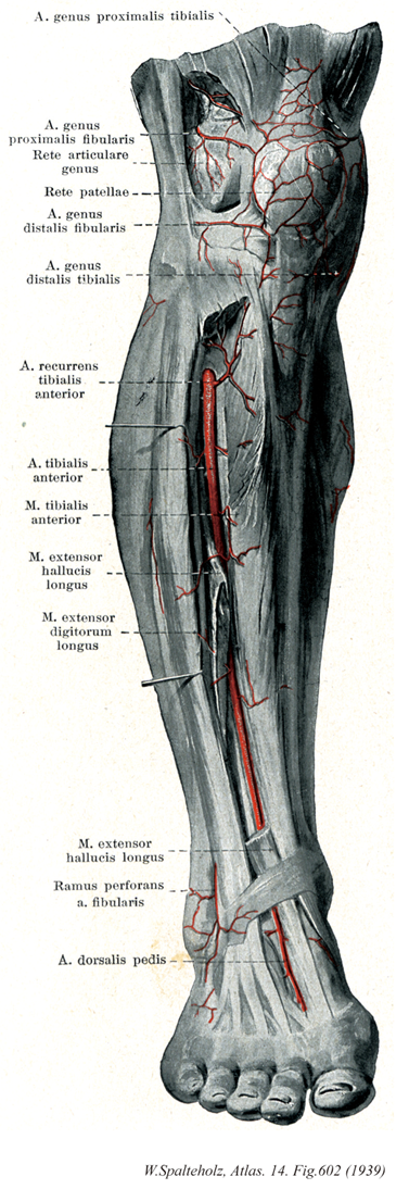
- 602_01【Superior medial genicular artery内側上膝動脈;脛側近位膝動脈 Arteria superior medialis genus; Arteria genus proximalis tibialis】 Artery passing beneath the tendon for the adductor magnus anteriorly to the genicular anastomosis.
→(内側上膝動脈は腓腹筋内側頭の上縁を通って前方へ向かい、大内転筋腱の深層に出て膝関節動脈網へ。一部は下行膝動脈と吻合枝、また筋枝を内側広筋に与える。しばしば弱小となり、このときは下行膝動脈関節枝によって代償される。)
- 602_02【Superior lateral genicular artery外側上膝動脈;腓側近位膝動脈 Arteria superior lateralis genus; Arteria genus proximalis fibularis】 Artery passing above the lateral femoral condyle and beneath the tendon of the biceps femoris anteriorly to the genicular anastomosis.
→(外側上膝動脈は腓腹筋の外側頭の上縁を通り、外側広筋に分布した後、膝関節動脈網へ。)
- 602_03【Genicular anastomosis膝関節動脈網;膝関節網 Rete articulare genus】 Arterial plexus mainly on the anterior side of the knee joint.
→(膝関節動脈網は膝関節の前面で、関節包の表面にある密な動脈網。これからたくさんの枝を関節の内部へ送る。この動脈網に加わる枝は、下行膝動脈、内および外上膝動脈、内および外下膝動脈、前および後脛骨反回動脈。)
- 602_04【Patellar anastomosis膝蓋動脈網 Rete patellare】 Special arterial plexus situated on the patella.
→(膝蓋動脈網は膝蓋骨のまわりの皮下組織にある動脈網で、膝関節動脈網の表層の一部と見なされる。膝蓋骨の上縁に沿って横走し、大腿四頭筋の表層にある動脈弓と、その下縁に沿って横走し、膝蓋靱帯の後面の脂肪組織の中にある動脈弓とが区別される。)
- 602_05【Inferior lateral genicular artery外側下膝動脈;腓側遠位膝動脈 Arteria inferior lateralis genus; Arteria genus distalis fibularis】 Artery passing beneath the lateral head of the gastrocnemius and beneath the fibular collateral ligament to the genicular anastomosis.
→(外側下膝動脈は腓腹筋外側頭および膝関節の外側側副靱帯の深層を通って膝関節の前面に出て、膝関節動脈網へ。)
- 602_06【Inferior medial genicular artery内側下膝動脈;脛側遠位膝動脈 Arteria inferior medialis genus; Arteria genus distalis tibialis】 Artery passing beneath the medial head of the gastrocnemius and tibial collateral ligament to the genicular anastomosis.
→(内側下膝動脈は、はじめ腓腹筋内側角深層を膝窩筋の上縁に沿って下内方へ走り、次いで脛骨内側顆の下方で内側側副靱帯の深層を通って前方にまわり、膝関節動脈網へ。)
- 602_07【Anterior tibial recurrent artery前脛骨反回動脈 Arteria recurrens tibialis anterior】 Artery passing through the tibialis anterior to the genicular anastomosis.
→(前脛骨反回動脈は本幹が下腿骨間膜を貫いてその前面に出た直後に分岐する。上行して膝関節動脈網と膝蓋動脈網へ。)
- 602_08【Anterior tibial artery前脛骨動脈 Arteria tibialis anterior】 It extends from its origin at the inferior border of the popliteus to the inferior border of the inferior extensor retinaculum. After penetrating the interosseous membrane, it lies between the tibialis anterior and extensor digitorum longus, then between the tibialis anterior and extensor hallucis longus.
→(前脛骨動脈は、腋窩の遠位部、すなわち膝窩筋の下縁の高さで、膝窩動脈が二分して生ずる枝の一つ。分岐後、下腿骨間膜の上部を越え、骨間膜の前面に出て下行する。経過中に下腿の前外側にある筋とその付近に枝を送る。動脈はとくに上部と下部で膝関節と足関節との周囲の膝蓋動脈網に小枝を送り吻合する。前脛骨動脈は、足関節のすぐ上方で表層に現れ、足関節の前側で前脛骨筋の腱の外側に沿って走り、足背で足背動脈となる。)
- 602_09【Tibialis anterior muscle前脛骨筋 Musculus tibialis anterior】 o:Lateral surface of tibia, interosseous membrane, deep fascia of leg. i: Medial aspects of medial cuneiform and first metatarsal. Dorsiflexion and supination of foot. I: Deep fibular nerve.
→(前脛骨筋は脛骨外側顆、脛骨外側面(近位2/3)、下腿筋膜および筋間膜から起始する。第1中足骨と第1楔状骨あたりの足底部に停止する。収縮中に筋腹は脛骨近位1/3の骨縁上に突出する。その腱は脛骨遠位1/3にかけて形成され、伸筋支帯の下を通って足の内側縁へ至る。その腱鞘は伸筋支帯より近位に始まり、距腿関節の関節腔のレベルにまで伸びている。腱鞘は前脛骨筋腱の遠位部および近位部浅層をおおい、中間部を包んでいる。前脛骨筋と長趾伸筋に対する近位の筋枝は深腓骨神経から同神経がまだ腓骨筋群を容れる部位を通っている内に分かれる。深腓骨神経が長趾伸筋を貫通してから遠位の筋枝が両筋の各々に行き(通常2条の)筋枝が母趾の伸筋へ行く。)
- 602_10【Extensor hallucis longus muscle長母趾伸筋;長母指伸筋(足の) Musculus extensor hallucis longus】 o: Interosseous membrane and fibula, i: Distal phalanx of great toe. Dorsiflexion of foot and great toe. I: Deep fibular nerve.
→(長母趾伸筋は腓骨内側面と骨間膜(中間2/4,3/4の部)の起始部では隣り合う2つの筋によって完全におおわれている。長母趾伸筋の腱は上伸筋支帯の直下で浅層を走り末節骨に着く。また、一部は足背筋膜をもたない母趾基節骨にも付く。長母趾伸筋の腱鞘は内果のレベルでようやく始まるが、ずっと遠位へ伸び、第1中足骨底あるいは頭まで至る。)
- 602_11【Extensor digitorum longus muscle長趾伸筋;長指伸筋(足の) Musculus extensor digitorum longus】 o: Lateral condyle of tibia, interosseous membrane, fibula, deep fascia of leg. i: Dorsal aponeurosis of the second through fifth toes. Dorsiflexion and pronation of foot. Extension of toes. I: Deep fibular nerve.
→(長趾伸筋は脛骨外側顆、腓骨前縁および骨間膜の狭い部から起こり、第2~5趾の足背腱膜へ至る。足背腱膜はその基本構造においては手指の手背腱膜と同じである(つまり、各腱の側縁束は末節骨に、中央束は中節骨に終わる)。足背筋膜は趾の部で完全に区分できるとは限らない。骨間膜の腱は通常基節骨にしか停止せず、虫様筋の腱索は第2~5趾の中節骨や末節骨に達するとは限らないので、第2~5趾の各関節を能動的に伸展することはしばしば困難となる。母指末節骨のみは長母趾伸筋の作用によって背屈することが可能である。)
- 602_12【Perforating branch of fibular artery貫通枝;穿通枝(腓骨動脈の) Ramus perforans (Arteria fibularis)】 It pierces the interosseous membrane immediately above the malleolus and passes to the lateral malleolar network and dorsum of foot.
→(腓骨動脈の貫通枝は外果の上方約5cmの付近で分岐し、ただちに下腿骨間膜を貫いて下腿の前面に出て、前外果動脈と吻合する。)
- 602_13【Dorsalis pedis artery; Dorsal artery of foot足背動脈 Arteria dorsalis pedis】 Continuation of the anterior tibial artery on the dorsum of foot. After crossing under the tendon of the extensor hallucis longus and passage of the extensor retinaculum, it lies lateral to the tendon where it is palpable.
→(足背動脈は前脛骨動脈よりつづいて、距腿関節の前面から起こり、足背の内側縁を下行して、母趾と第2趾の間にある第1中足骨間隙の近位で、第1背側中足動脈と深足底枝に分かれておわる。前脛骨動脈が弱小化して、代償的に発達した腓骨動脈の貫通枝が、前外果動脈を経て足背動脈に接続することが約7%に出現する。)