Spalteholz HANDATLAS DER ANATOMIE DES MENSCHEN VON WERNER SPALTEHOLZ
メニューは解剖学(TA)にリンクしてあります。図の番号をクリックすると下記の説明へ、右側の用語をクリックすると解剖学(TA)にジャンプします。
609 


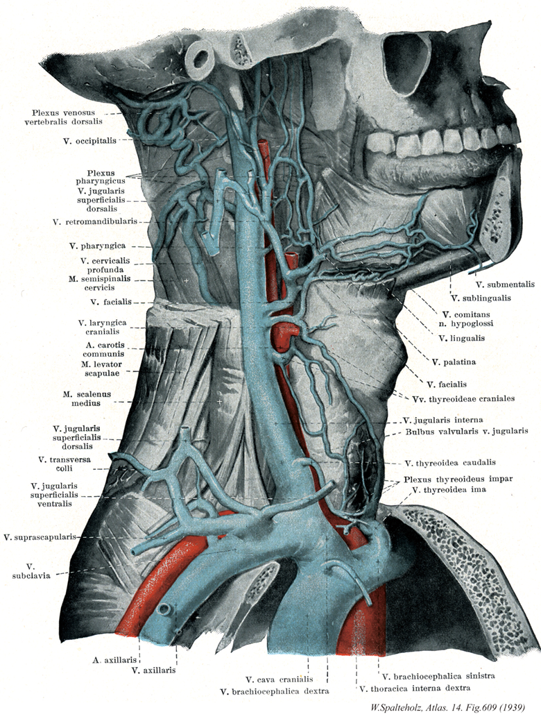
- 609_01【Posterior external vertebral venous plexus後外椎骨静脈叢;後椎骨静脈叢 Plexus venosus vertebralis externus posterior; Plexus venosi vertebrales dorsales】 Venous plexus lying posterior to the vertebral arches.
→(前外椎骨静脈叢および後外椎骨静脈叢は椎体の前面にあり、椎体よりの静脈を集める前外椎骨静脈叢と、椎骨の背面で追究の外表面にあり、主として脊柱管の内部よりの血液を集める後外椎骨静脈叢がある。これらの静脈叢は頚椎でよく発達し、椎骨の両側で互いに交通する。)
- 609_02【Occipital vein後頭静脈 Vena occipitalis】 Vein beginning in the venous plexus of the scalp. It frequently opens into the vertebral vein or also into the internal or external jugular vein.
→(後頭静脈は頭皮の静脈網から起こり、多くは椎骨静脈に、ときにはまた内頚静脈や外頚静脈にもひらく。)
- 609_03【Pharyngeal venous plexus咽頭静脈叢 Plexus venosus pharyngeus; Plexus pharyngeus】 Venous plexus on the pharyngeal muscles.
→(咽頭静脈叢は咽頭静脈を通って内頚動脈にはいる咽頭の後外側壁上の静脈叢。)
- 609_04【External jugular vein外頚静脈;外側浅頚静脈 Vena jugularis externa; Vena jugularis superficialis dorsalis】 Vein lying between the platysma and supetficial layer of cervical fascia and usually emptying into the subclavian vein. It is fed by the following veins.
→(外頚静脈は側頚部の皮下静脈であり、頚部のみならず頭部の表在性静脈血を集める。後耳介静脈と下顎後静脈が合して下顎角の後方ではじまり、広頚筋におおわれて胸鎖乳突筋の表面を斜めに下行し、大鎖骨上窩で鎖骨下静脈にそそぐ。下顎後静脈前枝を介して内頚静脈と連絡しているので、これら2静脈ならびに鎖骨下静脈とともに胸鎖乳突筋を斜めに取り囲む動脈輪を形成している。受け入れる静脈根は後頭静脈、後外頚静脈、頚横静脈と肩甲上静脈、前頚静脈である。)
- 609_05【Retromandibular vein下顎後静脈 Vena retromandibularis】 It extends from the union of several branches in front of the ear to the facial vein.
→(下顎後静脈は下顎頚の内側つまり耳管腺内で浅側頭静脈と顎静脈が合してはじまり、顔面静脈と合して内頚静脈に開口する。下顎後静脈は前枝と後枝と二分かれた形で耳下腺の下面から出る。そののち前枝は顔面静脈と合流する。後枝は後耳介静脈と合流し、外頚静脈を形成する。浅側頭静脈は表在性の静脈で、中側頭静脈、顔面横静脈を受け入れる。深在性の顎静脈は側頭下窩に広がる翼突筋静脈叢にはじまる。この静脈叢は顎動脈の分布域から血液を集め、中硬膜静脈などの硬膜静脈、深側頭静脈、前耳介静脈、耳下腺静脈、顎関節静脈、鼓室静脈、茎乳突孔静脈などを受け入れる。)
- 609_06【Pharyngeal veins咽頭静脈 Venae pharyngeae】 Veins from the pharyngeal plexus.
→(咽頭静脈は咽頭壁の咽頭静脈叢から出る。)
- 609_07【Deep cervical vein深頚静脈 Vena cervicalis profunda; Vena colli profunda】 Vein accompanying the deep cervical artery beneath the semispinalis capitis and cervicis.
→(深頚静脈は深頚動脈に伴行する。頭半棘筋と頚半棘筋に被われる。)
- 609_08【Semispinalis cervicis muscle; Cervical semispinalis muscle頚半棘筋 Musculus semispinalis cervicis; Musculus semispinalis colli】 o: Transverse processes ofT6-T2. i: Spinous processes of C6-C2. 1: Posterior rami of spinal nerves of C3-C6.
→(頚半棘筋の起始は上位6個の胸椎横突起、下位4個の頚椎関節突起。停止は第2~5頚椎棘突起。機能として脊柱の伸展、側方屈曲。頭、肋骨、骨盤の伸展。神経支配は下部3本の頚神経後枝。動脈は後肋間動脈の筋枝、後頭動脈の下行枝、肋頚動脈の深頚枝から受ける。胸および頚半棘筋はしばしば一単位を形成する。多裂筋と同様に、起始筋束は数個の停止筋束に分かれる。)
- 609_09【Facial vein顔面静脈 Vena facialis】 Vein beginning at the medial angle of eye that lies behind the facial artery and then beneath the submandibular gland.
→(顔面静脈は顔面動脈の分布域である顔面浅部からの静脈を集める。顔面静脈は内眼角から始まり(眼角静脈)、顔面動脈の後ろに沿って斜めに下方に走り、内・外頚動脈、舌下神経との浅側を後下方に向かい、舌骨の高さで内頚静脈または外頚静脈にそそぐ。顔面静脈は吻合に富み、また顔面の深部の静脈や頭蓋内の静脈(硬膜静脈洞)とも連絡している。たとえば、顔面静脈は内眼角の付近で、眼窩内の上眼静脈の根もと吻合し、さらに頭蓋腔内の顔面静脈洞とも連絡する。また、鼻や上唇の近くでも深部の静脈と連絡する。)
- 609_10【Superior laryngeal vein上喉頭静脈 Vena laryngea superior】 Companion vein of the superior laryngeal artery that drains into the superior thyroid vein.
→(上喉頭静脈は上喉頭動脈に伴行し上甲状腺静脈にひらく。)
- 609_11【Common carotid artery総頚動脈 Arteria carotis communis】 Artery of the neck without any branches. It runs on both sides of the trachea and larynx and passes deep to the sternocleidomastoid. It arises on the right from the brachiocephalic trunk and on the left from the aortic arch.
→(総頚動脈は頭部に血液を送る血管の主幹。右は腕頭動脈の枝、左は大動脈弓の上行部より出る。そのため左総頚動脈は右のものよりも4~5cm長い。総頚動脈は枝を出さず、気管・喉頭の両側を上行し、甲状軟骨上縁の高さで音叉のような形をなし内・外頚動脈に分かれる。分岐部の後側には頚動脈小体が存在する。また分岐部のないし内頚動脈始部の壁はやや薄く膨隆しており(頚動脈洞)、舌咽神経の枝を介し血圧を感受するという。)
- 609_12【Levator scapulae muscle; Levator scapular muscle肩甲挙筋 Musculus levator scapulae】 o:Posterior tubercles of cervical vertebrae C1-C4. i: Superior angle of scapula. Raises the scapula; rotates the inferior angle of the scapula medially. I: Dorsal scapular nerve.
→(肩甲挙筋は、上位4つの頚椎の横突起から、斜角筋と板状筋の間で起こる。起始部は外側頚三角にに、細い筋個体としてみえる。この筋は僧帽筋で被われ、斜めに下行して、肩甲骨上角および、肩甲棘よりも上の肩甲骨内側縁に停止する。)
- 609_13【Scalenus medius muscle; Middle scalene muscle中斜角筋 Musculus scalenus medius】 o:Transverse processes of C2-C7. i: First rib posterior to the groove for the subclavian artery. Elevation of the first rib and lateral flexion of the neck. I: Cervical plexus and brachial plexus (C4-C8).
→(中斜角筋はもっともよく発達した悌子状の筋で、C3-7横突起(前、後結節間の溝)に起始をもち、しばしば環椎と軸椎から起こる副束を持つ。この筋は第1肋骨に鎖骨下大静脈溝の背外側で停止し、ときに線維束の一部が第2肋骨外側外面に付くこともある。参考:斜角筋群は主に吸息筋として働き頚椎を動かす作用はむしろ従であるという。)
- 609_14【Transverse cervical veins頚横静脈 Venae transversae cervicis; Venae transversae colli】 Veins accompanying the transverse cervical artery.
→(頚横静脈は頚横動脈に伴行し外頚静脈下部にそそぐ。)
- 609_15【Anterior jugular vein前頚静脈;前浅頚静脈 Vena jugularis anterior; Vena jugularis superficirlis ventralis】 It commences at the level of the hyoid bone and, after crossing under the stemocleidomastoid, often drains into the external jugular vein.
→(前頚静脈は下顎部の静脈を集めて舌骨付近にはじまり、正中傍部皮下を下行して頚部下端に達し、外側に曲がって外頚静脈又は鎖骨下静脈に注ぐ。しばしば左右が合して正中線を下行し頚正中静脈をなす。左右の前頚静脈は胸骨上隙で交通して頚静脈弓をつくり、またしばしば胸鎖乳突筋前縁に沿って流れる静脈(頚斜頚静脈)を介して外頚静脈と交通する。)
- 609_16【Suprascapular vein肩甲上静脈 Vena suprascapularis】 Usually two veins accompanying the suprascapular artery.
→(肩甲上静脈は肩甲上動脈に伴行するもので多くは2本あり外頚静脈下部に注ぐ。)
- 609_17【Subclavian vein鎖骨下静脈 Vena subclavia】 Vein lying between the anterior scalene muscle and sternocleidomastoid. It extends from the internal jugular vein to the lateral border of the first rib.
→(鎖骨下静脈は第一肋骨の外側縁からの腋窩静脈の直接の続き。鎖骨の内側端で内頚静脈と合して腕頭静脈を形成するまでの部分を指す。前方は鎖骨と鎖骨下筋に、後方は前斜角筋を隔てて鎖骨下動脈に接し、下方は第1肋骨上面の鎖骨顆上脈溝に接する。まれに動脈と伴行して前斜角筋の後方を通ることがある。枝として①胸筋枝、②背側肩甲静脈、③胸肩峰静脈がある。)
- 609_18【Axillary artery腋窩動脈 Arteria axillaris】 Continuation of the subclavian artery that reaches the inferior border of the pectoralis major.
→(腋窩動脈は鎖骨下動脈よりつづく上肢の動脈の本幹で、第1肋骨外側縁の高さで鎖骨下動脈よりつづいてはじまり、大胸筋の停止腱、あるいは大円筋の停止腱の高さで上腕動脈に移行する。これに通常3部を区分し、第1部は、小胸筋の上縁より上方にある部分で、前面は大胸筋鎖骨部に被われ、後方と外側は腕神経叢に接する。第2部は、小胸筋の後面にあたる部分で、この部で腋窩動脈は腕神経叢に貫くため、その後面、内側面、外側面をそれぞれ腕神経叢の後神経束、内側神経束および外側神経束に達している。第3部は、小胸筋の下縁より下方にある部分で、前面は正中神経に、外側は筋皮神経と烏口腕筋に、内側は尺骨神経を介して腋窩静脈に、そして後面は橈骨神経と腋窩神経を介して肩甲下筋と広背筋の停止腱に接する。枝としては、①肩甲下肢、②最上胸動脈、③胸肩峰動脈、④外側胸動脈、⑤肩甲下動脈、⑥前上腕回旋動脈、⑦後上腕回旋動脈)
- 609_19【Axillary vein腋窩静脈 Vena axillaris】 Continuation of the subclavian vein. It extends from the lateral border of the first rib to the inferior border of the tendon of the pectoralis major.
→(腋窩静脈は上肢の静脈を集める。大胸筋の下縁の高さで上腕静脈からつづいておこり腋窩動脈の内側に沿って走り、第1肋骨の高さで鎖骨顆上脈に注ぐ。枝として①外側胸静脈、②胸腹壁静脈、③乳輪静脈叢がある。)
- 609_20【Superior vena cava上大静脈 Vena cava superior; Vena cava cranialis】
→(上大静脈は上半身の血液を集める静脈で、上縦隔の中で左右の腕頭静脈が合してはじまり、途中で奇静脈を受け入れながら上行大動脈の右側を下行して右心房にそそぐ。)
- 609_21【Right brachiocephalic vein右腕頭静脈 Vena brachiocephalica dextra】
→()
- 609_22【Submental veinオトガイ下静脈 Vena submentalis】 Companion vein of the submental artery. It anastomoses with the sublingual and anterior jugular veins.
→(オトガイ下静脈はオトガイ下動脈に伴行する。舌下静脈および前頚静脈と吻合。)
- 609_23【Sublingual vein舌下静脈 Vena sublingualis】 Larger vein coursing lateral to the hypogiossal nerve.
→(舌下神経の側方にあり、やや太い。 (Feneis))
- 609_24【Vena comitans of hypoglossal nerve舌下神経伴行静脈 Vena comitans nervi hypoglossi】 Vein accompanying the hypogiossal nerve.
→(舌骨舌筋の下および外側を舌下神経とともに走り、通常は舌下静脈に注ぐ。)
- 609_25【Lingual vein舌静脈 Vena lingualis】 Vein of the tongue that mostly lie near the lingual artery.
→(舌静脈は舌からの舌深静脈と舌背静脈、ならびに顎下腺、舌下腺からの舌下静脈が合して形成される。舌下静脈は舌下神経伴行静脈をも受ける。舌骨舌筋に対して舌動脈は内側を通るのに対して、舌静脈はその外側を通過する。内頚静脈または顔面静脈へ注ぐ。)
- 609_26【External palatine vein外口蓋静脈;口蓋静脈 Vena palatina externa; Vena palatina】 Conveys blood from the lateral tonsillar region of the palate and pharyngeal wall to the facial vein.
→(外口蓋静脈は口蓋扁桃の側方部と咽頭壁の血液を顔面静脈へ運ぶ。)
- 609_27【Superior thyroid vein上甲状腺静脈 Vena thyroidea superior; Vena thyroidea cranialis】 Companion vein of the superior thyroid artery. It empties into the facial or internal jugular vein.
→(上甲状腺静脈は甲状腺上部におこり上甲状腺動脈に伴行する。上喉頭静脈ならびに胸鎖乳突筋静脈を受け入れる。胸鎖乳突筋静脈は内頚静脈に直接そそぐこともある。またしばしば甲状腺下部におこる中甲状腺静脈がみられる。)
- 609_28【Internal jugular vein; Jugular vein内頚静脈 Vena jugularis interna】 Main vein of the neck that extends from the jugular foramer to the venous angle.
→(内頚静脈は脳、顔と頚の浅層からの血液を集める。この大きな静脈は、後頭蓋窩の後静脈孔で、S状静脈洞から直接つながって始まり、内頚動脈についで総頚動脈に沿って下行し、鎖骨下静脈と合して腕頭静脈に終わる。上端と下端では肥大しており、それぞれ頚静脈上球ならびに頚静脈下球とよばれる。内頚静脈に注ぐ根として蝸牛小管静脈、咽頭静脈、舌静脈、上甲状腺静脈、顔面静脈、下顎後静脈がある。内頚静脈と鎖骨下静脈とが合流するところを静脈角angulus venosusといい、左側の静脈角には右胸管が開口し、右側の静脈角の近くには右リンパ幹が注いでいる。)
- 609_29【Inferior bulb of jugular vein頚静脈下球;内頚静脈弁膜球 Bulbus inferior venae jugularis; Bulbus valvularis venae jugularis internae】 Dilatation at the end of the internal jugular vein that is closed off cranially by a valve.
→(頚静脈下球は内頚静脈下端の膨大部で、その頭側は弁で閉じられている(Feneis)。)
- 609_30【Inferior thyroid vein下甲状腺静脈 Vena thyroidea inferior】 Vein passing from the unpaired thyroid plexus beneath the thyroid gland to the left (occasionally also right) brachiocephalic vein.
→(下甲状腺静脈は甲状腺下縁の不対甲状腺静脈叢から出る。同名の下甲状腺動脈には伴行しない。)
- 609_31【Unpaired thyroid venous plexus不対甲状腺静脈叢 Plexus venosus thyroideus impar】 Venous plexus situated anterior to the trachea and below the caudal border of the thyroid gland.
→(不対甲状腺静脈叢は甲状腺下縁の下方で気管前方にある静脈叢。)
- 609_32【Thyroid ima vein最下甲状腺静脈 Vena thyreoidea ima】
→()
- 609_33【Left brachiocephalic vein左腕頭静脈 Vena brachiocephalica sinisra】
→()
- 609_34【Right internal thoracic vein右内胸静脈 Venae thoracicae internae dexter】
→()
- 609_34a【Internal thoracic veins; Internal mammary veins内胸静脈 Venae thoracicae internae】 Veins accompanying the internal thoracic artery. They are often present as two veins as far as the third costal cartilage, after which they pass as a single vein medial to the artery.
→(内胸静脈は内胸動脈の伴行静脈であり、腹部皮下の静脈網である腹皮下静脈にはじまり、上腹壁静脈となって横隔膜前端を貫き筋横隔静脈と合して内胸静脈として上行し、経過中に前肋間静脈を受け入れる。)