Spalteholz HANDATLAS DER ANATOMIE DES MENSCHEN VON WERNER SPALTEHOLZ
メニューは解剖学(TA)にリンクしてあります。図の番号をクリックすると下記の説明へ、右側の用語をクリックすると解剖学(TA)にジャンプします。
679


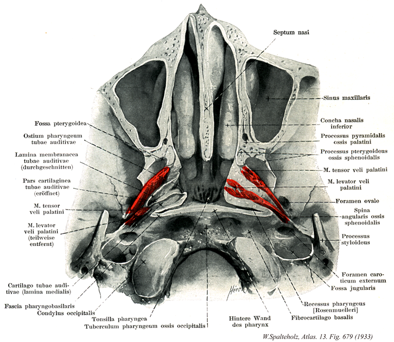
- 679_00【Pharynx咽頭 Pharynx】 Airway and food passageway; 1416 cm long. It extends from the vault of pharynx to the beginning of the esophagus in front of the sixth cervical vertebra.
→( 咽頭は上端で咽頭円蓋となりその前方で後鼻孔を介して鼻腔に、その直下で口峡を介して口腔につながる。下方は第6~7頚椎、または輪状軟骨下端の高さで食道に移行し、途中、第二頚椎の高さで咽頭前壁に喉頭口が開口する。したがって、咽頭は呼吸器系と消化器系が交叉している。前後にやや扁平な管で、肝の内腔が咽頭腔である。咽頭の後壁は単純であるが、前・側壁は発生時に鰓弓と関連が深く、生体での構造が複雑となる。咽頭を上から下へ、鼻部・後部・後頭部の三つに分ける。鼻部は鼻腔につづき、燕下時に軟口蓋が挙上すると、消化管から遮断される。したがって鼻部は気道に属するとみなされることが多い。後部は口峡を経て口腔につづき、軟口蓋と舌根とが前方上下に位置する。後頭部は前壁に後頭の後壁となる。咽頭の下端は食道に連続する。鼻部には耳管が開き、その開口部を耳管咽頭口という。この周囲では咽頭壁にかなり凹凸がみられる。耳管隆起は耳管軟骨により、挙筋隆起は口蓋帆挙筋により生ずる。耳管咽頭ヒダは耳管咽頭筋の足行き一致する。耳管隆起の後方のくぼみは咽頭陥凹とよばれる。鼻部の天井は頭蓋底直下にあたり、この部分を咽頭円蓋という。喉頭部では、舌根の後下方に喉頭蓋が突き出す後頭口の両側、すなわち後頭の側壁と咽頭の側壁の井田は梨状陥凹とよばれる。ここは燕下時に食物の通路となる。この部に上喉頭神経・動脈による後頭神経ヒダを認める。咽頭壁は、最上部では前方鼻腔へ通じる部分を除き、頭蓋底に付着する。頭蓋底近くでは、咽頭壁は筋層を欠き、結合織性の壁となす。これを咽頭頭底板という。咽頭の粘膜上皮、他では重層扁平上皮である。咽頭線は粘膜全体に分布する。)
- 679_01【Pterygoid fossa翼突窩 Fossa pterygoidea】 Depression between the lateral and medial plates of the pterygoid process for the medial pterygoid muscle.
→(翼状突起の内側板、外側板は後方に開いた翼突窩をつくる。ここから内側翼突筋が起始する。)
- 679_02【Pharyngeal opening of pharyngotympanic tube耳管咽頭口 Ostium pharyngeum tubae auditivae; Ostium pharyngeum tubae auditoriae】 Funnel-shaped or slitlike opening of the auditory tube above the levator eminence at the level of the inferior nasal meatus 1 cm in front of the posterior wall of the pharynx.
→(口蓋帆拳筋の根元の上方で、後咽頭壁より1cm前方の下鼻道へ開く漏斗状の開口部。 (Feneis))
- 679_03【Membranous lamina of pharyngotympanic tube膜性板(耳管の) Lamina membranacea tubae auditivae】 Membranous part in the wall of the cartilaginous part of auditory tube.
→(耳管軟骨部の膜性部。 (Feneis))
- 679_04【Cartilaginous part of nasal septum; Cartilaginous part of pharyngotympanic tube軟骨部(鼻中隔の) Pars cartilaginea tubae auditivae】 Part of the nasal septum between the membranous and bony parts.
→()
- 679_05【Tensor veli palatini muscle口蓋帆張筋 Musculus tensor veli palatini】 o: Spine of sphenoid bone, scaphoid fossa, and anterior (lateral) lip of cartilaginous part of auditory tube, i: After changing direction at the pterygoid hamulus, its fibers merge with the palatine aponeurosis, stiffening the anterior (lateral) wall of the membranous lamina of auditory tube and tensing the soft palate. I: Mandibular nerve.
→(口蓋帆張筋は舟状窩、蝶形骨大翼下面の細い帯および耳管の膜性外壁から起始する。翼突窩のレベルで口蓋帆張筋はすでに腱に移行し、腱は翼突鈎をめぐって方向を転じて、水平に口蓋腱膜へ放射する。口蓋帆張筋は燕下swallowing or deglutionの時に耳管を開く作用がある。)
- 679_06【Levator veli palatini muscle口蓋帆挙筋 Musculus levator veli palatini】 o: Petrous part of temporal bone in front of the inferior opening of the carotid canal. Inferior border of the cartilaginous auditory tube, i: Palatine aponeurosis. It draws the soft palate backward and upward, also moving the dorsomedial cartilaginous part of auditory tube when the pharyngeal opening of auditory tube is opened. I: Vagus nerve.
→(口蓋帆挙筋は、口蓋帆張筋の後に位置し、側頭骨錐体部下面で頚動脈管の前部および耳管軟骨から起始する。口蓋帆挙筋は耳管に沿って斜め前・下方へ走り、軟口蓋にはいる。両側の口蓋帆挙筋の腱線維はからみ合って、高さが調節できる筋性ワナを形成する。)
- 679_07【Cartilage of tube; Cartilage of pharyngotympanic tube耳管軟骨 Cartilago tubae auditivae; Cartilago tbae auditoriae】 Auditory tube cartilage that appears hook-shaped in cross-section. It decreases in height posterolaterally and is composed of elastic cartilage only in the angle between its two laminae.
→(横断面では鈎状の軟骨。外側後方へ低くなり、両軟骨板のなす角の部分のみ弾性軟骨でできている。 (Feneis))
- 679_08【Medial lamina of cartilage of pharyngotymanic tube内側板(耳管軟骨の) Lamina medialis cartilagis tubae auditivae】 Wider, medial sheet of cartilage.
→(幅の広い内側に面する板。 (Feneis))
- 679_09【Pharyngobasilar fascia咽頭頭底板 Fascia pharyngobasilaris; Membrana pharyngobasilaris】 Uppermost, nonmuscular, membranous part of the pharynx. It attaches the wall of the pharynx to the base of the cranium. Corresponds to the submucosa
→(咽頭壁のうち筋のない最上部の結合組織壁。)
- 679_10【Occipital condyle; *Condyle後頭顆 Condylus occipitalis】 Spherical eminence for articulation with the atlas.
→(後頭骨下面にある2つの細長い卵形をした関節面を有する高まりが後頭顆である。これは第1頚椎の上関節窩と関節する。)
- 679_11Luschka, Glands of, Tonsil of【Pharyngeal tonsil咽頭扁桃 Tonsilla pharyngealis; Tonsilla pharyngea】 Tonsil located in the vault of pharynx.
→(咽頭扁桃は咽頭鼻部天井の粘膜下組織の集まりにより形成される物であり、口蓋扁桃と同じく、小児期に最も発達して思春期以後には退縮傾向を示す。)
- 679_12【Pharyngeal tubercle; Pharngeal tubercle of occipital bone咽頭結節(後頭骨の) Tuberculum pharyngeum】 Small protuberance on the inferior aspect of the basilar part of the occipital bone that provides attachment to the pharyngeal raphe.
→(後頭骨の底部の下面は筋の付着部となるため全般に粗で、その中央に小さい咽頭結節がある。咽頭結節は咽頭後壁の咽頭縫線が着く所である。その両側には上咽頭収縮筋、そのさらに側方には頭長筋、前頭直筋が着く。)
- 679_13【Nasal septum鼻中隔 Septum nasi】 Nasal partition consisting of bony, cartilaginous, and membranous parts.
→(鼻中隔は軟骨性および骨性の要素から成る。鼻中隔軟骨は後突起を出して骨性の隔壁を補完している。鼻中隔軟骨は両側で大鼻翼軟骨内側脚が重なって外鼻孔の内側の境界をつくる。骨性の隔壁(骨鼻中隔)は篩骨垂直板、蝶形骨稜ならびに鋤骨からなる。)
- 679_14Highmore, Antrum of【Maxillary sinus上顎洞;ハイモア腔 Sinus maxillaris】 It measures over 3 cm vertically and sagittally and 2.5 cm in the frontal plane. Its floor is usually at least 1 cm below the floor of the nose and its lowest point is usually at the level of the first molar.
→(上顎洞は上顎体中にある最大の副鼻腔で、その形はだいたいにおいて上顎体の形に一致するが、尖端を外上方、すなわち頬骨突起の方に出しているので錐体状に近く、その底は鼻腔面にむく。ここにはなはだ大きい上顎洞裂孔があるが、完全な頭蓋ではこの裂孔は口蓋骨の垂直板、篩骨の鈎状突起および下鼻甲介の上顎、篩骨稜突起によりその一部がふさがれて著しく小さくなる。(生体では、さらに鈎状突起まで鼻粘膜に被われるため、中鼻甲介の下の半月裂孔に開く小さな開口を残すのみとなる。)上顎洞はその前壁が最も厚く、つぎは後壁、上壁の順で内側壁が最も薄い。下壁は歯槽突起に入り、場所によってその厚さが異なるが、大臼歯および小臼歯の歯根をおおう部、とくに第1、第2臼歯の付近で最も薄く、それらの歯根はしばしば洞に達する。また、下壁には歯槽中隔の為に多くの骨の高まりやくぼみを見るのを常とする。なお、上顎洞の前後稜壁には多くの細い歯槽溝または歯槽管および歯槽孔が見られる。『ハイモア洞』:イギリスの自然科学者Nathaniel Highmore (1613-1685)の名を冠するが、レオナルド・ダ・ビンチがすでに観察している。ハイモアは、この他にも精巣縦隔(Highmore's body)に名を残している。)
- 679_15【Inferior nasal concha下鼻甲介 Concha nasalis inferior】 The independent lower nasal concha, which is attached to the lateral nasal wall.
→(中鼻甲介と殆ど同じ形状でこれより大きく、その下方で鼻腔外側壁に付着する1対の独立した小骨で、縁が湾曲した薄い海綿状骨板で、鼻腔の側壁にあり、中鼻道と下鼻道を分ける。篩骨、涙骨、上顎骨、口蓋骨とで関節をなす。下鼻甲介の内側面は鼻腔内に向かってふくらんだ粗面である。下縁は中鼻甲介のように外側に少し巻いている。上縁に涙骨突起、上顎突起、および篩骨突起がある。海綿状骨板とその肥厚した粘膜骨膜で、熱交換のための広範な海綿状の血管床を含む)
- 679_16【Pyramidal process of palatine bone錐体突起(口蓋骨の) Processus pyramidalis (Os palatinum)】 The inferoposterior end of the perpendicular plate of the palatine bone, which is inserted in the pterygoid notch.
→(垂直板の下部は水平板より矢状径が広くなり、水平板より後に大きく突出する錐体突起となって蝶形骨翼状突起の翼突切痕にはまる。)
- 679_17【Pterygoid process of sphenoid bone翼状突起(蝶形骨の) Processus pterygoideus (Ossis sphenoidalis)】
→(翼状突起は蝶形骨の両側で蝶形骨体と蝶形骨大翼との間の下面から下方に向かい、頭蓋底面に対してほぼ直角に出る突起で、外側板と内側板からなり、その前面は口蓋骨垂直板および上顎体後部に接する。翼状突起の根部は前後に走る翼突管で貫かれる。)
- 679_18【Foramen ovale of sphenoid bone卵円孔(蝶形骨の) Foramen ovale (Ossis sphenoidalis)】 Opening for the passage of the mandibular nerve anteromedial to the foramen spinosum.
→(卵円孔は大翼の後内側端に位置し、三叉神経の下顎神経の通路の開口で、棘孔の内前方にある。海綿静脈洞と翼突静脈叢を連絡することがある。)
- 679_19【Spine of sphenoid bone蝶形骨棘;角棘 Spina ossis sphenoidalis; Spina angularis】 Inferiorly projecting tip of the greater wing of the sphenoid.
→(蝶形骨の大翼の外側縁と後縁と合する部の下面からは下方に向かう鋭い蝶形骨棘がでる。蝶形骨棘には蝶下顎靱帯、翼棘突靱帯がつき、口蓋帆張筋の上部が起こる。)
- 679_20【Styloid process of temporal bone茎状突起(側頭骨の) Processus styloideus (Ossis temporale)】 Long bony process in front of the stylomastoid foramen. It is a relic of the hyoid arch.
→(茎状突起は錐体下面の後外側端から前下方へ向かう細長い突起である。その長さは1~5cmで、茎突下顎靱帯、茎突舌骨靱帯、茎突喉頭筋などの起点となる。茎状突起の根部の前面は茎状突起鞘で被われる。なお、茎状突起は舌骨と関係ある第2鰓弓軟骨の一部が骨化したも野である。)
- 679_21【External opening of carotid canal頚動脈管外口;外口(頚動脈管の) Apertura externa (Canalis caroticus); Foramen caroticum exernum】 Opening in the external cranial base between the jugular foramen and the musculotubal canal.
→(頚静脈下の前内側には大きい頚静脈管外口がある。この頚静脈管外口の前から口蓋帆張筋の一部が起こる。)
- 679_22【Jugular fossa頚静脈窩;頚窩 Fossa jugularis】 Widening of the jugular foramen that contains the superior bulb of the jugular vein.
→(錐体下面の後縁に近い中部には弓状の大きく深い頚静脈窩がる。頚静脈上球を容れる。)
- 679_23Rosenmüller, Fossa of【Pharyngeal recess咽頭陥凹 Recessus pharyngeus】 Nasopharyngeal niche situated posterolateral to the auditory tube.
→(『ローゼンミュラー窩』とも呼ばれる。耳管隆起の後上方には、鉛直位の深窩、すなわち咽頭陥凹がある。ローゼンミューラー Rosenmueller, Johann Christian (17771-1820) ドイツの解剖学者。ローゼンミュラー腺(涙腺、鼡径輪リンパ腺)、ローゼンミュラー窩(鼻咽腔の外側の小陥凹)を記述。)
- 679_24【Basal fibrocartilage頭底線維軟骨;頭蓋底線維軟骨 Fibrocartilago basialis】
→()