Spalteholz HANDATLAS DER ANATOMIE DES MENSCHEN VON WERNER SPALTEHOLZ
メニューは解剖学(TA)にリンクしてあります。図の番号をクリックすると下記の説明へ、右側の用語をクリックすると解剖学(TA)にジャンプします。
914


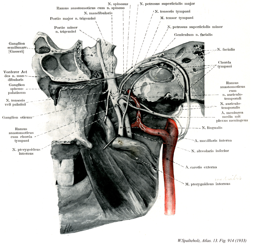
- 914_01【Meningeal branch of mandibular nerve; Nervus spinosus硬膜枝(下顎神経の) Ramus meningeus (Nervus mandibularis); Nervus spinosus】 Nerve passing through the foramen spinosum, accompanying the two branches of the middle meningeal artery. It supplies the dura mater, part of the sphenoidal sinus, and the mastoid cells.
→(下顎神経の硬膜枝は頭蓋を出るとすぐ分かれて棘孔を通って再び頭蓋腔に入り、上顎神経の硬膜枝とともに、中硬膜動脈に沿って脳硬膜に分布する知覚枝で、なお蝶形骨大翼乳突蜂巣の内部にも線維を与える。)
- 914_02【Communicating branch with meningeal branch; Communicating branch of glossopharyngeal nerve with meningeal branch硬膜枝との交通枝;下顎神経の硬膜枝との交通枝 Ramus communicans cum ramo meningeo; Ramus communicans nervus glossopharyngei cum ramo meningeo nervus vagi】 Branch communicating with the meningeal branch of mandibular nerve.
→()
- 914_03【Mandibular nerve; Mandibular division of trigeminal nerve [Vc; V3]下顎神経[三叉神経第3枝] Nervus mandibularis [Vc; V3]】 Third division of the trigeminal nerve that passes through the foramen ovale into the infratemporal fossa. It contains sensory fibers and motor fibers for the muscles of mastication.
→(三叉神経節からの感覚線維と運動根が卵円孔で結合してできる三叉神経の第三枝で最も太く、この神経は三叉神経節から出てただちに蝶形骨大翼の卵円孔を通って側頭下窩に出て硬膜、咀嚼筋、頬粘膜、耳介、外耳道付近その他へ枝を与えた後、舌神経、下歯槽神経の2終枝に分かれる。)
- 914_04【Sensory root of trigeminal nerve感覚根;知覚性根;知覚根;大部(三叉神経の);三叉神経根 Radix sensoria; Portio major; Radix nervi trigemini】 Sensory part of the nerve that emerges caudally from the pons and extends to the trigeminal ganglion.
→(三叉神経の知覚根は体性感覚線維で三叉神経の大部に相当し、橋に入り、三叉神経主感覚核と三叉神経脊髄路核に分布する。)
- 914_05【Motor root of trigeminal nerve運動根;運動性根;小部(三叉神経の) Radix motoria; Portio minor】 Root emerging from the trigeminal nerve toward the skull vertex and then passing under the trigeminal ganglion, innervating the muscles of mastication.
→(三叉神経の運動根は三叉神経の小根で、三叉神経運動核から出ている線維からなる。大きい知覚根の内側に位置して、橋から出て下顎神経に接続し咀嚼筋へ運動と固有受容の線維を送る。すなわち第一鰓弓に由来する筋で4種の咀嚼筋、おとがい舌骨筋、顎二腹筋の前腹、鼓膜張筋および口蓋帆張筋である。)
- 914_06Gasserian ganglion【Trigeminal ganglion三叉神経節;半月神経節 Ganglion trigeminale; Ganglion semilunare】 Crescent-shaped equivalent of a spinal ganglion of the trigeminal nerve lying in an outpouching in the subarachnoid space (trigeminal cavity) above the foramen lacerum on the medial, anterior surface of the petrous temporal bone.
→(ガッセル神経節またはガッサー神経節とよばれる。三叉神経の大きい扁平な知覚神経節で、中頭蓋窩の正中部分に沿った静脈洞に密接して脳硬膜の三叉神経腔にある。オーストラリアの解剖学者Johann Laurentius Gasser (1723-1765頃)によって報告された。彼自身についてはよく判っていない。)
- 914_07Meckel's ganglion【Pterygopalatine ganglion翼口蓋神経節;蝶口蓋神経節 Ganglion pterygopalatinum; Ganglion sphenopalatinum】 Ganglion measuring 4-5 mm that lies lateral to the sphenopalatine foramen in the pterygopalatine fossa. It contains cells for the postganglionic parasympathetic fibers to the lacrimal gland and the small nasal and palatine glands.
→(『メッケル神経節』ともよばれる。翼口蓋上部に位置する副交感神経節で、ここからの節後線維は涙腺などに分布する。上顎神経に付属する神経節で翼口蓋窩に位置し上顎神経の内側にある。形は不規則扁平でその頚は4~6mmである。毛様体神経節とおなじく3根ある。そのなかで知覚根は翼口蓋神経節由来で、一部はこの神経節で終わるが、一部はそのままこれを通って鼻腔および口蓋に分布する。運動根は顔面神経から出る大錐体神経で上唾液核から出て中間神経に入った副交感線維からなり、交感根は上顎神経節から出て内頚動脈神経叢を通ってくる抗感染位からなる深錐体神経である。大錐体神経および深錐体神経は、破裂孔の頭底軟骨を貫いて頭蓋下面に達して合し、翼突管神経として翼突管中を前進して翼口蓋神経節に入る。翼口蓋神経節から出る神経は主として鼻腔後部、口蓋および眼窩の一部などに分布して、知覚および植物神経線維を与える。ドイツの解剖学者・産科医Johann Friedrich Meckel (1714-1774)によって、1748年に記載された。メッケル憩室を発見したJohann F. Meckel (1781-1833)は彼の孫にあたる。)
- 914_08【Nerve to tensor veli palatini口蓋帆張筋神経 Nervus musculi tensoris veli palatini】 Branch supplying the tensor veli palatini. It sometimes arises from the nerve to the medial pterygoid.
→()
- 914_09Arnold's ganglion【Otic ganglion耳神経節 Ganglion oticum】 Flattened ganglion that varies in shape. It lies directly beneath the foramen ovale medial to the mandibular nerve and contains cells of postganglionic parasympathetic fibers to the parotid gland.
→(卵円孔の直下で三叉神経第3枝である下顎神経の内側に接して存する楕円形の副交感神経節で、その節後線維は分泌促進性で耳下腺へ分布する。根として下顎神経の枝(運動根)、中硬膜動脈を包む交感神経叢の枝(交感根)および舌咽神経、鼓室神経叢を経てきた小錐体神経(知覚および副交感根)がある。これより出る枝には口蓋翼張筋神経、鼓膜張筋神経、耳介側頭神経との交通枝、鼓索神経との交通枝、下顎神経の硬膜枝との交通枝。)
- 914_10【Communicating branch with chorda tympani; Communicating branch of glossopharyngeal nerve with chorda tympani鼓索神経との交通枝(舌咽神経の) Ramus communicans cum chorda tympani; Ramus communicans nervus glossopharyngei cum chorda tympani】 Sensory branch that communicates with the chorda tympani.
→()
- 914_11【Nerve to medial pterygoid内側翼突筋神経 Nervus pterygoideus medialis; Nervus musculi pterygoideus medialis】 Motor branch supplying the medial pterygoid. Small branches pass to the tensor veli palatini and tensor tympani.
→()
- 914_12【Greater petrosal nerve; Parasympathetic root of pterygopalatine ganglion大錐体神経;翼口蓋神経節の副交感神経根;大浅錐体神経 Nervus petrosus major; Radix parasympathica pterygopalatini; Nervus petrosus superficialis major】 Nerve leaving CN VII at the geniculate ganglion as a bundle of parasympathetic fibers. It reaches the anterior surface of the petrous pyramid, passes through the foramen lacerum, and travels with the deep petrosal nerve in the pterygoid canal to the pterygopalatine ganglion.
→(膝神経節から出て錐体の前上面を前にすすみ、破裂孔の軟骨を貫いて頭蓋底外面に出て、交感神経性の深錐体神経と合して翼突管神経をなし、翼口蓋神経節に入る。)
- 914_13【Nerve to tensor tympani鼓膜張筋神経 Nervus musculi tensoris tympani; Nervus tensoris tympani】 Branch supplying the tensor tympani that sometimes also supplies the medial pterygoid.
→()
- 914_14Eustachian muscle【Tensor tympani muscle鼓膜張筋 Musculus tensor tympani】 Muscle lying in the canal for the tensor tympani above the auditory tube. Its tendon runs laterally at nearly a right angle around the processus cochleariformis and attaches to the base of the handle of malleus. I: Mandibular nerve.
→(耳管上方の鼓膜張筋半管中にある。腱はサジ状突起でほぼ直角に外側へまがり、ツチ骨柄の底部へつく。神:鼓膜張筋神経。 (Feneis))
- 914_15【Lesser petrosal nerve; Parasympathetic root of otic ganglion小錐体神経;耳神経節の副交感神経根;小浅錐体神経 Nervus petrosus minor; Radix parasympathica ganglii otici】 Nerve containing parasympathetic fibers from the glossopharyngeal nerve. It arises from the tympanic plexus, penetrates the anterior wall of the petrous part of temporal bone, and emerges from the middle cranial fossa through the sphenopetrosal fissure. Its fibers synapse in the otic ganglion.
→(鼓室神経叢よりでて耳神経節へいたる副交感神経神経。錐体前壁を貫き、蝶錐体裂を通り、卵円孔の下、下顎神経の内側で耳神経節へ入る。 (Feneis))
- 914_16【Geniculum nerve; Geniculum of facial nerve顔面神経膝;顔面神経外膝 Geniculum nervi faciales】 Genu of the facial nerve located just beneath the anterior wall of the petrous part of temporal bone where the facial nerve changes direction from anterolateral to posterolateral.
→(顔面神経線維は外転神経核の周囲を回って向きを変えるところを顔面神経膝とよんでいる(内神経節)。)
- 914_17【Facial nerve [VII]顔面神経[脳神経VII] Nervus facialis [VII]】 Nerve arising from the second pharyngeal arch. It emerges from the brain at the pontocerebellar angle between the pons and inferior olive and passes with the vestibulocochlear nerve to the petrous part of the temporal bone, which it exits via the stylomastoid foramen. It supplies the muscles of facial expression.
→(顔面神経は第七脳神経である。狭義の顔面神経と中間神経とを合わせたもので、混合神経である。その主部をなす狭義の顔面神経は運動神経で、起始核たる顔面神経核は延髄上部から橋背部にかけてあり、これから出る神経は橋の後縁で脳を去り、内耳神経とともに内耳道に入り、その底で内耳神経と分かれ、内耳神経と分かれ、顔面神経管孔を経て顔面神経管に入り、間もなく殆ど直角をなして後外側に曲がる。この曲がるところは鼓室前庭窓の後上で顔面神経膝といい、ここに膝神経節がある。ついで弓状に後下方へ走り、茎乳突孔を通って頭蓋底外面に出て耳下腺中に入り、耳下腺神経叢を作った後、つぎつぎに多くの枝を出して広頸筋およびこれから分化したすべての浅頭筋(表情筋)、茎突舌骨筋、顎二腹筋後腹、アブミ骨筋などに分布する。以上の運動神経線維とは別に、膝神経節中の神経細胞から出る味覚神経線維が集まって、舌下腺および顎下腺に至る副交感性の分泌線維とともに中間神経を作り、広義の顔面神経の一部をなす。膝神経節細胞は偽単極性で、神経細胞より出る一条の突起はただちに分かれて、末梢および中枢の2枝となる。中枢枝は顔面神経に密接しつつ内耳道を経て脳に入って孤束核に終わり、末梢枝は、いわゆる上唾液核から出て舌下腺、顎下腺に至る副交感性の分泌腺にとともにいわゆる鼓索神経を作り、途中で再び分泌線維と分かれて舌神経に入り、舌体に分布して味覚を司る。)
- 914_18【Chorda tympani; Chorda tympani nerve鼓索神経 Chorda tympani】 Bundle of parasympathetic fibers to the submandibular ganglion and sensory fibers from the taste buds of the anterior two-thirds of the tongue. As a recurrent nerve it traverses the tympanic cavity, running between the malleus and incus, then continues through the petrotympanic fissure (glaserian fissure) or sphenopetrosal fissure to join the lingual nerve.
→(顔面神経管下端の近くで分かれ、鼓索神経小管を通って鼓室に入り、鼓膜の内面でキヌタ骨長脚とツチ骨柄との間を通って前進し、錐体鼓室裂を通って頭蓋底外面に出た後、後耳介神経と中硬膜動脈との内側を前進し、鋭角をなして舌神経に合する。味覚神経線維を舌神経に送り、顎下腺、舌下腺の分泌神経線維を顎下神経節に送るものである。)
- 914_19【Communicating branches with facial nerve; Communicating branches of auriculotemporal nerve with facial nerve顔面神経との交通枝(耳介側頭神経の) Rami communicantes cum nervus faciali; Rami communicantes nervus auriculotemporalis cum nervus faciali】 Branches communicating with the facial nerve. They convery parasympathetic fibers from the otic ganglion via the facial nerve to the paratid gland.
→(耳神経節よりでる副交感性の線維を顔面神経を通り耳下腺へ送る。 (Feneis))
- 914_20【Auriculotemporal nerve耳介側頭神経 Nervus auriculotemporalis】 Nerve usually encircling the middle meningeal artery. It sends a small branch to the temporomandibular joint and then ascends between the ear and superficial temporal artery to the skin of the temple.
→(中硬膜動脈を間にはさむ2根をもって始まり、下顎骨の関節突起の内側を通って後に向い、つぎに弓状をえがいて外上方に曲がり、耳下腺の下で浅側頭動脈の後側に達し、つぎに多くの枝に分かれて耳介前側および側頭部の皮膚に分布する。)
- 914_21【Middle meningeal artery中硬膜動脈 Arteria meningea media】 Artery passing medial to the lateral pterygoid and through the foramen spinosum into the middle cranial fossa, where it distributes vessels between the dura mater and bone.
→(中硬膜動脈は顎動脈より起こり外側翼突筋の内側を通り棘孔から中頭蓋窩に入り、そこで岩様部枝、腹硬膜枝、上鼓室動脈、前頭枝、頭頂枝に分枝する。上記の部位と終末枝を通って前頭蓋窩と中頭蓋窩に分布し、後頭動脈の硬膜枝、上行咽頭動脈、眼動脈、涙腺動脈、茎乳突孔動脈、顎動脈の腹硬膜枝、深側頭動脈と吻合する。)
- 914_22【Lingual nerve舌神経 Nervus lingualis】 Branch of the mandibular nerve curving anteriorly between the lateral and medial pterygoid into the floor of the mouth where it lies next to the wisdom tooth immediately beneath the mucosa.
→(下顎神経[CN V3]の終枝の一つで内側翼頭筋と外側翼突筋との間を通って前下方にすすみ、内側翼突筋の前縁に達して弓状に曲がり、つぎに口腔底に沿って顎下腺および顎舌骨筋の上を前に走ってしたの外側縁に至り、下顎骨体中央部の内側で多くの枝に分かれてしたの中に入り、舌の前3分の2と口腔底の粘膜に分布して、その知覚および味覚を司る。舌神経はその基部の近くで顔面神経の枝である鼓索神経と結合して、これから味覚神経線維および顎下腺と舌下腺への分泌線維を受け、また末端で舌下神経の枝と結合する。)
- 914_23【Maxillary artery顎動脈;上顎動脈;内顎動脈 Arteria maxillaris; Arteria maxillaris interna】 Thicker terminal branch of the external carotid artery. It lies beneath the temporomandibular joint and behind the ramus of mandible, running laterally or medially from the lateral pterygoid to the pterygopalatine fossa.
→(顎動脈は外頚動脈の最大の終枝である。下顎頚の後で起こり、咀嚼筋を通り、下顎枝の内側(側頭下窩)を前に走って翼口蓋窩に入る。顎動脈は顔面・頭部の深部(脳硬膜・鼓室・咀嚼筋・上顎骨・下顎骨・歯・歯肉・口蓋・鼻腔など)に広く分布する動脈で、その経過中に多くの枝を出している。顎動脈は外側翼突筋の外側(すなわち表層)を走る場合が多いが(94%)、外側翼突筋の内側(すなわち下層)を走る例も6%の頻度で見られる。また、顎動脈が頬神経の下層を通る例も24%にみられる。顎動脈に伴行するべき静脈が、太い単一の血管ではなく、静脈叢の形になっているのは、咀嚼運動の際の咀嚼筋の収縮瘤によって静脈壁が圧迫されて「欝血」congestionを起こすのを防ぐためである。)
- 914_24【Inferior alveolar nerve下歯槽神経 Nervus alveolaris inferior】 Thickest branch of the mandibular nerve containing sensory and motor fibers. It enters the mandibular canal through the mandibular foramen approximately 1 cm posterior to the lingual nerve.
→(下顎神経の終糸の一つで舌神経の後側に出て、下歯槽動脈に伴って下顎孔を通って下顎管に入るが、その直前に顎舌骨筋神経を出す。下顎管内では数枝に分かれ、これが歯槽下で結合して下歯神経叢を作り、下顎の歯および下顎骨の骨膜や歯肉に分布する。)
- 914_25【External carotid artery外頚動脈 Arteria carotis externa】 It extends from the carotid bifurcation to its terminal division into the superficial temporal and maxillary arteries posterior to the neck of mandible.
→(外頚動脈は主として前頚部と顔面に分布する動脈で、甲状軟骨上縁の高さで総頚動脈から分かれておこり、顎二腹筋後腹と茎突舌骨筋の内側を通り、耳下腺におおわれて下顎後窩を上行し、下顎頚の高さで顎動脈と浅側頭動脈の2終枝に分かれる。分枝は次のとおりである。①上甲状腺動脈、②上咽頭動脈、③舌動脈、④顔面動脈、⑤後頭動脈、⑥後耳介動脈、⑦浅側頭動脈、⑧顎動脈)
- 914_26【Medial pterygoid muscle内側翼突筋 Musculus pterygoideus medialis; Musculus pterygoideus internus】 o: Pterygoid fossa and the maxillary tuberosity. i: Pterygoid tuberosity on inner side of the angle of the mandible, passing obliquely downward and backward. Synergist of the temporal and masseter muscles. I: Mandibular nerve.
→(内側翼突筋は蝶形骨の翼突窩で起始して、下顎角内面に停止する。したがって、この筋は、下顎骨の外面側を走る咬筋浅部と同様な走行方向で下顎骨の内側面を走る。両筋は作用方向は同一であり、したがって協力筋である。)