Spalteholz HANDATLAS DER ANATOMIE DES MENSCHEN VON WERNER SPALTEHOLZ
メニューは解剖学(TA)にリンクしてあります。図の番号をクリックすると下記の説明へ、右側の用語をクリックすると解剖学(TA)にジャンプします。
942


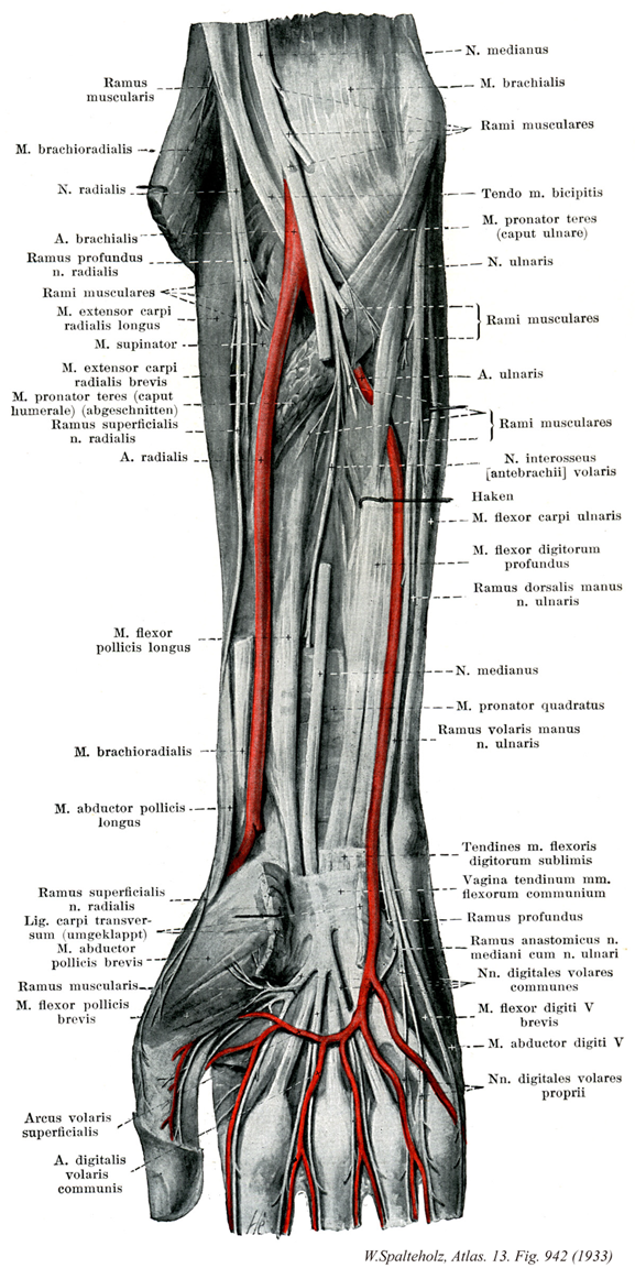
- 942_00【Forearm前腕;マエウデ Antebrachium】
→(上肢の肘と手首の間の部分。)
- 942_01【Muscular branches of radial nerve筋枝(橈骨神経の) Rami musculares (Nervus radialis)】 Motor branches to the triceps, anconeus, brachioradialis, and extensor carpi radialis longus.
→(橈骨神経の筋枝は上腕三頭筋、肘筋、腕橈骨筋および長橈側手根伸筋へいたる運動枝。)
- 942_02【Brachioradialis muscle腕橈骨筋 Musculus brachioradialis】 o: Lateral supracondylar ridge of humerus, lateral intermuscular septum, i: Radial styloid process. Flexes the forearm from the intermediate position between pronation and supination. I: Radial nerve.
→(腕橈骨筋は橈骨の外側縁に位置し、上腕骨の外側縁、外側上顆の上と外側上腕筋間中隔から起始する。腕橈骨筋は橈骨の茎状突起基底部に停止する。)
- 942_03【Radial nerve橈骨神経 Nervus radialis】 Nerve arising from the posterior cord (usually with fibers from C5-T1) spiraling in the groove for the radial nerve around the posterior side of the humerus, then continuing laterally between the brachialis and brachioradialis as well as the extensor carpi radiales. It divides at the elbow into deep and superficial branches.
→(橈骨神経は後神経束より起こる(多くはC5~Th1)。腋窩の中で腋窩動脈の背面をを通り、上腕深動脈とともに上腕骨の後面にいたる。橈骨神経溝の中で骨の近く上腕骨の外側縁を回り、外側筋間中隔を貫き、肘関節の上で屈側にでる。関節のすぐ遠位で知覚性の浅枝と太い運動性の深枝に分かれる。前者は前腕では腕橈骨筋を誘導する筋として使い、後者は回外筋に入り、螺旋状に曲がって前腕の伸側を下降する。橈側神経の腕のすべての伸筋を支配し、3つの皮枝、下外側上腕皮神経、後上腕皮神経、および後前腕皮神経は、腕の伸側の皮膚(三角筋部を除く)と手の皮膚(尺側縁、第4,5指、および第2、3指の末節を除く)を支配する。肘関節部で浅枝と深枝に分かれる。上腕三頭筋と肘筋への神経枝は、“橈骨神経管”の入り口の前と入り口部において、すでに橈骨神経から分岐している。)
- 942_04【Radial artery橈骨動脈 Arteria radialis】 Continuation of the brachial artery from its bifurcation, or first branch (embryological). It runs on the radial side between the brachioradialis and flexor carpi radialis over the pronator teres to the wrist (pulse palpation site). From there it ascends behind the trapezoid to the lateral aspect of the dorsum of hand where it courses to palmar. It extends between the two heads of the first dorsal interossei to reach to the deep palmar arch.
→(橈骨頚の高さで上腕動脈より分岐する動脈。前腕の橈側(外側)に沿って手根部まで下行し、次いで手根の外側(母指側)をまわって背側に達し、母指と第2指の中手骨間隙にある第1背側骨間筋の両頭の間を通って手掌に出る。前腕近位部では腕橈骨筋におおわれて走るが、遠位部では表在性になり、皮膚と前腕筋膜のおおわれるのみとなる。ことに手根部の近くではきわめて浅く、またその深層は橈骨下端に接するため、この部で脈拍を触れるのが容易である。この部は橈側手根筋のすぐ外側にあたる。手根から手背にいたる経路は、長母指外転筋、短母指伸筋、そして長母指伸筋のしたで、いわゆるtabatiereを通る。)
- 942_05【Deep branch of radial nerve深枝(橈骨神経の) Ramus profundus nervus radialis】 Deep branch supplying the extensor muscles of the forearm. It penetrates the supinator muscle, and supplies it and all extensor muscles (except the extensor carpi radialis longus) as well as the abductor pollicis longus.
→(橈骨神経の深枝は橈骨神経共通幹が腋窩で浅枝と深枝とに分かれる。深枝は回外筋を貫いてこの筋および前腕の心筋に分布する。最終的には後骨間神経となり骨間膜を進んで前腕1/3に達する。)
- 942_06【Extensor carpi radialis longus muscle長橈側手根伸筋 Musculus extensor carpi radialis longus】 o: Lateral supracondylar ridge of humerus, lateral intermuscular septum, i: Base of second metacarpal. Flexes the elbow joint. Dorsiflexion and radial abduction of the wrist joint. I: Radial nerve.
→(長橈側手根伸筋は腕橈骨筋起始部の下方の上腕骨外側縁と外側上腕筋間中隔から起こり、第2中手骨底に停止する。筋服の上縁は腕橈骨筋で被われているが、筋は外側上顆で外側に曲がり、短橈側手根伸筋の近位部を被っている。)
- 942_07【Supinator muscle回外筋 Musculus supinator】 o: Lateral epicondyle of humerus, radial collateral ligaments, anular ligament of radius, and supinator crest of ulna, i: Anterior surface of radius. Supination. I: Radial nerve.
→(回外筋は上腕骨の外側上顆、肘関節の外側腱索と尺骨の回外筋稜から起こる。薄い筋板は橈骨の外側を回り、後面から、橈骨粗面と円回内筋の停止部との間の橈骨の前面に付く。橈骨神経深枝はこの筋の近位辺縁近くで筋に入り、ラセン状の“回外筋管”を通って筋の遠位に抜ける。)
- 942_08【Extensor carpi radialis brevis muscle短橈側手根伸筋 Musculus extensor carpi radialis brevis】 o: Lateral epicondyle of humerus, anular ligament of radius, i: Base of third metacarpal. Dorsiflexion of the hand.
→(短橈側手根伸筋は外側上顆、橈骨輪状靱帯および総指伸筋と本筋とを分けている結合組織中隔から起こる。短い腱は第3中手骨の茎状突起につく。長および短橈側手根伸筋の腱は橈骨の外側縁を下方に進み、長母指外転筋と短母指外転筋の筋腹と交叉し、伸筋支帯のしたの第2腱区画を通る。)
- 942_09【Pronator teres muscle円回内筋 Musculus pronator teres】 Muscle that attaches on the pronator tuberosity of the radius. It flexes the elbow joint, acting as a pronator. I: Median nerve.
→(円回内筋の上腕頭は、前腕の浅層の屈筋群とともに上腕骨の内側上顆と内側上腕筋間中隔から起こる。発達の弱い尺骨頭は鈎状突起と尺骨粗面の間の尺骨内側面から起こる。円回内筋は尺骨と橈骨の上を斜走し、橈骨の前縁を回って、回外筋の停止部の下方の橈骨の前縁を回って、回外筋の停止部の下方の橈骨外側面につく。円回内筋は腕橈骨筋とともに肘窩の遠位側の境界となる。正中神経は円回内筋の上腕頭と尺骨頭の間を通る。)
- 942_10【Humeral head of pronator teres muscles上腕頭;上腕骨頭(円回内筋の) Caput humerale (Musculus pronatoris teretis)】 o: Medial epicondyle of humerus, medial intermuscular septum.
→(円回内筋の上腕頭は内側上顆から起こる部分。)
- 942_11【Superficial branch of radial nerve浅枝(橈骨神経の) Ramus superficialis nervus radialis】 Superficial cutaneous branch that runs along the brachioradialis with the radial artery, and then crosses under its accompanying muscle to reach the dorsum of hand and the fingers.
→(橈骨神経の浅枝は深枝とともに橈骨神経の最終枝で、腕橈骨筋に沿って橈骨動脈とともに走り、母指・示指・中指・第四指外側半の背面近位部の皮膚、および手背近位部の皮膚に分布する。)
- 942_12【Flexor pollicis longus muscle長母指屈筋(手の) Musculus flexor pollicis longus】 o: Anterior surface of the radius, distal to the radial tuberosity. i: Base of distal phalanx of thumb. Flexes hand and phalanges of thumb. Radial abduction. I: Median nerve.
→(長母指屈筋は系統発生学的には独立した深指屈筋の一部である。その起始部は橈骨粗面から方形回内筋の上縁までの橈骨の前面、および骨間膜にまで広がっている。その腱は手根間を通り、短母指屈筋の2頭の間に入り込み、母指の末節骨底に付く。)
- 942_13【Abductor pollicis longus muscle長母指外転筋 Musculus abductor pollicis longus】 o: Posterior surface of radius, ulna, and interosseous membrane. i: Base of first metacarpal. Radial abduction and dorsiflexion of the metacarpophalangeal joint of the thumb. I: Radial nerve.
→(長母指外転筋と短母指伸筋は1つの発生的、機能的単位を形成している:筋腹はしばしば形態学的に1つとなる。これらの筋は橈骨の背側面(2/4と3/4の間)と前腕骨間膜から起こる。羽状筋である長母指外転筋は回外筋の起始部下方からも起こり、また尺骨にも起始部をもつ。長母指外転筋は第1中手骨底に、短母指伸筋は母指基節骨底に付く。短母指伸筋の腱線維は長母指伸筋の腱終末に融合し、弱い指背腱膜を形成する。2つの筋は長および短橈側手根伸筋の腱と急角度で交叉し、これらの腱は橈骨の遠位端の背側の橈骨溝に進み、第1腱区画(手首の屈曲軸の掌側)を通過する。)
- 942_14【Transverse carpal ligament (flexor retinaculum)横手根靱帯(屈筋支帯) Ligamentum carpi transversum】
→()
- 942_15【Abductor pollicis brevis muscle短母指外転筋 Musculus abductor pollicis brevis】 o: Scaphoid, flexor retinaculum. i: Thumb, proximal phalanx, radial sesamoid bone, dorsal aponeurosis. Abducts and flexes the thumb. I: Median nerve.
→(短母指外転筋は表層にあり、母指対立筋をほぼ完全におおっている。筋は屈筋支帯と舟状骨結節から起こり、母指基節関節包に埋め込まれている橈側種子骨、基節骨底の外側縁ならびに指背腱膜に付く。正中神経(C8とTh1)から支配を受ける。)
- 942_16【Flexor pollicis brevis muscle短母指屈筋(手の) Musculus flexor pollicis brevis】 Same attachment site as abductor pollicis brevis. Flexion, abduction, adduction, and opposition at the metacarpophalangeal joint of the thumb.
→(短母指屈筋は短母指外転筋の内側に位置する。その起始部は長母指屈筋の腱で浅頭と深頭に分けられ、浅頭は屈筋支帯に、深頭は橈側遠位手根骨から起こる(2頭は発生的に異なる起源である)。短母指屈筋の終末腱は短母指外転筋の腱と融合し、短母指外転筋と並んで橈側種子骨、母指の基節骨ならびに指背腱膜に付く。浅頭は正中神経に、深頭は尺骨神経から支配を受ける(C8とTh1)。)
- 942_17【Superficial palmar arch; Superficial palmar arterial arch浅掌動脈弓;浅掌弓 Arcus palmaris superficialis】 It lies on the long flexor tendons. Its main tributary is from the ulnar artery: it anastomoses with the radial artery.
→(浅掌動脈弓は長指屈筋腱の表層で外反した母指の先端から手掌に引いた線のあたりにある手の動脈弓。尺骨動脈の本幹のつづきとみなされ、これに橈側から橈骨動脈の浅掌枝が加わる。ときに正中動脈がこれに加わることがある。手掌腱膜の直下で屈筋支帯や浅指屈筋の表層にあり、指に向かって凸の動脈弓で、総掌側枝動脈と固有掌側指動脈を出す。)
- 942_18【Common palmar digital arteries総掌側指動脈 Arteriae digitales palmares communes】 They pass toward the fingers as three or four arteries from convex palmar arch.
→(総掌側指動脈は通常3本で、浅掌動脈弓より出て浅指屈筋と虫様筋の表層を前進し、それぞれ相当する掌側中手動脈を合したのち、指の基部で二分して第2~5指の対向縁に分布する固有掌側指動脈となる。)
- 942_19【Median nerve正中神経 Nervus medianus】 Nerve formed by the medial and lateral cords.
→(内側および外側神経束よりなる(C6-T1)。(Feneis))
- 942_20Casserio's muscle【Brachialis muscle上腕筋 Musculus brachialis】 o: Anterior surface of the humerus below the deltoid tuberosity. i: Tuberosity of ulna. Flexes the elbow joint. I: Musculocutaneous nerve.
→(上腕筋は上腕骨前面の三角筋粗面(均一な三角筋-上腕筋系の骨付着部と考えらえている)より遠位部で起こり、尺骨粗面に停止する。[臨床]上腕筋は、上腕骨上に直に接しているため、筋を上腕骨に圧迫するような外力が加わるとか、上腕骨の(顆上)過伸展骨折(伸展骨折)の際に、骨折端によって穿通され、容易に損傷される。損傷した筋組織の部位に生じる結合組織の瘢痕は、収縮し、上腕筋の短縮が起こることがある。このような場合、腕は肘関節を伸展することが不可能になる。)
- 942_21【Muscular branches of median nerve筋枝(正中神経の) Rami musculares (Nervus medianus)】 Branches supplying the pronator teres, flexor carpi radialis, palmaris Iongus, and flexor digitorum superficialis.
→(正中神経の筋枝は大部分肘関節のあたりで出て円回内筋および前腕の屈筋中、尺側手根屈筋と深枝屈筋の尺側部以外の筋に分布する。)
- 942_22【Biceps brachii tendon; Tendon of biceps brachii muscle上腕二頭筋腱 Tendo musculus bicipitis brachii】
→()
- 942_22a【Biceps brachii muscle上腕二頭筋 Musculus biceps brachii】 Two-headed muscle that attaches on the radial tuberosity and extends with the aponeurosis brachii toward the ulna to blend into the antebrachial fascia. It acts in elbow joint flexion and forearm supination. I: Musculocutaneous nerve.
→(上腕二頭筋は、長頭が関節上結節に起始し、短頭は烏口突起に起始する。二頭筋の長頭(長いのは腱の部分のみ)は上腕骨頭を越え、結節間滑膜鞘に包まれて、結節間溝へ入る。共通の筋腹の終止腱は、肘窩の奥で、橈側粗面に停止する。腱性の帯である上腕二頭筋腱膜は終止腱から分かれ、前腕筋膜に放散している。肘関節を屈曲すると、上腕二頭筋は特に突出する。なぜならば、この筋は関節から離れて、上腕筋によって前に押し出されるからである。機能として肘関節に作用して前腕をまげる。上腕前面に力こぶをつくる。筋腹の内外両側の溝をそれぞれ内側二頭筋溝および外側二頭筋溝という。前者を尺側皮静脈、後者を橈側皮静脈が走る。長頭の件は滑膜に包まれながら肩関節腔を貫く。また上腕骨の結節間溝を通るところでは、結節間滑液鞘に包まれる。)
- 942_23【Ulnar head of pronator teres muscle尺骨頭;尺側頭(円回内筋の) Caput ulnare (Musculus pronatoris teretis)】 o: Coronoid process.
→(円回内筋の尺骨頭は鈎状突起から起こる部分。)
- 942_24【Ulnar nerve尺骨神経 Nervus ulnaris】 Nerve arising from the medial cord that initially travels in the medial bicipital groove, pierces the medial intermuscular septum of the arm, and, after traversing the groove for the ulnar nerve, penetrates the flexor carpi ulnaris.
→(腕神経叢の枝であり、上腕の内側後部を下り肘頭の内(尺)側に達してから前面に近づき、尺側手根筋と深指屈筋(尺骨半)への筋枝を出したのち前腕を下りながら途中で手背尺側半の皮膚に分布する背側指神経および手掌尺側半の皮膚に分布する一つの皮枝を出す。手掌部に達した尺骨神経の本幹は短掌筋、小指外転筋、短小指屈筋、小指対立筋、尺側の虫様筋、短母指屈筋の深頭、母指内転筋、および骨間筋への筋枝を出すほか、総掌側指神経とその末梢側のつづきである固有掌側指神経になり小指および薬指の表面をおおう皮膚に分布する。)
- 942_25【Muscular branch of median artery筋枝(尺骨神経の) Ramus muscularis (Nervus medianus)】
→()
- 942_26【Ulnar artery尺骨動脈 Arteria ulnaris】 Branch arising from the bifurcation of the brachial artery. It runs beneath the pronator teres toward the ulna, then accompanies the flexor carpi ulnaris on the flexor digitorum profundus to the wrist; from there it runs radially from the pisiform with the ulnar nerve to the palm, where it forms the superficial palmar arch.
→(尺骨動脈は上腕動脈の2終枝の一つ。肘関節の少し遠位で分岐し、このあたりでは橈骨動脈より太い。前腕の近位1/2における走行は深在性で、円回内筋・橈側手根屈筋・長掌筋・浅指屈筋の深層を斜め尺側に向かって下行し、前腕のほぼ中央でその尺側縁の浅層に出る。これからあと、尺骨神経と尺骨静脈(2条)に伴行して深指屈筋の表層で、これと尺側手根屈筋および浅指屈筋との間を手根にむけて下行する。次いで豆状骨の橈側を通って手掌に入り、屈筋支帯の表層で浅掌動脈弓をつくっておわる。)
- 942_27【Anterior interosseous nerve; Volar interosseus of median nerve前骨間神経;前前腕骨間神経;掌側前腕骨間神経 Nervus interosseus antebrachii anterior; Nervus interosseus antebrachii volaris】 Nerve arising at the elbow from the posterior side of the median nerve and running on the interosseous membrane. Distribution area: wrist joint, intercarpal joints, flexor pollicis Iongus, flexor digitorum profundus (radial portion), and pronator quadratus.
→(前前腕骨間神経は円回内筋の高さで分かれて同名の動脈とともに前腕骨間膜の掌側を下り、長母指屈筋、深指屈筋橈側頭、方形回内筋に分布するほか、橈骨と尺骨の骨間膜に枝を出す。)
- 942_28【Flexor carpi ulnaris muscle尺側手根屈筋 Musculus flexor carpi ulnaris】 Muscle that attaches on the pisiform, via the pisohamate ligament on the hamate, and via the pisometacarpal ligament on the fifth metacarpal. Flexes and abducts the hand toward the ulna.
→(尺側手根屈筋は屈筋群の尺側外縁を形成している。上腕頭は上腕骨の内側上顆と内側上腕筋間中隔から起こる。尺骨頭は肘頭、尺骨後縁の近位2/3と前腕筋膜から起こる。2頭は腱性の帯で結合しており、この下を尺骨神経が前腕の屈側に向かう。筋の腱は(尺骨神経、尺骨動・静脈の)尺側前腕路の内側の境をなす。この腱は手根管を通らず、豆状骨に停止した後、豆鈎靱帯と豆中手靱帯で有鈎骨と第5中手骨に至る。筋の腱中に種子骨として豆状骨の停止部があるため、回転軸からの距離が増し、尺側手根屈筋は手掌を曲げるのに有利なトルク(回転力)を得られる。)
- 942_29【Flexor digitorum profundus muscle深指屈筋 Musculus flexor digitorum profundus】 o: Upper twothirds of anterior surface of ulna, i: Bases of the distal phalanges of the second through fifth fingers. Flexes wrist and interphalangeal joints. I: Median and ulnar nerves.
→(深指屈筋は尺骨(近位2/3)の前面と内側面、骨間膜、および前腕筋膜の広い範囲から起きる。その大きな筋腹は尺骨の前内側面を包み、浅指屈筋の滑走面となっている。4つの腱は手根管で互いに並び、浅指屈筋の腱を貫通し、第2~第5指の末節骨底に達する。)
- 942_30【Dorsal branch of ulnar nerve; Branch of ulnar nerve to dorsum of hand手背枝(尺骨神経の);尺骨神経手背枝 Ramus dorsalis nervus ulnaris】 Cutaneous branch passing between the distal and middle one-third of the forearm deep to the flexor carpi ulnaris to the dorsum of hand.
→(尺骨神経手背枝は手首の近位で尺骨神経から分枝し、手背内側半、小指近位部、第4指内側面に分布する。)
- 942_31【Pronator quadratus muscle方形回内筋 Musculus pronator quadratus】 o: Distal one-fourth of the anterior surface of ulna, i: Distal onefourth of the anterior surface of radius. Forearm pronation. 1: Median nerve.
→(方形回内筋は尺骨と橈骨の下1/4の前面で両者を結合しているので、尺骨の起始部は前縁を少し回って内側面に達している。)
- 942_32【Palmar branch of ulnar nerve掌枝;掌側枝;手掌枝(尺骨神経の);尺骨神経掌枝 Ramus palmaris nervus ulnaris; Ramus volaris manus; Ramus superficialis (Nervus ulnaris)】 Nerve arising in the distal one-third of the forearm, piercing the deep fascia, and supplying the skin of the ulnar side of the palm.
→(尺骨神経の掌枝は前腕遠位部で分枝し、手掌の動脈に伴行して手に進入し、小枝の皮膚・第4指内側半・その近辺の手掌部の皮膚に分布する。)
- 942_33【Flexor digitorum superficialis tendons; Tendons of flexor digitorum superficial muscle浅指屈筋腱 Tendines musculi flexor digitorum superficialis】
→()
- 942_33a【Flexor digitorum superficialis muscle浅指屈筋 Musculus flexor digitorum superficialis; Musculus flexor digitorum sublimis】 Muscle that attaches on the middle phalanx of the second through fifth fingers. Flexes the wrist and proximal interphalangeal joints. 1: Median nerve.
→(浅指屈筋は2頭筋で、前述した屈筋よりやや深部に位置するので、これらの筋は浅指屈筋を部分的におおっている。上腕尺骨頭は上腕骨の内側上顆の浅屈筋群の起始部と鈎状突起から起きる。橈骨頭は円回内筋の停止部下方の橈骨の前面の細長い部分から起こる。その終末腱は第2~第4中節骨に付く。浅指屈筋は屈筋支帯近くで広がり、2個の浅筋膜(中指と薬指へ)と2個の伸筋腹(示指と小指へ)に不完全に分かれる。それらの腱は手根管を通り、基節骨の上で2分し、その間を深指屈筋の腱が末節腱に通り抜ける(そこで、浅指屈筋を“被貫通屈筋”、深指屈筋を“貫通屈筋”と呼ぶ。腱は第2~第5中節骨の両側縁に付く。浅指屈筋の深層腱線維は最初に(近位部で)分かれる。骨に直背側部分を形成する。この管(鞘)の壁側は長軸方向に走る浅指屈筋の分岐した腱で作られている。不完全で短い手掌部分は腱の近位浅層の不分岐部でつくられる。)
- 942_34【Common flexor sheath of hand: Common synovial sheath for flexor muscles指屈筋の総腱鞘;総指屈筋腱鞘(手の);尺側滑液包 Vagina communis tendinum musculorum flexorum manus; Vagina tendinum musculorum flexorum digitorum communium】 Common tendon sheath for the two long flexor tendons of the fingers.
→(尺側滑液包ulnar bursaともよばれる。手の指屈筋の総腱鞘は浅・深指屈筋また長母指屈筋の腱を包み、中手骨頭から末節骨骨底までの範囲に広がる。成人では長母指屈筋の腱鞘は母指の腱鞘と交通枝し(76%)、指屈筋の総腱鞘は時として小指の腱鞘と連なる(12%)。ステッドマンでは尺骨滑液包となっている。)
- 942_35【Deep branch of ulnar nerve深枝;深掌枝(尺骨神経の) Ramus profundus nervus ulnaris】 Deep branch of the ulnar nerve. It curves around the hamulus, supplies the hypothenar muscles, the interossei, the two ulnar lumbricals, adductor pollicis, and the deep head of flexor pollicis brevis.
→(尺骨神経の深枝は尺骨動脈の深掌枝や深掌動脈弓に伴行して手首の関節、第3、第4虫様筋、掌側・背側骨間筋、母趾内転筋、短母趾屈筋の深頭に分布する。)
- 942_36【Communicating branch with ulnar nerve; Communicating branch of anterior interosseus nerve with ulnar nerve尺骨神経との交通枝(正中神経と) Ramus communicans cum nervus ulnari; Ramus communicans nervus interossei antebrachii anterior cum nervus ulnari】
→(正中神経と尺骨神経との交通枝)
- 942_37【Common palmar digital nerves of median nerve総掌側指神経(正中神経の) Nervi digitales palmares communes】 Nerves traveling toward the spaces between the first and fourth fingers and then dividing.
→(正中神経の総掌側指神経は3本あって、さらに母指と示指との間に行くものは母指橈側、尺側および示指橈側の3本、示指と中指との間にいくものは示指尺側と中指橈側の2本、中指と薬指との間にいくものは中指尺側と薬指橈側の2本の固有掌側指神経に分かれ、全体として薬指を境として橈側の手掌および指縁の皮膚に分布する。このほか、短母指外転筋、短母指屈筋の一部、母指対立筋および橈側虫様筋などにも枝を与える。)
- 942_38【Flexor digiti minimi brevis muscle of hand短小指屈筋(手の) Musculus flexor digiti minimi brevis manus】 o: Hook of hamate, flexor retinaculum. i: Palmar surface of the base of the proximal phalanx. Flexion at the metacarpophalangeal joint. I: Ulnar nerve.
→(短小指屈筋は通常明確な境界がなく、橈側に付いており、屈筋支帯と有鈎骨鈎から起こる。停止部で小指外転筋と融合する。この筋は小指の中手指節関節の屈筋である。尺骨神経の深枝(C8とTh1)から支配を受ける。)
- 942_39【Abductor digiti minimi muscle of hand小指外転筋(手の) Musculus abductor digiti minimi manus; Musculus abductor digiti quinti】 o:Calcaneus and plantar aponeurosis. i: Lateral surface of proximal phalanx of fifth toe. Plantar flexion and abduction of fifth toe. Forms the lateral margin of foot. I: Lateral plantar nerve.
→(小指外転筋は豆状骨、豆鈎靱帯および屈筋支帯から起きる。筋は小指の基節骨底の尺側縁に付き、指背腱膜へも放散する。この筋の一部は小指の伸筋腱膜へも入り込む。機能的にはこの筋は純粋な外転筋である尺骨神経の深枝(C8とTh1)から支配を受ける。)
- 942_40【Proper palmar digital nerves固有掌側指神経 Nervi digitales palmares proprii】 Terminal branches of the common palmar digital nerves. They supply the skin of the palmar side of the radial 3'A fingers and the skin of the dorsal side of the 2'A radial distal phalanges.
→(固有掌側指神経は総掌側指神経の終糸。掌側では橈側3と1/2指の皮膚を、背側では橈側2と1/2指の皮膚を支配する。)