Spalteholz HANDATLAS DER ANATOMIE DES MENSCHEN VON WERNER SPALTEHOLZ
メニューは解剖学(TA)にリンクしてあります。図の番号をクリックすると下記の説明へ、右側の用語をクリックすると解剖学(TA)にジャンプします。
077


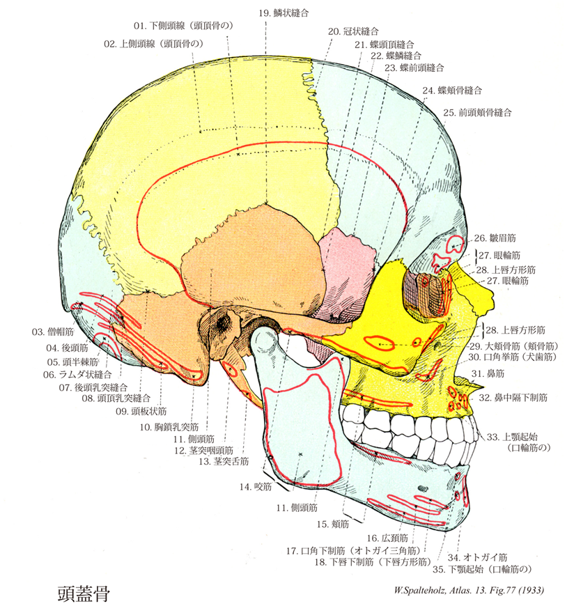
- 077_00【Bones of cranium; Skull bones頭蓋骨;ズガイコツ Ossa cranii】
→(頭蓋は15種23個の骨、すなわち10種16個の頭蓋骨および5種7個の顔面骨とにより形成されている。頭蓋骨は中枢神経系および感覚器に接する部分を形成する骨格で後頭骨(1個)、蝶形骨(1個)、側頭骨(1対2個)、頭頂骨(1対2個)、前頭骨(1個)、篩骨(1個)、下鼻甲介(1対2個)、涙骨(1対2個)、鼻骨(1対2個)、及び鋤骨(1個)である。頭蓋を構成する骨の分類には諸学者による見解の相違があり、後頭骨、蝶形骨、側頭骨、頭頂骨、前頭骨の5種7個を脳頭蓋とし、他の10種16個を顔面骨とする意見もある。)
- 077_01【Inferior temporal line of parietal bone下側頭線;側頭線(頭頂骨の) Linea temporalis inferior; Linea temporalis (Ossis parietalis)】 Curved line giving origin to the temporal muscle.
→(頭頂結節の下方に上下2本の弓状の線が認められるが、下の線を下側頭線といい側頭筋の着くところである。)
- 077_02【Superior temporal line of parietal bone上側頭線;筋膜側頭線(頭頂骨の) Linea temporalis superior; Linea temporalis fascialis (Ossis parietalis)】 Curved line for attachment of the temporal fascia. It forms the superior border of the temporal plane.
→(頭頂骨の上側頭線は頭頂骨上の2本の曲線のうちの上方の線。側頭筋膜が付着する。)
- 077_03【Trapezius muscle僧帽筋 Musculus trapezius】 Muscle that consists of three parts that act together to position the scapula and clavicle, draw both toward the vertebral column, and brace the shoulder girdle. I: Accessory nerve; brachial plexus C2-C4.
→(背部第1層にみられる扁平な菱形の筋で背部上半部を占める。僧帽筋は上肢の運動の時に肩甲骨を動かす重要な筋である。とくに上腕の外転のときに、肩甲骨を後内側に引くと同時に下角を外側に回し、関節窩が上外側を向くようにする。僧帽筋は下行部、横走部、上行部に分けられる。[臨床]僧帽筋の完全麻痺(副神経と上部腕神経の同時の傷害)の場合、肩は健側よりも深く位置するようになる項肩線は弓状を呈さず、乱れる。肩甲骨は正中線より、はるかに離され、関節窩は前下方を向く。肩は(肩甲挙筋の)弱いエネルギーにより持ち上げることが出来るにすぎず、わずかに(菱形筋により)後方にもたらされるにすぎない。腕の外側への挙上は大きく減少する。腕は通常水平面まで外転され得ない。腕の前方への挙上は(前鋸筋による肩甲骨の回転により)ほとんど制限さされないが、矢状面での挙上は強く妨げられる。副神経のみが傷害された場合、僧帽筋の下行部の機能は(上頚神経の付随的支配により)種々の程度に保存される。肩甲骨の位置の変化はそれほど著明ではない。しかし、腕を横または後へ挙上することは、ちょうどその程度に応じて制限される。)
- 077_04【Occipital belly of occipitofrontalis muscle後腹(後頭前頭筋の);後頭筋 Venter occipitalis (Musculus occipitofrontalis); Musculus occipitalis】 Portion of the occipitofrontalis muscle that passes from the highest nuchal line into the epicranial aponeurosis. Antagonist of the frontal belly.
→(人類では帽状腱膜の移動性は少ないので前頭筋は主に眉を上げるためだけである。また後頭筋はその一般に退化的でその働きも弱い。)
- 077_05【Semispinalis capitis muscle頭半棘筋;横突後頭筋 Musculus semispinalis capitis; Musculus transversooccipitalis】 o: Transverse processes of T6-C3. i: Inferior to the superior nuchal line. I: Posterior rami of spinal nerves of C1-C5.
→(頭半棘筋の起始は上位6個の胸椎と第7頚椎の横突起、第4~6頚椎関節突起。停止は後頭骨の上下項線間の項平面。機能として脊柱の伸展、側方屈曲。頭、肋骨、骨盤の伸展。神経支配は第1~6頚椎。動脈は後肋間動脈の筋枝、後頭動脈の下行枝、肋頚動脈の深頚枝から受ける。頭半棘筋は頚部の板状筋に完全におおわれ頚最長筋と頭最長筋の内側にある。固有背筋の外側筋群を形成する筋原基から大部分形成される。それ故に、この筋は脊髄神経後枝の内側枝ばかりでなく、外側枝の支配も受ける。この筋は複合羽状型であり、不完全に狭い内側筋束と、線維質の外側筋束に分化し、両者とも中間腱を所有する(内側筋束はときどき2つ)。)
- 077_06【Lambdoid sutureラムダ状縫合;ラムダ縫合;人字縫合 Sutura lambdoidea】 Suture that unites the occipital bone with the two parietal bones.
→(後頭骨と左右頭頂骨の間の縫合。ギリシャ文字のラムダ(λ)の形から命名された。矢状縫合がぶつかる点をラムダlambdaといい、胎生期に小泉門があった場所である。(イラスト解剖学))
- 077_07【Occipitomastoid suture後頭乳突縫合 Sutura occipitomastoidea】 Continuation of the lambdoid suture to the cranial base.
→(後頭乳突縫合は後頭骨と側頭骨乳突部の間。)
- 077_08【Parietomastoid suture頭頂乳突縫合 Sutura parietomastoidea】 Posterior suture that unites the parietal bone and the mastoid process of the temporal bone.
→(頭頂乳突縫合は頭頂骨と乳様突起の間で、後方にある縫合。)
- 077_09【Splenius capitis muscle頭板状筋 Musculus splenius capitis】 Portion of the splenius extending to the head. o:Spinous processes of T3-C4. i: Lateral part of the superior nuchal line and mastoid process.
→(頭板状筋の起始は頚靱帯の下半分、第7頚椎と上位3~4個の胸椎棘突起。停止は側頭骨の乳様突起と上項線の外側部。機能として共同で頭と頚の伸展と側方屈曲をしかつ頭を少し回旋する。神経支配は中および下頚神経の後枝の外側枝。動脈は後頭動脈下行枝の筋枝、頚横動脈の浅枝から受ける。)
- 077_10【Sternocleidomastoid muscle胸鎖乳突筋 Musculus sternocleidomastoideus】 o: Two-headed muscle arising from the sternum and clavicle, i: Mastoid process; superior nuchal line. Rotates the face to the contralateral side and bends the head to the ipsilateral side. Bilateral contraction elevates the face. I: Accessory nerve, cervical plexus (C1-C2).
→(胸鎖乳突筋は側頚部にある強大な斜めに縦走する浅層の筋。胸骨柄前面と鎖骨の胸骨端から2頭をもっておこり、両頭は合して強い筋腹をつくって後上方に走り、乳様突起および後頭骨の上項線につく。作用は複雑で、両側が同時に働くとオトガイを上げて後頭部を片側が働けば頭を対側にまわすが、その浅オトガイが対側に向かって上り、頭は逆に同側に傾く。支配神経は副神経外枝と頚神経叢筋枝(C2, C3)であり、したがって僧帽筋と同系の筋である。また、第6咽頭弓に発生する鰓弓筋で、鎖骨上窩を囲む2頭(胸骨頭と鎖骨頭)をもって始まる。胸骨頭は胸骨柄の上縁から、鎖骨頭は鎖骨の胸骨端から起こる。筋膜は頚筋膜浅葉に鞘状に包まれており、斜め上方に向かって幾分螺旋状に回転しながら頚部外側面を横切り、よく発達した腱となって乳様突起と上項線に停止する。筋の表面は、起始部で腹側に、停止部で外側に向く。参考:副神経外枝の僧帽筋枝は、外枝がこの筋に入る前に分かれることと、筋内で分かれて再び外に現れることがある。胸鎖乳突筋はドイツ語ではKopfnicker(頭をこっくりとうなずかせる筋)と呼ばれるが、これは作用の点からは正しくない。この筋が片側だけ収縮すると、頭はその側へ傾き反対側を振り向いて、あたかも「首をかしげる」状態になる。また両側の物が同時に収縮すると、頭を胴体にめり込ませるように働くのえある。Musculus sternocleidomastoideusというラテン名はあまりにも長たらしいので、米英では多少簡略化してsternomastoid muscleともよぶ。片側の胸鎖乳突筋が先天的に短い場合、または出産時の外傷などによって瘢痕化して短縮すると、この筋の作用を考えればすぐわかるように、頭は病側へ傾くと共に健側にねじれたままの状態になるこれを斜径torticolis, wryneck(性格には筋性斜径)といい、かなり頻度の高いものである。略語(SCM))
- 077_11【Temporalis muscle; Temporal muscle側頭筋 Musculus temporalis】 o:Inferior temporal line, infratemporal crest, temporal fascia [temporal fossa], i: Its fibers converge at the coronoid process and continue inferiorly to the level of the occlusal plane and near the pterygomandibular raphe. It raises and retracts the mandible, and fixes the pharynx during swallowing. I: Mandibular nerve.
→(側頭筋は扇状になって側頭窩および側頭筋膜から起始する。筋線維は収斂して、頑丈な腱をもって下顎骨筋突起に付着する。付着腱は上方へ伸びて筋肉内にまで達する。側頭筋は頬骨弓下を通過して、その付着部に達する。その筋線維がかなりの長さであるので、筋はかなりの収縮可能性を有するし、かつ純粋な“咬むための筋”である。歯をかみ合わせると、側頭筋の収縮を耳介の上方で触れることができる。側頭部をコメカミというのは、コメをカムときに動くからである。)
- 077_12【Stylopharyngeus muscle茎突咽頭筋 Musculus stylopharyngeus】 o: Styioid process, i: It extends medially between the superior and middle constrictor muscles and reaches the wall of the pharynx, thyroid cartilage, and epiglottis. I: Glossopharyngeal nerve.
→(茎突咽頭筋は茎状突起の頭蓋底近くから起こり、上および中咽頭収縮筋の間を通って筋の内面に至り、口蓋咽頭筋の線維束とともに甲状軟骨に停止する。一部は咽頭蓋の粘膜下に終わる。)
- 077_13【Styloglossus muscle茎突舌筋 Musculus styloglossus】 o: Styloid process, i: Radiates from posterosuperior into the lateral part of the tongue and merges with the hyoglossus. It draws the tongue backward and upward. I: Hypoglossal nerve.
→(茎突舌筋は外舌筋の1つ。茎状突起(および茎突下顎靱帯)から放射して口蓋咽頭弓のレベルで舌に至る。茎突舌筋の線維の主部は舌縁で舌尖に向かって走り(筋の縦索)、個々の線維束は内側へ曲がり、横舌筋(内舌筋)の線維に付着する。)
- 077_14【Masseter muscle咬筋 Musculus masseter】 The most prominent masticatory muscle. It acts to close the mouth and, together with the temporal and medial pterygoid muscles, determines the level of masticatory force. It consists of the following two parts.
→(咬筋は最も浅層にある咀嚼筋である。浅部と深部の2部からなり、浅部は強い腱で頬骨弓の前3分の2の下縁と内面から起こり後下方に向かい、深部は頬骨弓の後3分の2の下縁に垂直に下り向かい下顎枝および下顎角の外面に付く。作用は下顎骨を引き上げて歯をかみ合わせる。咬筋は強大な筋で、歯をかみ合わせると、体表からみることができ、かつ触れることができる。)
- 077_15【Buccinator muscle頬筋 Musculus buccinator】 Muscle arising from the pterygomandibular raphe and adjacent areas of the maxilla and mandible to the height of the first molar teeth, and inserting into the orbicularis oris at the angle of the mouth. It forms the cheek, moves food from the oral vestibule between the dental arcades during mastication, prevents entrapment of the mucous membrane of the mouth, and is active during laughing and crying. I: Facial nerve.
→(頬筋は頬の筋性土台に該当し、口角部で口輪筋に付着する。頬筋は弓状に上顎骨歯槽突起の臼歯部、かつ下顎骨歯槽突起から起こる。上および下顎間は腱性の翼突下顎放線によって橋渡しされ、この放線もまた頬筋の起始である。上咽頭収縮筋の一部がこの放線の後部で起始する。口角付近で、線維索が交叉するので、頬の上方に位置する部分は下唇に広範囲わたって達することもあるし、達しないこともある頬筋は上顎の第2大臼歯のレベルで耳下腺管によって貫通され、しかも本筋は脂肪体からこれを隔てる浅筋膜(頬咽頭筋膜)を有する唯一の顔面筋である。頬筋は上・下歯列弓および頬粘膜間に入り込んだ植物片を再度歯列弓間に押し戻し、咀嚼および植物片のかたちづくりに重要な役割を果たしている。本筋は口腔前庭を圧縮して、空気あるいは液体を口裂を通してふき出す(泡をふき出す、口笛をふく、吐き出す:“トランペット吹きの筋”)。両側の頬筋の収縮はは口角の外側部をくぼませる。参考:この筋は頬粘膜に密に結合しているが、皮膚との間は脂肪組織で隔てられている。上顎第2大臼歯の高さで耳下腺管に貫かれる。)
- 077_16【Platysma muscle広頚筋 Platysma】 Cutaneous muscle that extends (with anatomical variations) from above the mandible to the thorax. I: Facial nerve,
→(広頚筋は前頚部にある薄い膜状の皮筋で、第2咽頭弓(舌骨弓)に発生した筋原基に由来し、しかも頚部にとどまった浅顔面筋である。他の全ての浅顔面筋は頭部に完全に移り表情筋をつくる。広頚筋は極めて薄い筋性の板で、皮膚の直下にある。下顎骨縁から第2(3)肋骨の高さに広がり、さらに遠く肩峰に達する。広頚筋は頚筋膜浅葉の上に広がっていて、ここを走る外頚静脈の上を通る。上方で、筋束は下顎骨と顔面皮膚に付着する。無数の筋線維が表情筋の線維索と交錯している。下方で、広頚筋はさまざまな長さの線維束となって皮下組織に放散し、一部は真皮結合組織内に終わる。左右の筋の内側部のさまざまな長さの線維束となって皮下組織に放散し、一部は真皮結合組織内に終わる。左右の筋の内側部の線維は通常オトガイ下で互いに交錯するが、下方に向かうにつれて、互いに離れ、前頚部の三角形をした正中面は広頚筋に被われずに残る。参考:頚筋中唯一の皮筋で表情筋と同系である。皮膚とは固く、頚筋膜浅葉とはゆるくつく。顔面部は下唇下制筋とつづく。)
- 077_17【Depressor anguli oris muscle口角下制筋;オトガイ三角筋 Musculus depressor anguli oris; Musculus triangularis】 Muscle that passes from the anterior and lateral margins of the mandible to the angle of mouth. I: Facial nerve.
→(口角下制筋は下顎骨下縁から口角まで収斂しながら走る。表層に位置する口角下制筋は口角を下方へ引き、かつ鼻唇溝の上部を伸展させる。)
- 077_18【Depressor labii inferioris muscle下唇下制筋;下唇方形筋 Musculus depressor labii inferioris; Musculus quadratus labii mandibularis】 Muscle that lies deep to the depressor anguli oris and passes from the platysma superiorly and medially to the lower lip. I: Facial nerve.
→(下唇下制筋は広頸筋から斜めに下唇へ向かって、下外側から上内側へ放射する。下唇下制筋は口角下制筋によって被われ、下唇を下方および側方へ引く(不快の表情)。起始と走行:下顎骨の前面でオトガイ孔の下付近から起こり、その外側部は口角下制筋に被われる。斜めに内上方にはしる。参考:筋束の一部は広頸筋から移行し、また神経支配も共通である。そのため口角を外下方に引くとき、広頸筋も同時に収縮する。)
- 077_19【Squamous suture鱗状縫合;頭頂側頭縫合 Sutura squamosa; Sutura parietotemporalis】 Platelike suture, e.g., at the temporal bone.
→(鱗状縫合は縫合の形態上の分類に使われるほか、側頭骨鱗部と頭頂骨との縫合をさすことがある。広い骨の傾斜面が重積することによって生じる一種の縫合で、連結する両方の骨縁が片刃のようにそり落とされた形をしている。(側頭骨鱗部と頭頂骨との縫合)。)
- 077_20【Coronal suture冠状縫合 Sutura coronalis】 Suture between the frontal bone and the two parietal bones.
→(前頭鱗と頭頂骨前頭縁の間の鋸状縫合で、頭蓋冠の前部を横に冠状に走る。この縫合は蝶形骨大翼と前頭骨との間に横たわる蝶前頭縫合に合流する。)
- 077_21【Sphenoparietal suture蝶頭頂縫合;蝶骨頭頂縫合 Sutura sphenoparietalis】 Continuation of the sphenofrontal suture beginning at the coronal suture.
→(蝶頭頂縫合は蝶形骨大翼と頭頂骨の間にある縫合。。)
- 077_22【Sphenosquamosal suture蝶鱗縫合;蝶骨鱗縫合 Sutura sphenosquamosa】 Suture between the squamous part of the temporal bone and the greater wing of the sphenoid.
→(蝶鱗縫合は側頭骨鱗部と蝶形骨大翼の間の縫合。)
- 077_23【Sphenofrontal suture蝶前頭縫合;蝶骨前頭縫合 Sutura sphenofrontalis】 Suture that gradually ascends posteriorly along the lateral aspect of the cranium, joining the greater wing of the sphenoid and the frontal bone. Interior cranium: suture that joins the frontal bone and the lesser wing of the sphenoid.
→(蝶前頭縫合は頭蓋の外側面で、蝶形骨大翼と前頭骨の間を孔へゆるやかに登る縫合線。頭蓋内面では、前頭骨と蝶形骨小翼の間にある。)
- 077_24【Sphenozygomatic suture蝶頬骨縫合;蝶骨頬骨縫合 Sutura sphenozygomatica】 Suture in the lateral wall of the orbit that joins the greater wing of the sphenoid and the zygomatic bone.
→(蝶頬骨縫合は蝶形骨大翼と頬骨の間にある眼窩外側壁の縫合。)
- 077_25【Frontozygomatic suture前頭頬骨縫合;頬骨前頭縫合 Sutura frontozygomatica; Sutura zygomaticofrontalis】 Suture at the lateral margin of the orbit between the frontal and zygomatic bones.
→(前頭頬骨縫合は前頭骨と頬骨の間。)
- 077_26Koyter's muscle【Corrugator supercilii muscle皺眉筋 Musculus corrugator supercilii; Musculus corrugator glabellae】 Muscle that passes laterally from the nasal part of the frontal bone into the skin of the eyebrow. It produces vertical wrinkling of the forehead. I: Facial nerve.
→(皺眉筋は前頭骨鼻部から起こり、後頭前頭筋の前頭腹の筋線維束内側縁部を貫いて、眉毛の外側で皮膚に付着する。御筋は皮膚を鼻根の方向へ引き寄せ、前頭に垂直方向の皺を作る)
- 077_27【Orbicularis oculi muscle; Orbicular muscle of eye眼輪筋 Musculus orbicularis oculi】 Ringlike sphincter muscle around the eye consisting of various parts. It acts to close the eyelids and support the flow of tears into the lacrimal sac and nose. I: Facial nerve.
→(眼輪筋の外側部にある(外側)眼瞼縫線は筋の後面と内側部のほかは不明瞭である。外眼角と頬骨を結ぶ外側眼瞼靱帯とは別物で、両者は内側部でゆるく結合するだけであるが、この縫線と靱帯はしばしば混同されている。外側部では眼輪筋と骨との結合がないので、眼を強く閉じると筋とともに皮膚が外眼角に引かれて目じりにヒダができるのである。なお、眼裂を開くのは主に上眼挙筋(眼筋)が行ない、大きく開くときは同時に前頭筋によって眉が上がることが多い。下眼瞼は弾力によって元に戻ると思われるが、その動きは小さい。まばたきのとき涙が吸い込まれることは、涙がこぼれそうになると無意識にまばたきを行うことからも判る。その機構として眼を閉じるとき眼輪筋の涙嚢部が涙嚢の壁を引いて広げるという節が有名である。しかし、一般に涙嚢部といわれる筋(Horner筋)が涙嚢と関係のないことは以前から記載があり(Eisler)涙の排出機構にも諸説があった。長嶋1954, 1955は涙道の圧をしらべ、瞼を開くときに涙小管腔が陰圧になって涙が吸い込まれ、瞼を閉じるとき涙小管は陽圧に涙嚢は陰圧になって涙は涙嚢に送られ、さらに瞼を開くとき涙嚢は陽圧となって鼻涙管に排出されるとした。その後の研究も合わせると、Horner筋は瞼閉じるとき涙小管を屈曲・圧迫するもので、これとは別に涙嚢を包む筋膜(涙嚢間膜)から起こるごく小さい筋束(Jones 1957)があって涙嚢を広げるという機構が有力である。このポンプ作用の主力はHoener筋と涙小管にあり、そのほか毛細管現象による吸引、重力による鼻腔への流下なども助けるという。なお、いわゆるHorner筋は涙小管を囲んで眼瞼縁に向かうが、Jonesの筋束は外輪筋眼瞼部の周辺部に合するもので、後者は従来見落とされていたが、または、涙嚢部と区別だれていなかったと思われる。)
- 077_28【Quadratus labii superioris muscle上唇方形筋 Musculus quadratus labii superioris】
→()
- 077_29【Zygomaticus major muscle; Greater zygomatic muscle大頬骨筋;頬骨筋 Musculus zygomaticus major】 Muscle extending from the zygomatic bone to the angle of the mouth and upper lip. I: Facial nerve.
→(大胸骨筋は頬骨弓から内側下方へ向かって上唇および口角まで走る。大頬骨筋は口角を引き挙げ、かつ口角を外方へ引く(狭義の“笑う筋”)。)
- 077_30【Levator anguli oris muscle口角挙筋;犬歯筋 Musculus levator anguli oris; Musculus caninus】 Muscle passing from the canine fossa to the angle of the mouth. I: Facial nerve.
→(口角挙筋は犬歯上方および眼窩下孔で犬歯窩から口角まで走る。)
- 077_31【Nasalis muscle; Nasal muscle鼻筋 Musculus nasalis】 Collective term for the following two muscles of the nose originating near the canine fossa. I: Facial nerve.
→(鼻筋は貧弱に発達しているにすぎない。鼻筋は上顎犬歯の歯槽壁から起始し、鼻翼(鼻部、外鼻孔の拡張化)および鼻背軟骨部(横部、外鼻孔狭小化)まで走る。両側の本筋は腱性板によって鼻背を横方向に横断して連結する。参考:鼻孔圧迫筋ともいう。)
- 077_32【Depressor septi nasi muscle鼻中隔下制筋;鼻起始(口輪筋の) Musculus depressor septi nasi; Origo nasalis musculus orbicularis oris】 Muscle that passes from the alveolar wall over the medial incisor tooth to the cartilaginous nasal septum and draws the tip of the nose inferiorly. I: Facial nerve.
→(鼻中隔下制筋は中切歯上方の歯槽突起から起始する薄い線維索ととして出現し、軟骨性鼻中隔の先端に付着し、且つ鼻尖を下方へ引く。)
- 077_33【Maxillary origin of orbicuaris oris muscle; Incisivus labii superioris muscle上顎起始;上唇切歯筋(口輪筋の) Origo maxillaris (Musculus orbicularis oris); Musculus incisivus labii superioris】
→()
- 077_34【Mentalis muscleオトガイ筋 Musculus mentalis】 Muscle that radiates from the mandible at the height of the roots of the incisor teeth into the skin of the chin (chin cleft). I: Facial nerve.
→(オトガイ筋は口角下制筋下にあり、下顎骨の側切歯の部分で起始する。オトガイ筋はオトガイ皮膚にむかって斜内側下方へ走り、オトガイのクボミをつくり、しかも下唇を口輪筋と一緒に前方へ突出させることに関与する(子供の口をとがらす状況)。参考:旧名は頤筋。収縮するとオトガイ部に小さい凸凹ができる。)
- 077_35【Mandibular origin of orbicuaris oris muscle; Incisivus labii inferioris muscle下顎起始(口輪筋の);下唇切歯筋 Origo mandibularis (Musculus orbicularis oris); Musculus incisivus labii inferioris】
→()